. 2022 May;12(5):e743.
doi: 10.1002/ctm2.743.

Targeting DCLK1 overcomes 5-fluorouracil resistance in colorectal cancer through inhibiting CCAR1/β-catenin pathway-mediated cancer stemness

Affiliations

Targeting DCLK1 overcomes 5-fluorouracil resistance in colorectal cancer through inhibiting CCAR1/β-catenin pathway-mediated cancer stemness

Lanqing Wang et al. Clin Transl Med. 2022 May.

Abstract

Background: To date, 5-fluorouracil-based chemotherapy is very important for locally advanced or metastatic colorectal cancer (CRC). However, chemotherapy resistance results in tumor recurrence and metastasis, which is a major obstacle for treatment of CRC.

Methods: In the current research, we establish 5-fluorouracil resistant cell lines and explore the potential targets associated with 5-fluorouracil resistance in CRC. Moreover, we perform clinical specimen research, in vitro and in vivo experiments and molecular mechanism research, to reveal the biological effects and the mechanism of DCLK1 promoting 5-fluorouracil resistance, and to clarify the potential clinical value of DCLK1 as a target of 5-fluorouracil resistance in CRC.

Results: We discover that doublecortin-like kinase 1 (DCLK1), a cancer stem cell maker, is correlated with 5-fluorouracil resistance, and functionally promotes cancer stemness and 5-fluorouracil resistance in CRC. Mechanistically, we elucidate that DCLK1 interacts with cell cycle and apoptosis regulator 1 (CCAR1) through the C-terminal domain, and phosphorylates CCAR1 at the Ser343 site, which is essential for CCAR1 stabilisation. Moreover, we find that DCLK1 positively regulates β-catenin signalling via CCAR1, which is responsible for maintaining cancer stemness. Subsequently, we prove that blocking β-catenin inhibits DCLK1-mediated 5-fluorouracil resistance in CRC cells. Importantly, we demonstrate that DCLK1 inhibitor could block CCAR1/β-catenin pathway-mediated cancer stemness and consequently suppresses 5-fluorouracil resistant CRC cells in vitro and in vivo.

Conclusions: Collectively, our findings reveal that DCLK1 promotes 5-fluorouracil resistance in CRC by CCAR1/β-catenin pathway-mediated cancer stemness, and suggest that targeting DCLK1 might be a promising method to eliminate cancer stem cells for overcoming 5-fluorouracil resistance in CRC.

Keywords: CCAR1; DCLK1; cancer stemness; chemoresistence; colorectal cancer; β-catenin.


Conflict of interest statement

The authors declare that they have no conflict of interest.

Figures
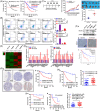
FIGURE 1
FIGURE 1
Doublecortin‐like kinase 1 (DCLK1) is correlated with 5‐fluorouracil resistance in colorectal cancer (CRC). (A) Kaplan–Meier plot of overall survival of CRC patients (n = 271) (a), and the 5‐fluorouracil sensitive patients (n = 163) and the 5‐fluorouracil resistant patients (n = 108) (b). (B) Apoptosis of 5‐fluorouracil resistant cells or parental cells. Cells were treated with 5‐fluorouracil for 48 h, then subjected to FITC‐annexin V/propidium iodide staining and analysed by FACS (n = 3). (C) The indicated cells were subcutaneously injected to nude mice, respectively (n = 6), then treated with 5‐fluorouracil twice a week for 3 weeks (a). The volumes of subcutaneous xenograft tumour were observed (b and c). (D) The genes differentially expressed in 5‐fluorouracil resistant cells or parental cells were detected by mRNA sequencing. (E) The interesting genes were verified by real‐time quantitative PCR (RT‐qPCR) (n = 3). (F) DCLK1 expression was analysed by Western blotting in the indicated cells and by immunohistochemistry in subcutaneous xenograft tumours. Survival analysis data were gained from the GEPIA2 database (http://gepia2.cancer‐pku.cn/#survival). (G) DCLK1 expression was analysed by immunohistochemistry in CRC tissues (n = 96). Kaplan–Meier plot of overall survival of CRC patients with high DCLK1 (n = 71) and low DCLK1 (n = 25). (H) Kaplan–Meier plot of overall survival and DCLK1 expression (immunohistochemical staining intensity) of 5‐fluorouracil sensitive (n = 49) and resistance (n = 44) patients. (I) Kaplan–Meier plot of overall survival of patients with DCLK1‐high or ‐low in 5‐fluorouracil sensitive and resistance patients, respectively. And DCLK1 expression (immunohistochemical staining intensity) of 5‐fluorouracil sensitive patients with DCLK1‐high (n = 29) and 5‐fluorouracil resistant patients with DCLK1‐high (n = 40) patients. Data are expressed as mean ± SD. *< .05, **< .01 and ***< .001
FIGURE 2
FIGURE 2
Doublecortin‐like kinase 1 (DCLK1) promotes cancer stemness and 5‐fluorouracil resistance in colorectal cancer (CRC). (A) Colony formation ability and self‐renewal activity of HCT116 cells transfected with the indicated shRNAs was determined by clonogenic assay (a) and tumoursphere‐forming assay respectively (b) (n = 3). Scale bar represents 200 μm. CD44+ and CD133+ cells were detected by FACS assay (c). (B) Colony formation ability and self‐renewal activity of HCT116 cells transfected with the indicated constructs was determined by clonogenic assay (a) and tumoursphere‐forming assay, respectively (b) (n = 3). Scale bar represents 200 μm. CD44+ and CD133+ cells were detected by FACS assay (c). (C) Apoptosis of HCT116 and SW480 cells transfected with the indicated constructs and treated with 5‐fluorouracil for 48 h, were subjected to FITC‐annexin V/propidium iodide staining and analysed by FACS (n = 3). (D) TUNEL staining (red) PCNA staining (purple) in HCT116 and SW480 cells. Cells were transfected with the indicated constructs and treated with 5‐fluorouracil for 48 h, then subjected to immunofluorescence staining. (E) HCT116 stable cell lines transfected with the indicated constructs were subcutaneously injected to nude mice, respectively (n = 6), then treated with 5‐fluorouracil twice a week for 3 weeks (a). The volumes of subcutaneous xenograft tumour were observed (b and c), and tumours were analysed by TUNEL staining (red) and PCNA staining (purple) (d). (F) Western blotting showed the knock‐down efficiency of DCLK1 by two different shRNAs in 5‐fluorouracil resistant cells (HCT116‐R and SW480‐R). (G) Apoptosis of HCT116‐R and SW480‐R cells transfected with the indicated shRNAs for 48 h and treated with 5‐fluorouracil for 24 h, then were subjected to FITC‐annexin V/propidium iodide staining and analysed by FACS (n = 3). (H) TUNEL staining (a maker of apoptosis, red) and PCNA staining (a maker of proliferation, purple) of HCT116‐R and SW480‐R cells. Cells were transfected with the indicated shRNAs and treated with 5‐fluorouracil for 48 h, then subjected to immunofluorescence staining. (I) HCT116 stable cell lines transfected with the indicated shRNAs were subcutaneously injected to nude mice, respectively (n = 6), then treated with 5‐fluorouracil twice a week for 3 weeks (a). The volumes of subcutaneous xenograft tumour were observed (b and c), and tumours were analysed by TUNEL staining (red) and PCNA staining (purple) (d). Data are expressed as mean ± SD. *< .05, **< .01 and ***< .001. PCNA, proliferating cell nuclear antigen; TUNEL, TdT‐mediated dUTP Nick‐End Labeling
FIGURE 3
FIGURE 3
Identifying the C‐terminal domain of doublecortin‐like kinase 1 (DCLK1) to interact with cell cycle and apoptosis regulator 1 (CCAR1). (A) Tandem Affinity Purification‐Mass Spectrometry analysis showed DCLK1‐binding proteins in HEK293T cells. The number of peptides for each protein identified was listed. (B) Gene Set Enrichment Analysis (GSEA) of single gene showed that DCLK1 was positively correlated to Wnt/β‐catenin signal pathway. The original mRNA expression data were gained from the TCGA database (https://portal.gdc.cancer.gov/projects/TCGA‐COAD), and the gene set pathway data were gained from the enrichment pathway collection of the KEGG database on the GSEA data analysis website (http://www.gsea‐msigdb.org/gsea/login.jsp). (C and D) HEK293T cells co‐transfected with Flag‐DCLK1 and Myc‐CCAR1 constructs for 24 h were lysed with NP40 buffer. Then cell lysates were collected and analysed by immunoprecipitation using S‐protein agarose beads or Myc agarose and Western blotting with the indicated antibodies (n = 3). (E) HCT116 cells were collected and conducted to immunoprecipitation using anti‐DCLK1 or anti‐CCAR1 antibodies, and then analysed by Western blotting (n = 3). (F) Immunofluorescence staining determined the location of DCLK1 (red) and CCAR1 (green) proteins. (G) The Duolink PLA probe experiment detected the interaction between DCLK1 and CCAR. (H) Schematic description of wild‐type DCLK1 (DCLK1‐WT) and DCLK1 deletion mutants (DCLK1‐MT) used in this study. (I) HEK293T cells co‐transfected with the indicated constructs encoding Flag‐DCLK1 and indicated CCAR1 mutants for 24 h were collected and lysed. Then the supernatants were conducted to immunoprecipitation using S‐protein agarose beads, and analysed by Western blotting with the indicated antibodies (n = 3). KEGG, Kyoto Encyclopedia of Genes and Genomes; TCGA, The Cancer Genome Atlas
FIGURE 4
FIGURE 4
Doublecortin‐like kinase 1 (DCLK1) positively regulates the stability of cell cycle and apoptosis regulator 1 (CCAR1). (A) The protein levels of DCLK1 and CCAR1. HCT116 cells were transfected with the indicated shRNAs or constructs for 48 h, and cell lysates were collected and analysed by Western blotting with the indicated antibodies (n = 3). Immunofluorescence staining was used to detect the expression of protein of DCLK (red) and CCAR1 (green). (B) HCT116 cells transfected with the indicated constructs were treated with or without MG132. Then whole cell lysates were collected and analysed by Western blotting (n = 3). (C) The half‐life of CCAR1. Left panel: HCT116 cells were transfected with the indicated constructs and harvested at the indicated points after cycloheximide (CHX) with or without MG132 treatment for Western blotting analysis. Right panel: quantification of the CCAR1 band intensity was shown (n = 3). (D) The half‐life of CCAR1. Left panel: HCT116 cells were transfected with the indicated shRNAs or constructs for 48 h. Cells were treated with 20 μg/ml of CHX and harvested at the indicated time points. The protein level of CCAR1 was detected by Western blotting. Right panel: quantification of the CCAR1 band intensity was shown (n = 3). (E) The ubiquitination of CCAR1. HEK293T cells transfected with the indicated shRNAs or constructs for 24 h. Cells were treated with MG132 (10 μM) for 4 h before harvesting. The lysates were incubated with Myc beads and then subjected to Western blotting (n = 3). (F) Representative immunohistochemical staining of DCLK1 and CCAR1 in the paired colorectal cancer (CRC) tissues (a). Linear regression analysis of the expression of DCLK1 and CCAR1 (b). Data are expressed as mean ± SD
FIGURE 5
FIGURE 5
Doublecortin‐like kinase 1 (DCLK1) phosphorylates cell cycle and apoptosis regulator 1 (CCAR1) at Ser343 site which is essential for CCAR1 stabilisation. (A) Schematic description of a catalytic domain of the serine/threonine kinase in the C‐terminal of DCLK1. (B) The half‐life of CCAR1. Up panel: HCT116 cells were transfected with the indicated constructs for 48 h. Cells were treated with 20 μg/ml of cycloheximide (CHX) and harvested at the indicated time points. The protein level of CCAR1 was detected by Western blotting. Down panel: quantification of the CCAR1 band intensity was shown. (C) The ubiquitination of CCAR1. HEK293T cells were transfected with the indicated constructs for 24 h, then treated with MG132 (10 μM) for 4 h before harvesting. The lysates were incubated with Myc beads and then subjected to Western blotting. (D) The conserved HP‐X‐Arg‐NB‐X‐Ser/Thr‐HP (R‐NB‐X‐S/T‐HP) motif in substrates for CaMKII (X, NB and HP represent any amino acid, non‐basic and hydrophobic amino acids, respectively, and Ser/Thr denote sites of phosphorylation). And the candidate serine sites in CCAR1. (E) HEK293T cells were transfected with the indicated constructs for 24 h were collected and then conducted to immunoprecipitation. These samples were analysed by Western blotting with the indicated antibodies. (F) The half‐life of wild‐type CCAR1 and CCAR1 mutants. Up panel: HCT116 cells were transfected with the indicated constructs for 48 h. Cells were treated with 20 μg/ml of CHX and harvested at the indicated time points. The protein level of CCAR1 was detected by Western blotting. Down panel: quantification of the CCAR1 band intensity was shown. (G) The ubiquitination of wild‐type CCAR1 and CCAR1 mutants. HEK293T cells were transfected with the indicated constructs for 24 h, then treated with MG132 (10 μM) for 4 h before harvesting. The lysates were incubated with Myc beads and then subjected to Western blotting. Data are expressed as mean ± SD
FIGURE 6
FIGURE 6
Doublecortin‐like kinase 1 (DCLK1) positively regulates β‐catenin signalling via cell cycle and apoptosis regulator 1 (CCAR1). (A) HCT116‐R cells were transfected with the indicated shRNAs for 48 h. Protein in nucleus or cytoplasm, and total RNA were collected. Western blotting was conducted to detect the indicated protein level, and real‐time quantitative PCR (RT‐qPCR) was performed to analyse the expression of β‐catenin target genes (n = 3). Immunofluorescence staining was used to detect the expression of DCLK (red) and β‐catenin (green). (B) HCT116 cells were transfected with the indicated constructs for 48 h. Protein in nucleus or cytoplasm, and total mRNA were collected. Western blotting was conducted to detect the indicated protein level, and RT‐qPCR was performed to analyse the expression of β‐catenin target genes (n = 3). Immunofluorescence staining was used to detect the expression of DCLK (red) and β‐catenin (green). (C) HCT116 and SW480 cells were co‐transfected with the indicated constructs and siRNAs for 48 h. Protein in nucleus or cytoplasm, and total RNA were collected. Western blotting was conducted to detect the indicated protein level, and RT‐qPCR was performed to analyse the expression of β‐catenin target genes (n = 3). (D) HCT116 and SW480 cells were transfected with the indicated constructs for 48 h, then treated with β‐catenin inhibitor (IWR‐1‐endo) for 24 h. Protein in nucleus or cytoplasm, and total RNA were collected. Western blotting was conducted to detect the indicated protein level, and RT‐qPCR was performed to analyse the expression of β‐catenin target genes (n = 3). Data are expressed as mean ± SD
FIGURE 7
FIGURE 7
Doublecortin‐like kinase 1 (DCLK1) promotes 5‐fluorouracil resistance by cell cycle and apoptosis regulator 1 (CCAR1)/β‐catenin signalling‐mediated cancer stemness. (A) Colony formation ability of HCT116 cells transfected with the indicated constructs for 48 h and treated with IWR‐1‐endo for 24 h, was determined by clonogenic assay (n = 3). (B) Self‐renewal activity of HCT116 cells transfected with the indicated constructs for 48 h and treated with IWR‐1‐endo for 24 h, was assessed by tumoursphere‐forming assay (n = 3). Scale bar represents 200 μm. (C) CD44+ cells and CD133+ cells of HCT116 cells transfected with the indicated constructs for 48 h and treated with/without IWR‐1‐endo for 24 h, was analysed by FACS assay. (D) Apoptosis of HCT116 and SW480 cells. Cells were transfected with the indicated constructs for 48 h, and treated with 5‐fluorouracil or 5‐fluorouracil + IWR‐1‐endo for 24 h, then subjected to FITC‐annexin V/propidium iodide staining and analysed by FACS (n = 3). (E) TUNEL staining (red) and PCNA staining (purple) in HCT116 and SW480 cells. The cells were transfected with the indicated constructs for 48 h, and treated with 5‐fluorouracil or 5‐fluorouracil + IWR‐1‐endo for 24 h, then subjected to immunofluorescence staining. (F) HCT116 stable cell lines transfected with the indicated constructs were subcutaneously injected to nude mice, respectively (n = 6), then treated with 5‐fluorouracil or 5‐fluorouracil + IWR‐1‐endo twice a week for 6 weeks (a). The volumes of subcutaneous xenograft tumour were observed (b–d). Data are expressed as mean ± SD. *< .05, **< .01 and ***< .001
FIGURE 8
FIGURE 8
Targeting doublecortin‐like kinase 1 (DCLK1) suppresses 5‐fluorouracil resistant colorectal cancer (CRC) cells. (A) HCT116 and SW480 cells were transfected with the indicated constructs for 48 h and treated with/without DCLK1 inhibitor (DCLK1‐IN‐1) for 24 h. Protein in nucleus or cytoplasm was collected. Western blotting was conducted to detect the indicated protein level. (B) Apoptosis of HCT116‐R and SW480‐R cells. Cells were treated with DCLK1‐IN‐1 for 24 h, then subjected to FITC‐annexin V/propidium iodide staining and analysed by FACS (n = 3). (C) TUNEL staining (red) and PCNA staining (purple) in HCT116‐R and SW480‐R cells. Cells were treated with DCLK1‐IN‐1 for 24 h, then subjected to immunofluorescence staining. (D) Apoptosis of HCT116 and SW480 cells. Cells were transfected with the indicated constructs for 48 h, and treated with 5‐fluorouracil or DCLK1‐IN‐1 for 24 h, then subjected to FITC‐annexin V/propidium iodide staining and analysed by FACS (n = 3). (E) TUNEL staining (red) and PCNA staining (purple) in HCT116 and SW480 cells. Cells were transfected with the indicated constructs for 48 h, and treated with 5‐fluorouracil or DCLK1‐IN‐1 for 24 h, then subjected to immunofluorescence staining. (F) HCT116 stable cell lines transfected with the indicated constructs were subcutaneously injected to nude mice, respectively (n = 6), then treated with 5‐fluorouracil or DCLK1‐IN‐1 twice a week for 5 weeks (a). The volumes of subcutaneous xenograft tumour were observed (b–d). (G) Representative immunohistochemical staining of DCLK1 and cell cycle and apoptosis regulator 1 (CCAR1) in subcutaneous xenograft tumours. Data are expressed as mean ± SD. *< .05, **< .01 and ***< .001
FIGURE 9
FIGURE 9
A proposed model shows the mechanism of doublecortin‐like kinase 1 (DCLK1)‐promoting 5‐fluorouracil resistance in colorectal cancer (CRC) through cell cycle and apoptosis regulator 1 (CCAR1)/β‐catenin pathway‐mediated cancer stemness. High level of DCLK1 leads to its binding to CCAR1 and phosphorylates CCAR1 at Ser343 site, which is essential for CCAR1 stabilisation. And subsequently it results in transcriptional activation of β‐catenin that promotes cancer stemness and 5‐fluorouracilresistance. However, DCLK1 inhibitor or β‐catenin inhibitor can block CCAR1/β‐catenin pathway‐mediated cancer stemness, which consequently suppresses 5‐fluorouracil resistant CRC cells

Similar articles

Cited by

References

    1. Bray F, Ferlay J, Soerjomataram I, Siegel RL, Torre LA, Jemal A. Global cancer statistics 2018: GLOBOCAN estimates of incidence and mortality worldwide for 36 cancers in 185 countries. CA Cancer J Clin. 2018;68:394–424. - PubMed
    1. Loupakis F, Cremolini C, Masi G, et al. Initial therapy with FOLFOXIRI and bevacizumab for metastatic colorectal cancer. N Engl J Med. 2014;371:1609–1618. - PubMed
    1. Tol J, Koopman M, Cats A, et al. Chemotherapy, bevacizumab, and cetuximab in metastatic colorectal cancer. N Engl J Med. 2009;360:563–572. - PubMed
    1. Overman MJ, Lonardi S, Wong KYM, et al. Durable clinical benefit with nivolumab plus ipilimumab in DNA mismatch repair‐deficient/microsatellite instability‐high metastatic colorectal cancer. J Clin Oncol. 2018;36:773–779. - PubMed
    1. Overman MJ, Kopetz S, McDermott RS, et al. Nivolumab ± ipilimumab in treatment (tx) of patients (pts) with metastatic colorectal cancer (mCRC) with and without high microsatellite instability (MSI‐H): CheckMate‐142 interim results. J Clin Oncol. 2016;34:3501.

Publication types

MeSH terms