. 2022 Jun;161(5):435-452.
doi: 10.1111/jnc.15612. Epub 2022 May 6.

Developmental, neurochemical, and behavioral analyses of ErbB4 Cyt-1 knockout mice

Affiliations

Developmental, neurochemical, and behavioral analyses of ErbB4 Cyt-1 knockout mice

Larissa Erben et al. J Neurochem. 2022 Jun.

Abstract

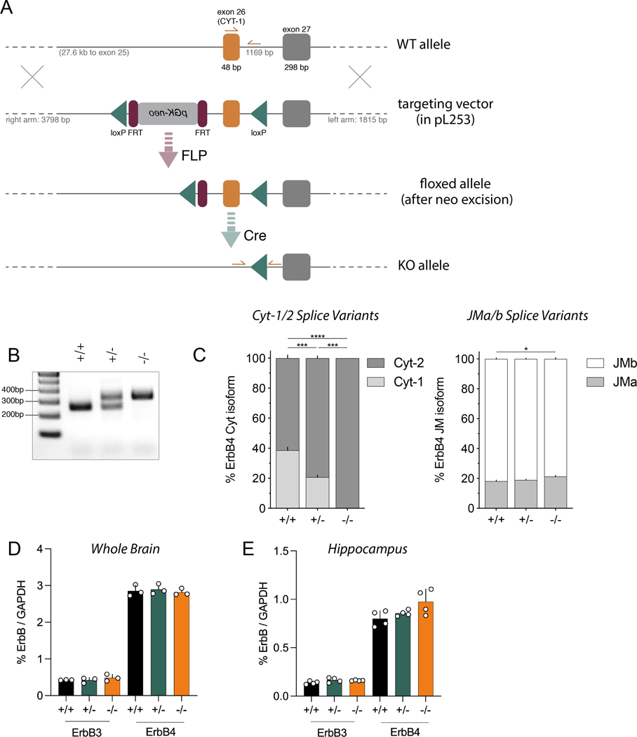
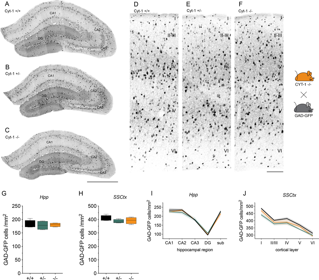
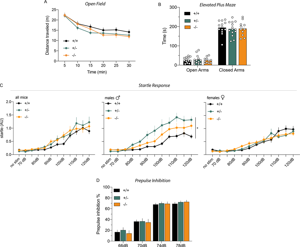
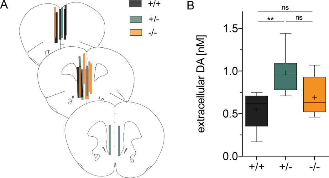
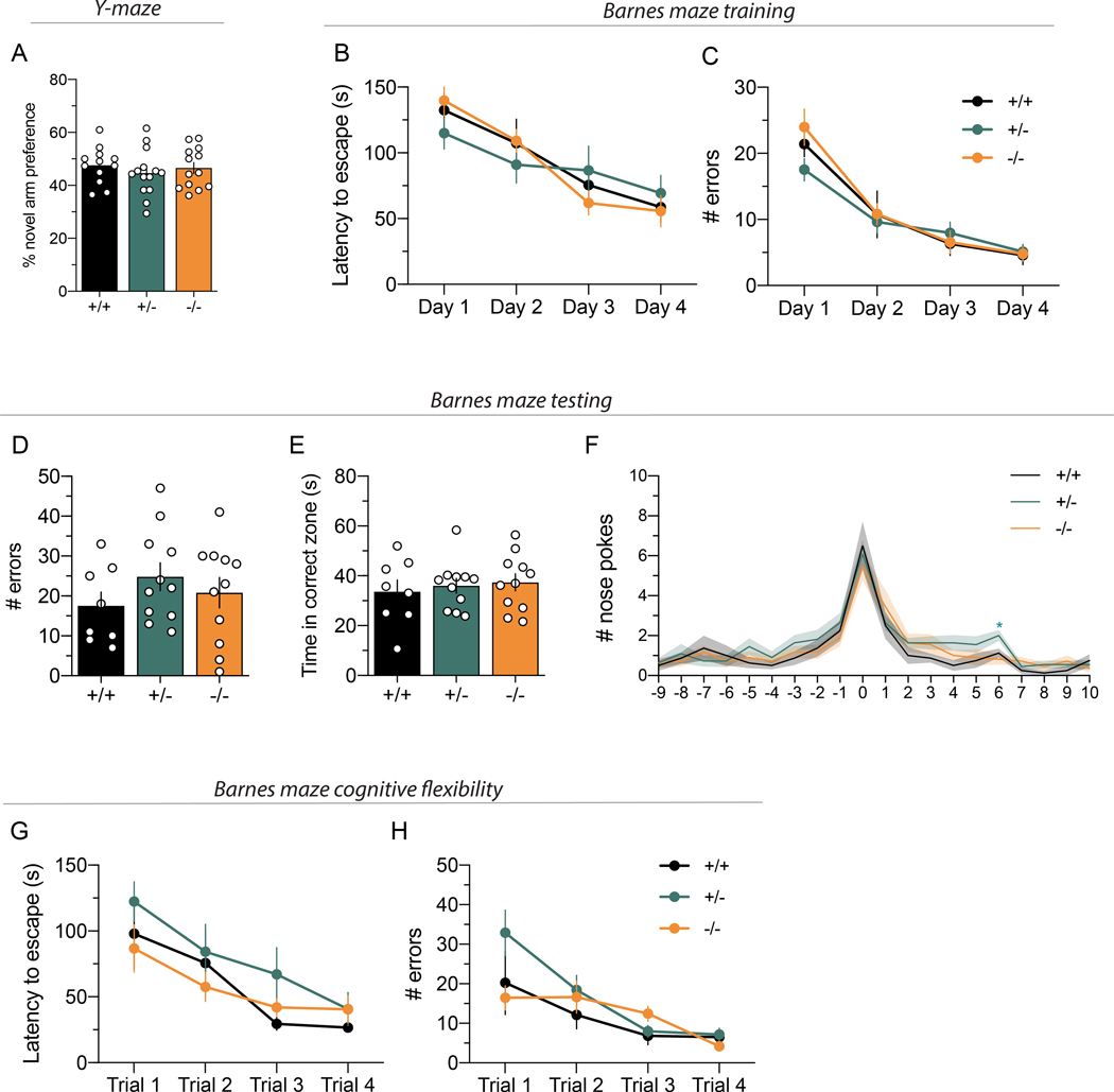
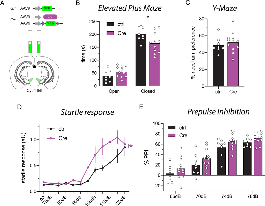
Neuregulins (NRGs) and their cognate neuronal receptor ERBB4, which is expressed in GABAergic and dopaminergic neurons, regulate numerous behaviors in rodents and have been identified as schizophrenia at-risk genes. ErbB4 transcripts are alternatively spliced to generate isoforms that either include (Cyt-1) or exclude (Cyt-2) exon 26, which encodes a cytoplasmic domain that imparts ErbB4 receptors the ability to signal via the phosphoinositide 3-kinase (PI3K)/protein kinase B (Akt) pathway. Although ErbB4 Cyt-1/2 isoforms have been studied in transfected cultured cells, their functions in vivo remain unknown. Here, we generated ErbB4-floxed (ErbB4-Cyt1fl/fl ) mice to investigate the effects of germline (constitutive) and conditional (acute) deletions of the Cyt-1 exon. Overall receptor mRNA levels remain unchanged in germline ErbB4 Cyt-1 knockouts (Cyt-1 KOs), with all transcripts encoding Cyt-2 variants. In contrast to mice lacking all ErbB4 receptor function, GABAergic interneuron migration and number are unaltered in Cyt-1 KOs. However, basal extracellular dopamine (DA) levels in the medial prefrontal cortex are increased in Cyt-1 heterozygotes. Despite these neurochemical changes, Cyt-1 heterozygous and homozygous mice do not manifest behavioral abnormalities previously reported to be altered in ErbB4 null mice. To address the possibility that Cyt-2 variants compensate for the lack of Cyt-1 during development, we microinjected an adeno-associated virus expressing Cre-recombinase (AAV-Cre) into the DA-rich ventral tegmental area of adult ErbB4-Cyt1fl/fl mice to acutely target exon 26. These conditional Cyt-1 KOs were found to exhibit behavioral abnormalities in the elevated plus maze and startle response, consistent with the idea that late exon 26 ablations may circumvent compensation by Cyt-2 variants. Taken together, our observations indicate that ErbB4 Cyt-1 function in vivo is important for DA balance and behaviors in adults.


Conflict of interest statement

Conflicts of Interest

Andrés Buonanno is a senior editor for the Journal of Neurochemistry. No additional conflicts of interest to report.

Figures

Figure 1.
Figure 1.. Generation of ErbB4 Cyt-1 KO mice and analysis of receptor splice variants.
A) Targeting strategy to generate Cyt-1 mutant mice (for more details see Methods). Exon 26 (orange), encoding the Cyt-1 cassette, was targeted by site-specific recombination in C57BL/6J-derived embryonic stem cells to insert flanking loxP sites (green). Subsequently, the FRT-pGK-neo-FRT selection cassette (magenta) was removed in mice harboring the targeted allele by crossing to a FLP deleter strain. The Cyt-1 exon was then ablated in ablated in germline by crossing to mice expressing Cre recombinase under control of the ubiquitously active EIIa promoter. B) Representative genotyping results using primers indicated in panel A. C) Relative levels of different ErbB4 splice variants in whole brain RNA quantified by Taqman qRT-PCR using specific primers for either Cyt-1/Cyt-2 (left) or JMa/JMb (right) isoforms (n=3/genotype). Cyt-1 transcripts were undetectable in Cyt-1 KOs and reduced by approximately half in heterozygotes, relative to wild-type littermates (% Cyt-1, genotypes +/+: 38.66 ± 2.11 %, +/−: 20.712 ± 1.42 % and −/−: 0.00 ± 0.00 %; n=3, one-way ANOVA F(2,6)=174.4, ****p<0.0001), whereas relative levels of JM splice variants were unchanged (genotypes +/+: 18.16 ± 0.70%, +/−: 18.97 ± 0.52% and −/−: 21.27 ± 0.78%; n=3, one-way ANOVA F(2,6)=5.725, *p=0.0407). (D) Amounts of ErbB3 and ErbB4 receptor transcripts in forebrain, relative to GAPDH, are the same between wild-type, Cyt-1 heterozygote and KO mice (% ErbB3/GAPDH; +/+: 0.426 ± 0.004%, +/−: 0.423 ± 0.042% and −/−: 0.485 ± 0.054% and % ErbB4/GAPDH; +/+: 2.85 ± 0.09%, +/−: 2.90 ± 0.08% and −/−: 2.82 ± 0.05%; n=3, one-way ANOVA, F(2,6)=0.2687, p=0.7731 and n=3, one-way ANOVA, F(2,6)=0.8039, p=0.4906). (E) Relative ErbB3 and ErbB4 receptor transcript levels in the hippocampus are also unchanged between wild-type, Cyt-1 heterozygote and KO mice (% ErbB3/GAPDH levels; +/+: 0.138 ± 0.009%, +/−: 0.165 ± 0.012 % and −/−: 0.161 ± 0.002%; n=4, one-way ANOVA, F(2,9)=2.67, p=0.1229) and (% ErbB4/GAPDH; +/+: 0.801 ± 0.041%, +/−: 0.857 ± 0.015% and −/−: 0.975 ± 0.066%; n=4, one-way ANOVA, F(2,9)=3.82, p=0.0628).
Figure 2.
Figure 2.. Expression of ErbB4 Cyt isoforms in hippocampal and cortical interneurons.
Expression of ErbB4 Cyt-1 and Cyt-2 splice variants was analyzed with exon-junction-specific ISH probes on coronal sections of adult mice using Basescope. A schematic of the coronal sections utilized is shown and highlights the approximate dorsal hippocampal (Hpp; pink shade) and primary somatosensory cortex (SSCtx; green) regions used for the analysis. A,B) Representative results for (A) Cyt-1 and (B) Cyt-2 in the Hpp (left) and SSCtx (right). C,D) Quantitative analysis of relative levels of Cyt splice variants in the Hpp (Cyt-1: 40.83 ± 0.9215%, Cyt-2: 59.17 ± 0.9215%, n=4; two-tailed Wilcoxon test, p=0.1250) and SSCtx (Cyt-1: 37.28 ± 1.276%, Cyt-2: 62.72 ± 1.276%, n=4; two-tailed Wilcoxon test, p=0.1250). Scale bars 500 μm in B, 100 μm in D.
Figure 3.
Figure 3.. GABAergic interneurons in the hippocampus and primary somatosensory cortex are unaltered in Cyt-1 mutant mice.
Representative images of GFP-expressing GABAergic interneurons in the (A-C) dorsal hippocampus and (D-F) primary somatosensory cortex of control (A,D), Cyt-1 heterozygote (B,E), and KO (C,F) adolescent mice (P30). GABAergic neurons were labeled by breeding ErbB4 Cyt-1 KOs to transgenic GAD67-GFP mice (see Methods). Quantification of GFP-labelled GABAergic interneurons/mm2 in the (G) hippocampus (genotypes +/+: 180.8 ± 7.73, +/−: 178.1 ± 8.21 and −/−: 180.1 ± 3.54 cells/mm2; n=4, Kruskal-Wallis test, p=0.9410) and (H) in SSCtx (genotypes +/+: 407.7 ± 10.71, +/−: 385.5 ± 7.10 and −/−: 392.0 ± 11.99 cells/mm2; n=4, one-way ANOVA F(2,9)=1.264, p=0.3283). I,J) Graphs showing (I) the Hpp subregional and (J) SSCtx layer-specific distribution of GFP-labeled GABAergic interneurons in ErbB4 Cyt-1 KO (orange), heterozygote (green) and control (black) mice (n=4; two-way ANOVA). Scale bars 500 μm in C, 100 μm in F. DG, dentate gyrus; sub, subiculum; CA1-CA3, cornu ammonis regions.
Figure 4.
Figure 4.. Behaviors altered in ErbB4 null mice are not affected in Cyt-1 KOs.
Performances on motor, affective (anxiety) and sensory-motor behavioral tasks known to be altered in ErbB4 null and PV-Cre; ErbB4fl/fl mice were analyzed in cohorts of adult Cyt-1 KO (orange), heterozygote (green) and control (black) mice. A) Open field: mice showed similar habituation to a novel environment and total distance travelled in 30 min (genotypes +/+: 101.7 ± 6.2, +/−: 91.3 ± 3.8, −/−: 85.6 ± 2.4 meters, Welch’s ANOVA, F(2,21.4)=3.197, p=0.0610, Dunnett’s multiple comparisons test +/+ vs. −/− p=0.0791, +/+ vs. +/− p=0.4167, +/− vs. −/− p=0.5085; n=10–14/genotype). B) Elevated plus maze: Cyt-1 KO, heterozygotes and control mice spent the same time (sec) in the open (genotypes +/+: 27.04 ± 3.87, +/−: 28.59 ± 6.41 and −/−: 25.07 ± 4.36) and closed (genotypes +/+: 194.5 ± 9.82, +/− 187.8 ± 9.81, −/− 189.7 ± 11.21; mixed-effects analysis, F(2,69)=0.1420, p=0.8679, 1 outlier value removed) arms of the plus maze. C) Startle response (arbitrary units; AU) to increasing auditory stimuli intensities (70–120 dB) in both sexes (left panel: n=17–18/genotype, mixed-effects analysis, F(22,532)=1.191, p=0.2494, 10 outlier values removed), male ErbB4 Cyt-1 KOs only (center panel: n=8–9/genotype, mixed-effects analysis, F(2,23)=5.321, p=0.0126*), and female KO mice only (right panel: n=9/genotype, mixed-effects analysis, F(2,24)=0.01569, p=0.9844). D) PPI: sensory-motor inhibitory responses were not significantly different between Cyt-1 KO, heterozygote and control mice (n=17–18/genotype, two-way ANOVA, F(2,49)=0.1582, p=0.8541).
Figure 5.
Figure 5.. Tonic extracellular DA levels are increased in Cyt-1 heterozygote mice
Extracellular DA levels were analyzed by unilateral no-net-flux microdialysis in the mPFC of freely moving control (black), heterozygote (green) and Cyt-1 KO (orange) mice. (A) Verification of microdialysis probe placement in the mPFC at bregma levels +2.22 mm, +1.98 mm and +1.78 mm in 50 μm-thick sections using Nissl staining. (B) Measurements for tonic extracellular DA levels in the mPFC of Cyt-1 mutants and controls (+/+ 0.546 ± 0.080 nM, +/− 0.977 ± 0.103 nM, +/+ 0.689 ± 0.085 nM, n=6–7/genotype, one-way ANOVA, F(2,17)=5.863, *p=0.0116; +/+ vs. −/− p=0.4876, +/+ vs. +/− **p=0.0094, +/− vs. −/− p=0.089).
Figure 6.
Figure 6.. Cyt-1 mutant mice have normal working and reference memory, and cognitive flexibility.
(A) Working memory, plotted as percentage for novel arm preference in the Y-maze, is normal in Cyt-1 mutant mice (−/−; orange) compared to heterozygote (+/−; green) and wildtype (+/+; black) littermates (n=12–14/genotype, % alternation +/+ 47.58 ± 2.00%, +/− 44.66 ± 2.32%, +/+ 46.54 ± 2.12%, one-way ANOVA, F(2,36)=0.4677 p=0.6302). (B-G) Spatial reference memory of Cyt-1 KO mice in the Barnes maze is comparable to heterozygote and wild-type littermates during (B,C) initial training over four days, (D-F) testing on day five (n=8–11/genotype) and (G) adaption to a new target location. (B) Latency to escape during training (n=8–11/genotype, two-way ANOVA, F(6,81)=1.580, p=0.1635). (C) Number of errors committed during training (n=8–11/genotype, mixed-effects analysis, F(6,80)=1.158, p=0.3373). (D) Number of errors during testing (+/+: 17.5 ± 3.5, +/−: 24.8 ± 3.5, −/−: 20.8 ± 3.8, one-way ANOVA, F(2,27)=0.9498, p=0.3994). (E) Time spent in the correct quadrant/zone during testing (+/+ 33.6 ± 4.7 s, +/− 36.0 ± 3.0 s, −/− 37.4 ± 3.4 s, one-way ANOVA, F(2,27)=0.2508, p=0.7799). (F) Nose pokes in individual holes during testing (0 – target, 10 – opposite; two-way ANOVA (no outlier exclusion), F(38,513)=0.8407, p=0.7396, Tukey’s multiple comparisons test main column effect, +/+ vs. +/− p=0.1450, +/+ vs. −/− p=0.8096, +/− vs. −/− p=0.3263) (G) Latency to escape (n=8–11/genotype, mixed-effects analysis, F(6,79)=0.5949, p=0.7335, 2 outlier values) and (H) number of errors after moving target to a new position (cognitive flexibility; n=8–11/genotype, mixed-effects analysis, F(6,78)=1.906, p=0.0903, 3 outlier values removed).
Figure 7.
Figure 7.. Deletion of the Cyt-1 exon in the VTA of adult mice results in behavioral abnormalities.
(A) Illustration of adeno-associated virus (AAV) injections into the VTA of ErbB4 Cyt-1fl/fl mice used to generate controls (AAV9-hSynI-GFP) and acute Cyt-1 KOs (AAV9-hSynI-Cre & AAV9-hSynI-DIO-GFP). (B-E) Controls (n=8) and acute Cyt-1 KOs (n=11) were tested in different behavioral paradigms. (B) Acute Cyt-1 KOs spend less time in closed arms of the elevated plus maze (two-way ANOVA, F(1,17)=4.421, p=0.0507, Sidak’s multiple comparison test ctrl vs. Cre, closed arms p=0.0234*, open arms p=0.4123). (C) No differences were found between cohorts in the Y-maze (unpaired two-tailed t-test, p=0.5380). (D) Acute Cyt-1 KOs exhibit an enhanced startle response relative to controls, (mixed-effects analysis, F(11,181)=1.874, p=0.0454*, ctrl vs. Cre p=0.0490*; 6 outlier values from a total of 209 values in 5 mice); however, (E) PPI does not differ between cohorts, (two-way ANOVA, F(3,51)=0.09446, p=0.9628, ctrl vs. Cre p=0.0601).

References

    1. Abe Y, Namba H, Zheng Y. and Nawa H. (2009) In situ hybridization reveals developmental regulation of ErbB1–4 mRNA expression in mouse midbrain: implication of ErbB receptors for dopaminergic neurons. Neuroscience 161, 95–110. - PubMed
    1. Andersson RH, Johnston A, Herman PA, Winzer-Serhan UH, Karavanova I, Vullhorst D, Fisahn A. and Buonanno A. (2012) Neuregulin and dopamine modulation of hippocampal gamma oscillations is dependent on dopamine D4 receptors. Proc Natl Acad Sci U S A 109, 13118–13123. - PMC - PubMed
    1. Bartolini G, Sanchez-Alcaniz JA, Osorio C, Valiente M, Garcia-Frigola C. and Marin O. (2017) Neuregulin 3 Mediates Cortical Plate Invasion and Laminar Allocation of GABAergic Interneurons. Cell Rep 18, 1157–1170. - PMC - PubMed
    1. Buonanno A. (2010) The neuregulin signaling pathway and schizophrenia: from genes to synapses and neural circuits. Brain Res Bull 83, 122–131. - PMC - PubMed
    1. Buonanno A. and Fischbach GD (2001) Neuregulin and ErbB receptor signaling pathways in the nervous system. Curr Opin Neurobiol 11, 287–296. - PubMed

Publication types