. 2022 May 6;13(1):2413.
doi: 10.1038/s41467-022-30080-6.

Ataluren binds to multiple protein synthesis apparatus sites and competitively inhibits release factor-dependent termination

Affiliations

Ataluren binds to multiple protein synthesis apparatus sites and competitively inhibits release factor-dependent termination

Shijie Huang et al. Nat Commun. .

Abstract

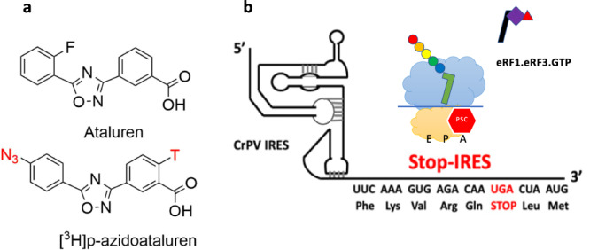
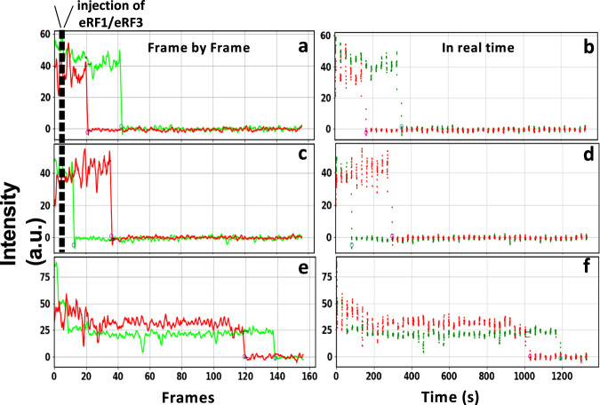
Genetic diseases are often caused by nonsense mutations, but only one TRID (translation readthrough inducing drug), ataluren, has been approved for clinical use. Ataluren inhibits release factor complex (RFC) termination activity, while not affecting productive binding of near-cognate ternary complex (TC, aa-tRNA.eEF1A.GTP). Here we use photoaffinity labeling to identify two sites of ataluren binding within rRNA, proximal to the decoding center (DC) and the peptidyl transfer center (PTC) of the ribosome, which are directly responsible for ataluren inhibition of termination activity. A third site, within the RFC, has as yet unclear functional consequences. Using single molecule and ensemble fluorescence assays we also demonstrate that termination proceeds via rapid RFC-dependent hydrolysis of peptidyl-tRNA followed by slow release of peptide and tRNA from the ribosome. Ataluren is an apparent competitive inhibitor of productive RFC binding, acting at or before the hydrolysis step. We propose that designing more potent TRIDs which retain ataluren's low toxicity should target areas of the RFC binding site proximal to the DC and PTC which do not overlap the TC binding site.


Conflict of interest statement

The authors declare no competing interests.

Figures

Fig. 1
Fig. 1. Targets of azidoataluren photoincorporation.
a Structures of ataluren and [3H]-AzAt. T refers to tritium. N3 is the azide which confers photolability on azidoataluren. b Stop-IRES mRNA encoding FKVRQStopLM. The cartoon depicts the Stop-POST5 complex containing FKVRQ-tRNAGln bound in the P-site adjacent to an empty A-site containing the UGA stop codon, and the incoming eRF1.eRF3.GTP complex which catalyzes cleavage of the ester bond linking FKVRQ to tRNAGln after binding to the A-site.
Fig. 2
Fig. 2. AzAt Photoaffinity Labeling.
a AzAt photoincorporation into Stop-POST5, the RNA fraction of Stop-POST5, 80S.IRES, and the RNA fraction of 80S.IRES. All of the labeling stoichiometries are normalized to that of Stop-POST5 at 600 µM AzAt, which was equal to 1.2/Stop-POST5. b AzAt photoincorporation into eRF1 both alone and complexed with eRF3.GDPNP. The values are normalized to the saturation labeling of isolated eRF1. c Inhibition of AzAt (250 µM) photoincorporation by the addition of either ataluren (1000 µM) or GJ072 (150 µM). d The mutation rate fold change for PAL vs. PRE samples for Stop-POST5 (red) and 80S.IRES complexes (blue) for the 22 sites most pertinent for ataluren function, and the asterisks indicates sites of particular interest (see text). e Saturation curves for photoincorporation into 18S-A1195, as measured by photoincorporation into Fragment I, vs. the sum of the photoincorporations into 26S A3093, A2669, and A2672, as measured by photoincorporation into Fragments II and III. f Location of A1195 within the 40S subunit containing bound eRF1. g Locations of A2669, A2672, and A3093 within the 60S subunit containing bound eRF1. All of the error bars in this Figure represent average deviations for n = 2 independent determinations, with the exception of the eRF1 labeling within the ternary complex in b, for which n=3. Source data for (ae) are provided as a Source Data file.
Fig. 3
Fig. 3. Typical traces showing the dissociation of Atto-647 labeled peptide (red) and Cy3-labeled tRNA (green) following eRF1/eRF3 injection.
In the time-lapse experiment the sample was briefly illuminated between fixed time intervals. Sample traces following eRF1/eRF3 injection. In a the peptide signal disappears prior to tRNA signal; in (c) the tRNA signal disappears prior to peptide signal. b and d Corresponding real-time scatter plots of the traces presented in (a) and (c) where each dot represents one frame. e Sample trace from a control experiment where only buffer was injected to obtain the photobleaching/spontaneous dissociation rate, which is clearly much slower than the rates seen in (a) and (c). f The real-time scatter plot of the trace presented in (e). Source data for (a), (c), and (e) are provided as a Source Data file.
Fig. 4
Fig. 4. Ataluren effects on RFC-dependent termination of polypeptide synthesis.
Cumulative distributions of (a) peptide and (b) tRNA dissociation times at indicated RFC concentrations, one also with 1 mM ataluren. Each cumulative distribution is constructed from ≥300 kinetic traces. Rates of dissociation of (c). peptide and (d). tRNA as a function of RFC concentration at different fixed ataluren concentrations. Error bars are ± s.e.m. for n = 250–750 trials. Normalized plots of ensemble experiments showing single exponential fits (solid lines) of decimated smoothed raw data (points) of atto647 pentapeptide release reaction measured by fluorescence anisotropy decay vs. time at 25 °C. e At indicated RFC concentrations. The control shows the near constancy of observed anisotropy in the absence of added RFC or ataluren. f At an RFC concentration of 0.0625 µM and varying ataluren concentrations. g The rates of dissociation of atto647 pentapeptide in ensemble experiments as a function of free RFC concentration at varying ataluren concentrations. Error bars are average deviation (a. d.) for n = 2–6 independent determinations. Values of n for each point are presented in Supplementary Table 5. h Ataluren inhibition of normalized rates of dissociation of atto647 pentapeptide as measured by single molecule (red) and plate reader (black) assays. [eRF1], 32 nM; [eRF3], 0.2 µM and 0.8 µM in the single molecule and ensemble assays, respectively. Error bars are ± s.e.m. n ≥ 250 trials (single molecule) and ± a.d., n = 2 independent determinations (plate reader). i Rates of dissociation of atto647 labeled and unlabeled pentapeptide as measured by millipore filtration, calculated using both filtrate and filter retained values. Error bars are ± s.d., n = 8 independent measurements for all points except for the 4 min measurements, for which n = 11. [eRF1], 0.2 µM; [eRF3], 0.8 µM; POST5, 0.05 µM. j Ataluren inhibition of peptide and tRNA release when added at different times (4–25 s) following RFC addition to Stop-POST5. [RFC], 0.08 µM; [Ataluren], 1 mM. Values at zero-time correspond to simultaneous addition of ataluren and RFC. Error bars are ±s.e.m. for n ≥ 200 trials. Source data for all panels are provided as a Source Data file.
Fig. 5
Fig. 5. Scatter plots of peptide and tRNA dissociation times.
Each point represents dissociation times from an individual ribosome. CC is correlation coefficient. a 2 µM RFC. b 16 nM RFC. Source data are provided as a Source Data file.
Fig. 6
Fig. 6. A simplified model for RFC catalysis of termination.
This model, consistent with all of our results, as well as with recent published results of others, posits that ataluren inhibition results from the cooperative binding of (n) molecules of ataluren to the pretermination complex P5 in competition with RFC binding. It also invokes a hypothetical complex C3 resulting from an at least partial rate-determining conformational change, following cleavage of the tRNA-peptide ester bond to account for the similarity in the rate constants of tRNA, peptide, and eRF1 release (see text). The question marks associated with possible eRF1 release are shown to indicate our current uncertainty as to whether eRF1 release is coordinated with either peptide or tRNA release, or proceeds independently of either.

Similar articles

Cited by

References

    1. Krawczak M, et al. Human gene mutation database-a biomedical information and research resource. Hum. Mutat. 2000;15:45–51. doi: 10.1002/(SICI)1098-1004(200001)15:1<45::AID-HUMU10>3.0.CO;2-T. - DOI - PubMed
    1. Campofelice A, et al. Strategies against nonsense: Oxadiazoles as Translational Readthrough-Inducing Drugs (TRIDs) Int. J. Mol. Sci. 2019;20:3329–3346. doi: 10.3390/ijms20133329. - DOI - PMC - PubMed
    1. Mort M, Ivanov D, Cooper DN, Chuzhanova NA. A meta-analysis of nonsense mutations causing human genetic disease. Hum. Mutat. 2008;29:1037–1047. doi: 10.1002/humu.20763. - DOI - PubMed
    1. Nagel-Wolfrum K, Möller F, Penner I, Baasov T, Wolfrum U. Targeting nonsense mutations in diseases with Translational Read-Through-Inducing Drugs (TRIDs) Biodrugs. 2016;30:49–74. doi: 10.1007/s40259-016-0157-6. - DOI - PubMed
    1. Keeling KM, Xue X, Gunn G, Bedwell DM. Therapeutics based on stop codon readthrough. Annu. Rev. Genom. Hum. Genet. 2014;15:371–394. doi: 10.1146/annurev-genom-091212-153527. - DOI - PMC - PubMed

Publication types

MeSH terms