. 2022 May 6;12(1):189.
doi: 10.1038/s41398-022-01939-5.

Magnetization transfer imaging alterations and its diagnostic value in antipsychotic-naïve first-episode schizophrenia

Affiliations

Magnetization transfer imaging alterations and its diagnostic value in antipsychotic-naïve first-episode schizophrenia

Du Lei et al. Transl Psychiatry. .

Abstract

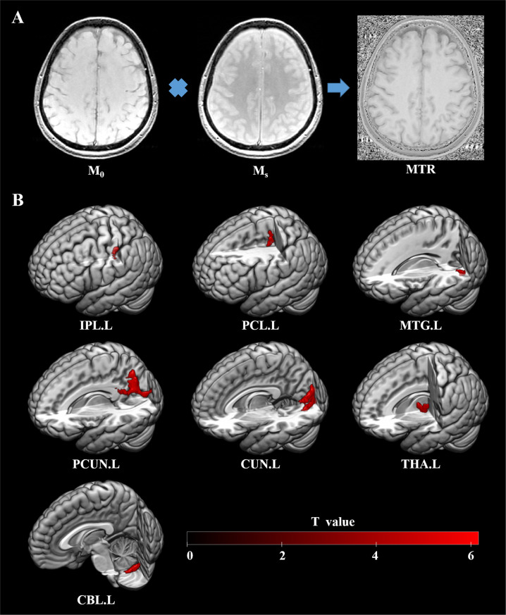
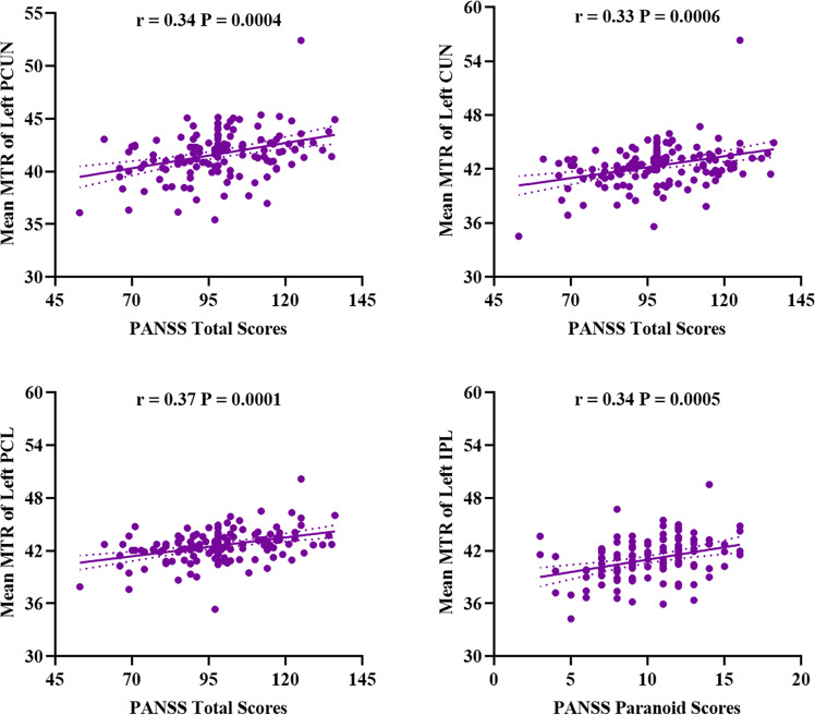
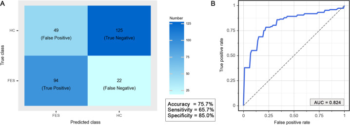
Magnetization transfer imaging (MTI) may provide more sensitivity and mechanistic understanding of neuropathological changes associated with schizophrenia than volumetric MRI. This study aims to identify brain magnetization transfer ratio (MTR) changes in antipsychotic-naïve first-episode schizophrenia (FES), and to correlate MTR findings with clinical symptom severity. A total of 143 individuals with antipsychotic-naïve FES and 147 healthy controls (HCs) were included and underwent 3.0 T brain MTI between August 2005 and July 2014. Voxelwise analysis was performed to test for MTR differences with family-wise error corrections. Relationships of these differences to symptom severity were assessed using partial correlations. Exploratory analyses using a support vector machine (SVM) classifier were conducted to discriminate FES from HCs using MTR maps. Model performance was examined using a 10-fold stratified cross-validation. Compared with HCs, individuals with FES exhibited higher MTR values in left thalamus, precuneus, cuneus, and paracentral lobule, that were positively correlated with schizophrenia symptom severity [precuneus (r = 0.34, P = 0.0004), cuneus (r = 0.33, P = 0.0006) and paracentral lobule (r = 0.37, P = 0.001)]. Whole-brain MTR maps identified individuals with FES with overall accuracy 75.5% (219 of 290 individuals) based on SVM approach. In antipsychotic-naïve FES, clinically relevant biophysical abnormalities detected by MTI mainly in the left parieto-occipital regions are informative about local brain pathology, and have potential as diagnostic markers.


Conflict of interest statement

JAS is a consultant for Vera Sci. All other authors declare no competing interests.

Figures

Fig. 1
Fig. 1. MTR in voxel-based differences between individuals with first-episode schizophrenia and healthy controls.
A Schematic illustration of MTR calculated on a voxel-by-voxel basis as MTR = 100 × (M0 – MS)/M0, where M0 and MS represent the signal intensities without and with the saturation pulse; B Significant clusters were identified in individuals with first-episode schizophrenia compared to healthy controls based on a voxel-level statistical threshold of P < 0.05 (family-wise error corrected, cluster size > 40). The color bar indicates T values. MTR magnetization transfer ratio, L left, CUN cuneus, PCUN precuneus, MTG middle temporal gyrus, IPL inferior parietal lobule, THA thalamus, CBL cerebellum, PCL paracentral lobule.
Fig. 2
Fig. 2. Scatter plots showing the correlations between PANSS total scores and regional mean MTR in first-episode schizophrenia.
Partial correlations (presented as r) were conducted between regional mean MTR and PANSS score, with age, sex, and education years as covariates. Findings were significant (presented as P) after correction for multiple comparisons using the Bonferroni correction. MTR magnetization transfer ratio, PCUN precuneus, CUN cuneus, IPL inferior parietal lobule, PCL paracentral lobule, PANSS Positive and Negative Syndrome Scale.
Fig. 3
Fig. 3. Classification performance based on whole-brain MTR maps.
A The confusion matrix of classification between FES and HC. B Receiver operating characteristic curve for a prediction model based on whole-brain MTR maps using support vector machine. MTR magnetization transfer ratio, FES first-episode schizophrenia, HC healthy controls, AUC area under receiver operating characteristic curve.

Similar articles

Cited by

References

    1. Dhindsa RS, Goldstein DB. Schizophrenia: from genetics to physiology at last. Nature. 2016;530:162–3. doi: 10.1038/nature16874. - DOI - PubMed
    1. Gong Q, Lui S, Sweeney JA. A selective review of cerebral abnormalities in patients with first-episode schizophrenia before and after treatment. Am J Psychiatry. 2016;173:232–43. doi: 10.1176/appi.ajp.2015.15050641. - DOI - PubMed
    1. Selemon LD, Goldman-Rakic PS. The reduced neuropil hypothesis: a circuit based model of schizophrenia. Biol Psychiatry. 1999;45:17–25. doi: 10.1016/S0006-3223(98)00281-9. - DOI - PubMed
    1. Wolff SD, Balaban RS. Magnetization transfer contrast (MTC) and tissue water proton relaxation in vivo. Magn Reson Med. 1989;10:135–44. doi: 10.1002/mrm.1910100113. - DOI - PubMed
    1. Antosik-Biernacka A, Peuskens H, De Hert M, Peuskens J, Sunaert S, Van Hecke P, et al. Magnetization transfer imaging in chronic schizophrenia. Med Sci Monit. 2006;12:Mt17–Mt21. - PubMed

Publication types

Substances