

The bifurcation angle is associated with the progression of saccular aneurysms
- PMID: 35523805
- PMCID: PMC9076676
- DOI: 10.1038/s41598-022-11160-5
The bifurcation angle is associated with the progression of saccular aneurysms
Abstract
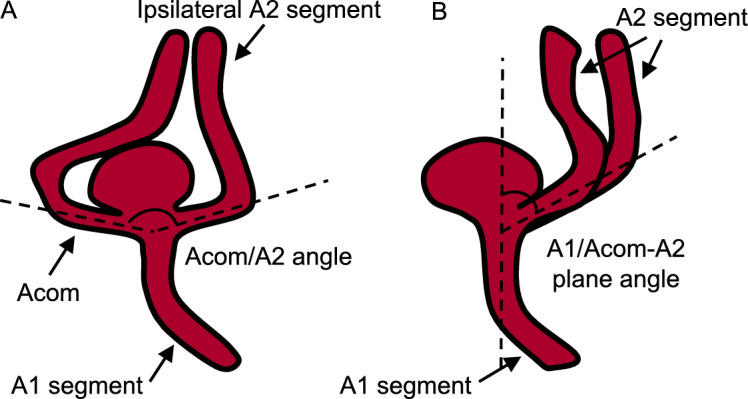
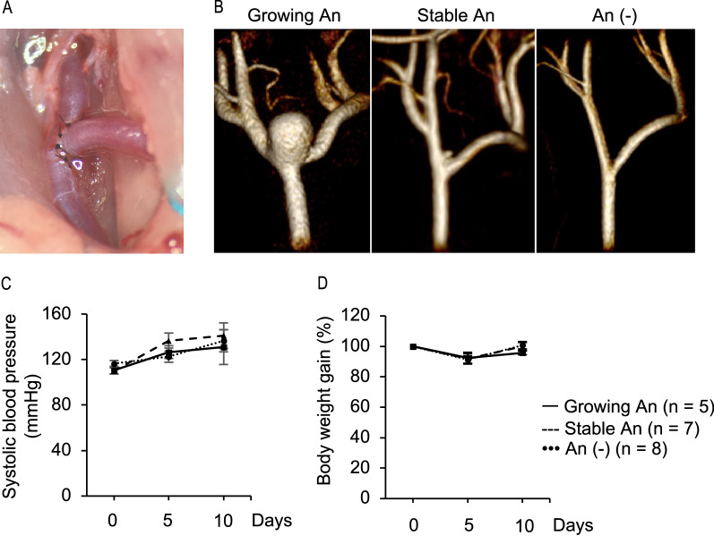
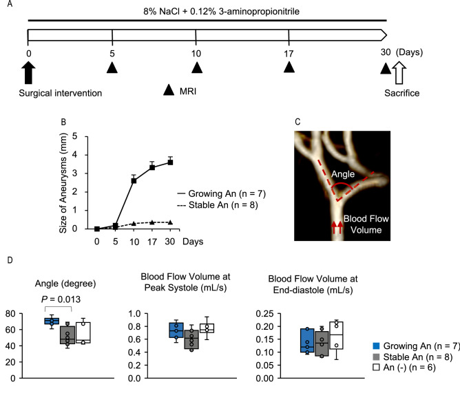
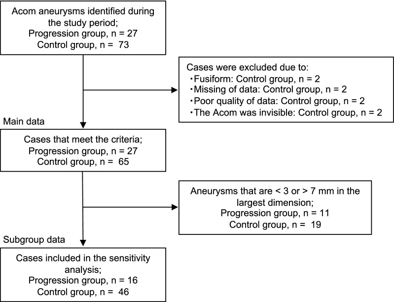
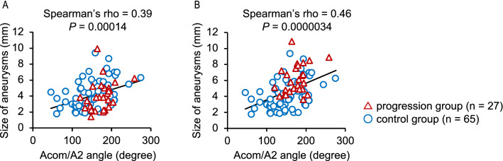
The role of the bifurcation angle in progression of saccular intracranial aneurysms (sIAs) has been undetermined. We, therefore, assessed the association of bifurcation angles with aneurysm progression using a bifurcation-type aneurysm model in rats and anterior communicating artery aneurysms in a multicenter case-control study. Aneurysm progression was defined as growth by ≥ 1 mm or rupture during observation, and controls as progression-free for 30 days in rats and ≥ 36 months in humans. In the rat model, baseline bifurcation angles were significantly wider in progressive aneurysms than in stable ones. In the case-control study, 27 and 65 patients were enrolled in the progression and control groups. Inter-observer agreement for the presence or absence of the growth was excellent (κ coefficient, 0.82; 95% CI, 0.61-1.0). Multivariate logistic regression analysis showed that wider baseline bifurcation angles were significantly associated with subsequent progressions. The odds ratio for the progression of the second (145°-179°) or third (180°-274°) tertiles compared to the first tertile (46°-143°) were 5.5 (95% CI, 1.3-35). Besides, the bifurcation angle was positively correlated with the size of aneurysms (Spearman's rho, 0.39; P = 0.00014). The present study suggests the usefulness of the bifurcation angle for predicting the progression of sIAs.
© 2022. The Author(s).
Conflict of interest statement
The authors declare no competing interests.
Figures
Similar articles
-
Presence of Anterior Communicating Artery Aneurysm Is Associated With Age, Bifurcation Angle, and Vessel Diameter.Stroke. 2018 Feb;49(2):341-347. doi: 10.1161/STROKEAHA.117.019701. Epub 2018 Jan 4. Stroke. 2018. PMID: 29301972
-
Middle cerebral artery bifurcation aneurysms are associated with patient age, sex, bifurcation angle, and vascular diameters.Sci Rep. 2023 Dec 21;13(1):22844. doi: 10.1038/s41598-023-50380-1. Sci Rep. 2023. PMID: 38129685 Free PMC article.
-
Risk Factors for the Rupture of Bifurcation Intracranial Aneurysms Using CT Angiography.Yonsei Med J. 2016 Sep;57(5):1178-84. doi: 10.3349/ymj.2016.57.5.1178. Yonsei Med J. 2016. PMID: 27401649 Free PMC article.
-
Widening and high inclination of the middle cerebral artery bifurcation are associated with presence of aneurysms.Stroke. 2014 Sep;45(9):2649-55. doi: 10.1161/STROKEAHA.114.005393. Epub 2014 Aug 12. Stroke. 2014. PMID: 25116869 Free PMC article.
-
Impact of bifurcation angle and inflow coefficient on the rupture risk of bifurcation type basilar artery tip aneurysms.J Neurosurg. 2018 Mar;128(3):723-730. doi: 10.3171/2016.10.JNS161695. Epub 2017 Mar 3. J Neurosurg. 2018. PMID: 28298037
Cited by
-
Aneurysm Formation at the Internal Carotid Artery Bifurcation Is Related to the Vascular Geometry of the Bifurcation.Brain Sci. 2024 Dec 12;14(12):1247. doi: 10.3390/brainsci14121247. Brain Sci. 2024. PMID: 39766446 Free PMC article.
-
True superficial temporal artery aneurysm: A case after extracranial-intracranial bypass surgery and a systematic review.Surg Neurol Int. 2022 Dec 9;13:573. doi: 10.25259/SNI_848_2022. eCollection 2022. Surg Neurol Int. 2022. PMID: 36600761 Free PMC article. Review.
