. 2022 May 6;13(1):2503.
doi: 10.1038/s41467-022-30181-2.

Regulation of A-to-I RNA editing and stop codon recoding to control selenoprotein expression during skeletal myogenesis

Affiliations

Regulation of A-to-I RNA editing and stop codon recoding to control selenoprotein expression during skeletal myogenesis

Yuta Noda et al. Nat Commun. .

Abstract

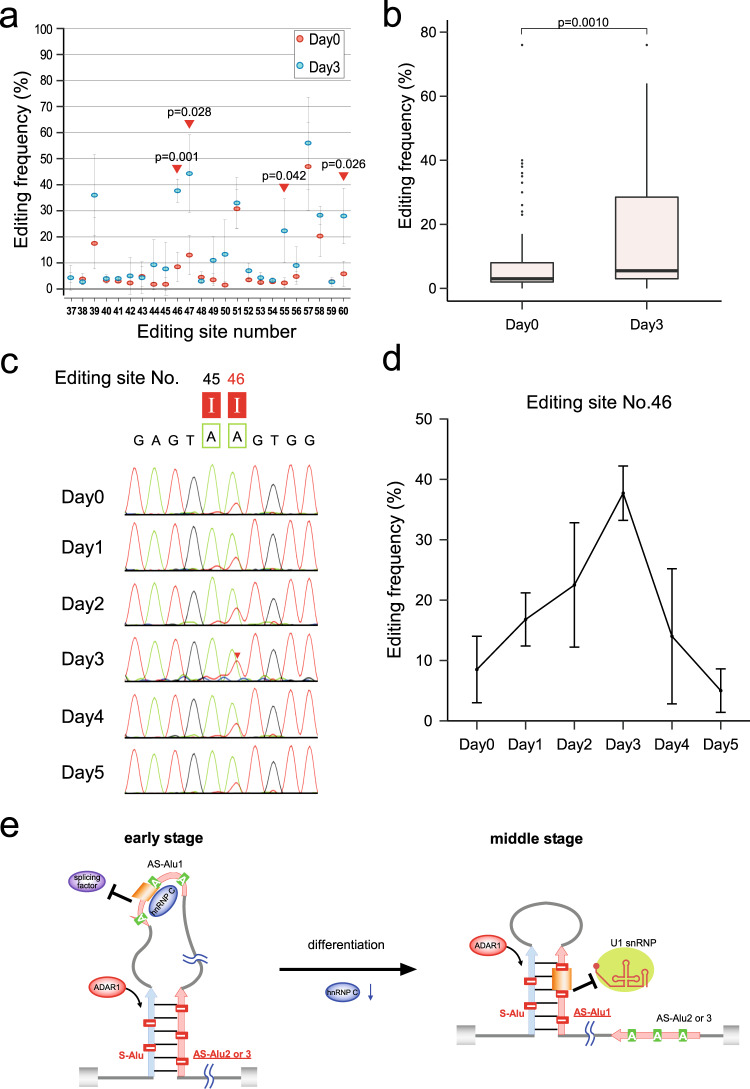
Selenoprotein N (SELENON), a selenocysteine (Sec)-containing protein with high reductive activity, maintains redox homeostasis, thereby contributing to skeletal muscle differentiation and function. Loss-of-function mutations in SELENON cause severe neuromuscular disorders. In the early-to-middle stage of myoblast differentiation, SELENON maintains redox homeostasis and modulates endoplasmic reticulum (ER) Ca2+ concentration, resulting in a gradual reduction from the middle-to-late stages due to unknown mechanisms. The present study describes post-transcriptional mechanisms that regulate SELENON expression during myoblast differentiation. Part of an Alu element in the second intron of SELENON pre-mRNA is frequently exonized during splicing, resulting in an aberrant mRNA that is degraded by nonsense-mediated mRNA decay (NMD). In the middle stage of myoblast differentiation, ADAR1-mediated A-to-I RNA editing occurs in the U1 snRNA binding site at 5' splice site, preventing Alu exonization and producing mature mRNA. In the middle-to-late stage of myoblast differentiation, the level of Sec-charged tRNASec decreases due to downregulation of essential recoding factors for Sec insertion, thereby generating a premature termination codon in SELENON mRNA, which is targeted by NMD.


Conflict of interest statement

The authors declare no competing interests.

Figures

Fig. 1
Fig. 1. ADAR1 and hnRNP C suppress Alu exonization of SELENON mRNA.
a Human SELENON gene with exons (boxes) and introns (lines). The Alu exon and SECIS are indicated by orange and magenta boxes, respectively. Sense and antisense strands of Alu elements are indicated by blue and red arrows, respectively. Int. and Term. represent initiation and termination codons. b Intronic region of SELENON including four Alu elements (green dashed box in a). Purple column represents A-to-I editing sites determined by ICE method. Pink column represents iCLIP sites for hnRNP C. c RNA sequence of AS-Alu1 in SELENON. A-to-I editing sites are boxed in red, with site numbers shown in Supplementary Fig. 5. hnRNP C binding site identified by iCLIP is boxed. The Alu exon sequence is colored in orange. 3′SS and 5′SS of the Alu exon are indicated by underbars. PTC (UGA) is indicated in red. d Alu exonization of SELENON in HeLa cells upon knockdown of each target mRNA. Control siRNA targeted luciferase. Upper panel shows RT-PCR products of a part of SELENON mRNA spanning exons 2 and 3. Lower panel shows the inclusion ratio (%), determined by RT-qPCR. Data are presented as mean values ±S.D. Statistical significance was determined by an unpaired two-tailed t test; n = 3 biologically independent samples. e Alu exonization of SELENON in HeLa cells upon double-knockdown of target mRNAs. Upper panel shows the inclusion and skipping isoforms with a schematical illustration. Lower panel shows the inclusion ratio (%), as described in d, above. f cDNA sequences of the Alu exon 5′SS in SELENON pre-mRNAs from HeLa cells treated with siRNAs targeting luciferase (control) and ADAR1 (KD). The editing sites are indicated. Consensus sequence of 5′SS (underlined) base-pairs with a part of U1 snRNA. The electropherograms for A, G, T, and C are colored green, red, gray, and blue, respectively. Ψ, pseudouridine. g Minigene constructs bearing the Alu exon (WT) or its editing-mimic mutant (AG). Skipping and inclusion isoforms from the minigene were detected by RT-PCR. This experiment was done once. Source data and unprocessed gel images are provided in Source Data file.
Fig. 2
Fig. 2. Stage-specific expression profiles and dynamic alteration of Alu exonization of SELENON during skeletal myogenesis of Hu5/KD3 cells.
a Experimental schedule for differentiation of Hu5/KD3 cells. b Phase-contrast images showing Hu5/KD3 cell differentiation from Day 0 to 5. This experiment was repeated more than three times independently with similar results. c Expression analyses of the differentiation markers (MYOG, MYH1), SELENON, hnRNP C, ADAR1, and GAPDH (control) by western blotting on the indicated days during differentiation of Hu5/KD3 cells. SELENON (full-length and short isoforms), hnRNP C2 and hnRNP C1, and p150 and p110 isoforms of ADAR1, are indicated by arrowheads. The samples derived from the same experiment and the gels/blots were processed in parallel. This experiment was repeated more than three times independently with similar results. d Distribution of Log2(fold-change) of expressed genes in RNA-seq data in Hu5/KD3 cells on each day of differentiation relative to Day 0, shown as violin and box plots. For the box plot, center lines indicate median, box limits indicate upper and lower quartiles, whiskers indicate 1.5× interquartile range and points indicate outliers; n = 58,278 genes. e Heat map of RNA-seq transcriptome analysis of differentiation markers, SELENON, hnRNP C, ADAR1, UGA/Sec recoding factors, and ER stress markers on the indicated days in differentiating Hu5/KD3 cells. Colors correspond to the per-gene z-score calculated from log10 fragments per million (FPM) reads mapped. f RT-qPCR to estimate the Alu exonization level of SELENON mRNAs during myoblast differentiation. Transcripts of each isoform were quantified using Hu5/KD3 total RNA from each day of differentiation. The graph represents the ratio between the inclusion and skipping isoforms. Data are presented as mean values ±S.D.; n = 3 biologically independent samples. Source data and unprocessed gel images are provided in the Source Data file.
Fig. 3
Fig. 3. Dynamic alteration of A-to-I RNA editing during myoblast differentiation.
a Alteration of A-to-I RNA editing frequency in AS-Alu1 from Day 0 (red) to 3 (blue). Data are presented as mean values ±S.D. The positions showing statistical significance [p < 0.05, determined by unpaired two-tailed t test; n = 4 (Day 0) and 3 (Day 3) biologically independent samples] are indicated by red triangles. Source data are provided in Source Supplementary Data 1. b The distribution of the A-to-I RNA editing frequency in AS-Alu1 on Days 0 and 3 is shown as box plots (center lines indicate median, box limits indicate upper and lower quartiles, whiskers indicate 1.5× interquartile range and points indicate outliers). Error bars denote S.D. Statistical significance was determined by unpaired two-tailed t test; n = 96 (Day 0) and 72 (Day 3). Source data are provided in Source Supplementary Data 1. c Sanger sequences of the 5′SS of the Alu exon in AS-Alu1 of SELENON mRNAs in differentiating Hu5/KD3 cells on the indicated days. The A-to-I RNA editing sites are boxed, with the site number shown above. d Frequency of editing site No. 46 in Hu5/KD3 cells on different differentiation days. Data are presented as mean values ±S.D.; n = 4 (Day 0 and Day 2), 5 (Day 1), and 3 (Day 3–5) biologically independent samples. Source data are provided in Source Supplementary Data 1. e Schematic model of conformational changes in SELENON pre-mRNA from the early-to-middle stage of differentiation. At the early stage, hnRNP C binds to AS-Alu1, and then S-Alu forms a double-stranded structure with AS-Alu2 or 3. A-to-I RNA editing occurs in this double strand. At the middle stage, the level of hnRNP C decreases. AS-Alu1 forms a double-stranded structure with S-Alu, which is recognized by ADAR1, which then performs A-to-I RNA editing in this double-stranded region, including the 5′SS of the Alu exon, thereby inhibiting U1 snRNA recognition. Source data and unprocessed gel images are provided in Source Data file.
Fig. 4
Fig. 4. Dynamic regulation of UGA/Sec recoding during myoblast differentiation.
a Constructs of the FLAG-SELENON expression vector and its mutants ΔSECIS and GGA. TM (transmembrane domain), EF-hand domain, and Sec residue in the catalytic domain are indicated. b Expression of FLAG-SELENON and its mutants in HEK293T cells, detected by western blotting with an anti-FLAG antibody. This experiment was done once. c Northern blotting of cytoplasmic tRNASec in total RNA obtained on each day of differentiation. Relative steady-state level of tRNASec on each day of differentiation compared with the level on Day 0 is shown below. Results were normalized relative to the band intensity of 5.8 S rRNA in the same sample. Data are presented as mean values ±S.D. Statistical significance was determined by unpaired two-tailed t test; n = 3 biologically independent samples. d Volcano plot of RNA-seq transcriptome data showing altered gene expression [log2(fold-change)], with p values, on Day 5 versus Day 0 of Hu5/KD3 cell differentiation. UGA/Sec recoding factors are indicated, and significantly differentially expressed genes (two-sided p value <0.05 determined by likelihood ratio test) are highlighted in red. e Expression analyses of GAPDH (control) and SEPSECS in differentiating Hu5/KD3 cells by western blotting on the indicated days. The expression level of SEPSECS was normalized to that of GAPDH, making the relative expression level on Day 0 = 1.00 (shown below). The samples derived from the same experiment and the gels/blots were processed in parallel. This experiment was repeated more than three times independently with similar results. f Subcellular localization of SEPSECS on the indicated days in differentiating Hu5/KD3 cells immunostained with an anti-SEPSECS antibody (green). Nuclei were stained with DAPI (blue) and myotubes were stained with an anti-myosin antibody (red). Magnification: ×100. Exposure time: 150 ms (Day 0) or 300 ms (Day 3 and 5). This experiment was done once. Source data and unprocessed gel image are provided in Source Data file.
Fig. 5
Fig. 5. Reduction of Sec-tRNASec formation at the late stage of myoblast differentiation.
a Outline of aminoacyl-tRNASec preparation for LC/MS analysis. Small yellow and red circles attached to amino acids represent alkyl and acetyl groups, respectively. IAA, iodoacetamide. Ac2O, acetic anhydride. aa, amino acid. b Secondary structure of human tRNASec harboring post-transcriptional modifications (highlighted in red). The symbols used to denote modifications are as follows: mcm5U, 5-methylcarboxymethyluridine; i6A, N6-isopentyladenosine.; Ψ, pseudouridine; m1A, 1-methyladenosine. Watson–Crick and GU pairs are indicated by solid lines and asterisks, respectively. c tRNASec from Day 0 and 5 of Hu5/KD3 differentiation was isolated and resolved by 10% denaturing PAGE. The excised bands for tRNASec are boxed in red. The shorter band indicated by the asterisk was found to be tRNAArg, which was co-isolated. This experiment was done once. The unprocessed gel image is provided in Source Data file. d Base peak mass chromatogram for tRNASec digested by RNase T1. Assigned fragments are numbered and listed in Supplementary Data 2. e Mass spectrometry analyses of amino acids attached to the CCA end of tRNASec isolated on Day 0 and 5 of Hu5/KD3 differentiation. Extracted ion chromatograms (XICs) show corresponding negative ions of the CCA trinucleotides, with amino acids indicated on the right-hand side of each chromatogram. The m/z value and charge state are shown on the right. Frequency was calculated from the relative peak intensities of the aminoacylated fragments. f Collision-induced dissociation spectrum of the CCA trinucleotides with Sec bearing alkylation (Alk) and acetylation (Ac). Product ions were assigned as indicated on the sequence. g Unique MS isotope distribution of Sec-attached CCA trinucleotides from the isolated tRNASec at Day 0. The chemical structure of the molecule is shown on the panel. The major natural isotopes of Se are shown on each spectrum.
Fig. 6
Fig. 6. Mechanisms underlying post-transcriptional regulation of SELENON expression during myoblast differentiation.
At the early stage, hnRNP C binds to AS-Alu1 of SELENON pre-mRNA and inhibits U2AF65 recognition, thereby suppressing Alu exonization. At the middle stage, the level of hnRNP C decreases, thereby releasing AS-Alu1, which forms a long double strand with S-Alu to be recognized by ADAR1. ADAR1 edits AS-Alu1, including the 5′SS of the Alu exon, thereby inhibiting U1 snRNA recognition to suppress Alu exonization. Sequential regulation of Alu exonization by the two consecutive mechanisms maintains SELENON expression from the early-to-middle stage of differentiation. At the late stage, the level of ADAR1 also decreases, thereby promoting Alu exonization and generating aberrant mRNA with PTC, followed by degradation by NMD. Moreover, at the middle-to-late stage, the levels of SEPSECS and other UGA/Sec recoding factors decrease to downregulate Sec-tRNASec formation, thereby suppressing UGA/Sec recoding. As a result, SELENON mRNA is subjected to degradation (probably by NMD) or translation to yield the truncated isoform of SELENON.

Similar articles

Cited by

References

    1. Calderón JC, Bolaños P, Caputo C. The excitation-contraction coupling mechanism in skeletal muscle. Biophys. Rev. 2014;6:133–160. doi: 10.1007/s12551-013-0135-x. - DOI - PMC - PubMed
    1. Tu MK, Levin JB, Hamilton AM, Borodinsky LN. Calcium signaling in skeletal muscle development, maintenance and regeneration. Cell Calcium. 2016;59:91–97. doi: 10.1016/j.ceca.2016.02.005. - DOI - PMC - PubMed
    1. Renganathan M, Messi ML, Delbono O. Dihydropyridine receptor-ryanodine receptor uncoupling in aged skeletal muscle. J. Membr. Biol. 1997;157:247–253. doi: 10.1007/s002329900233. - DOI - PubMed
    1. Aniansson A, Hedberg M, Henning GB, Grimby G. Muscle morphology, enzymatic activity, and muscle strength in elderly men: a follow-up study. Muscle Nerve. 1986;9:585–591. doi: 10.1002/mus.880090702. - DOI - PubMed
    1. Evans WJ. Skeletal muscle loss: cachexia, sarcopenia, and inactivity. Am. J. Clin. Nutr. 2010;91:1123S–1127S. doi: 10.3945/ajcn.2010.28608A. - DOI - PubMed

Publication types