. 2022 May 6;12(1):7415.
doi: 10.1038/s41598-022-11343-0.

Irisin protects cardiomyocytes against hypoxia/reoxygenation injury via attenuating AMPK mediated endoplasmic reticulum stress

Affiliations

Irisin protects cardiomyocytes against hypoxia/reoxygenation injury via attenuating AMPK mediated endoplasmic reticulum stress

Rongchuan Yue et al. Sci Rep. .

Abstract

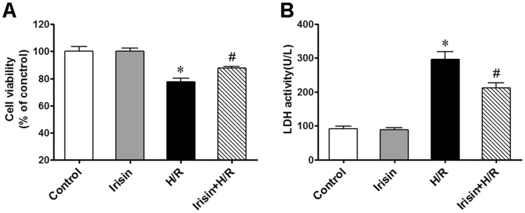
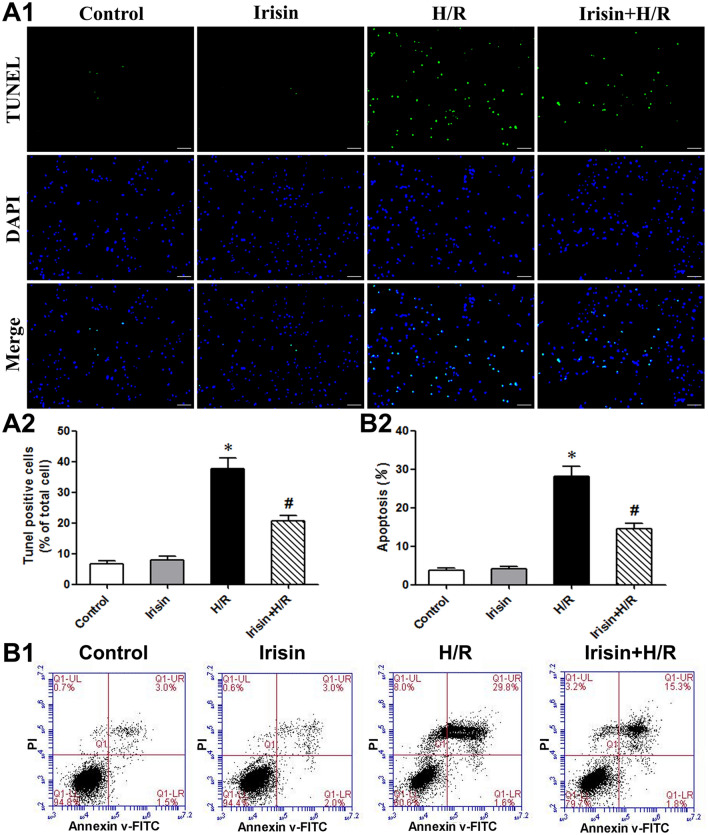
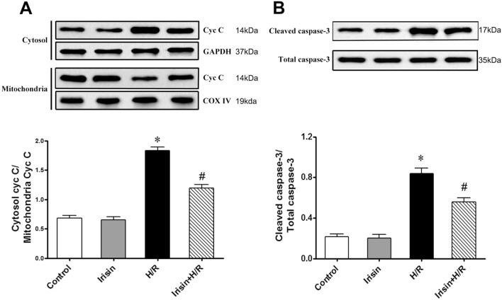
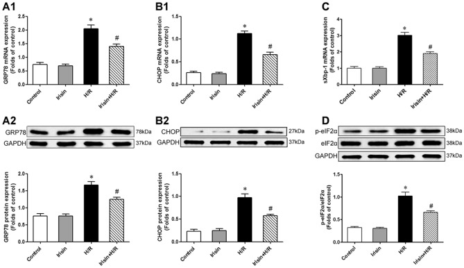
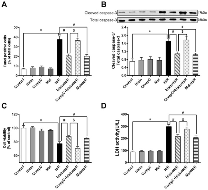
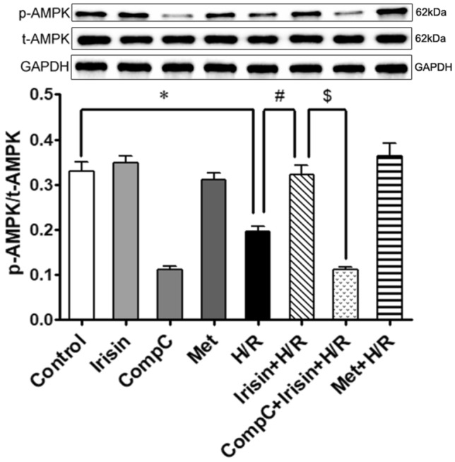
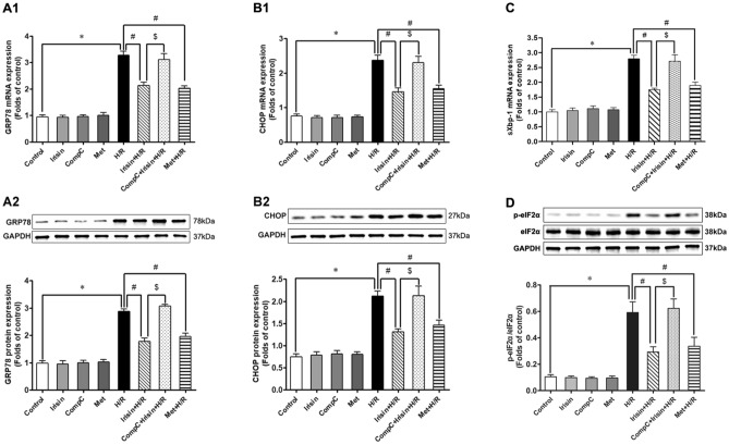
Endoplasmic reticulum (ER) stress plays a central role in myocardial ischemia/reperfusion (I/R) injury. Irisin has been reported to have protective properties in ischemia disease. In this study, we aimed at investigating whether irisin could alleviate myocardial I/R injury by ER stress attenuation. The in vitro model of hypoxia/reoxygenation (H/R) was established, which resembles I/R in vivo. Cell viability and apoptosis were estimated. Expressions of cleaved caspase-3, cytochrome c, GRP78, pAMPK, CHOP, and eIF2α were assessed by western blot. Our results revealed that pre-treatment with irisin significantly decreased cytochrome c release from mitochondria and caspase-3 activation caused by H/R. Irsin also reduced apoptosis and increased cell viability. These effects were abolished by AMPK inhibitor compound C pre-treatment. Also, GRP78 and CHOP expressions were up-regulated in the H/R group compared to the control group; however, irisin attenuated their expression. The pAMPK level was significantly decreased compared to the control, and this effect could be partly reversed by metformin pre-treatment. These results suggest that ER stress is associated with cell viability decreasing and cardiomyocytes apoptosis induced by H/R. Irisin could efficiently protect cardiomyocytes from H/R-injury via attenuating ER stress and ER stress-induced apoptosis.


Conflict of interest statement

The authors declare no competing interests.

Figures

Figure 1
Figure 1
Irisin attenuated H/R induced cell injury. Cell viability was determined by CCK-8 kit, pretreated with irisin(100 ng/ml) significantly alleviated cell viability decrease induced by H/R exposure (A). Irisin pre-treatment also significantly alleviated the release of LDH induced by H/R exposure (B). *P < 0.05 versus control group, #P < 0.05 versus H/R group, n = 6.
Figure 2
Figure 2
Irisin attenuated H/R-induced cardiomyocyte apoptosis. Representative images (A1) and quantification (A2) of cardiomyocytes in situ TUNEL assay. TUNEL-positive is green, and DAPI is blue. Scale bar: 200 mm. (B) Flow cytometry analysis of Annexin-V FITC and propidium iodide staining in cardiomyocytes. *P < 0.05 versus control group, #P < 0.05 versus H/R group, n = 6.
Figure 3
Figure 3
Irisin inhibited H/R-induced cytochrome c release and caspase-3 activation in cardiomyocytes. (A) Assessment of cytochrome c (Cyt c) in mitochondria and cytosol derived from cardiomyocytes subjected to H/R. COX IV: Cytochrome c oxidase subunit IV; GAPDH: glyceraldehyde-3-phosphate dehydrogenase. (B) Caspase-3 activation in cardiomyocytes subjected to H/R. *P < 0.05 versus control group, #P < 0.05 versus H/R group, n = 6.
Figure 4
Figure 4
H/R treatment resulted in excessive ER stress and CHOP upregulation. The GRP78, CHOP, sXbp-1, and eIF2α expressions were assessed by PCR or Western blot analysis. (A) Effects of irisin on H/R induced GRP78 mRNA (A1) and protein (A2) expression. (B) Effects of irisin on H/R induced CHOP mRNA (B1) and protein (B2) expression. (C) Effects of irisin on H/R induced sXbp-1 mRNA expression. (D) Effects of irisin on H/R induced eIF2α phosphorylation. *P < 0.05 versus control group, #P < 0.05 versus H/R group, n = 6.
Figure 5
Figure 5
Irisin blunted H/R induced apoptosis via an AMPK-mediated manner. To investigate the effect of the AMPK signal pathway in cardioprotection of irisin, the AMPK inhibitor compound C was used to pretreat cardiomyocytes(20 μM, 1 h) before irisin pre-treatment, and the AMPK activator metformin pretreats (2.5 mM, 1 h) group was used to make a comparison of apoptosis (A), caspase-3 activities (B), cell viability (C), and LDH activity (D). *P < 0.05 versus control group, #P < 0.05 versus H/R group, $P < 0.05 versus Irisin + H/R group, n = 6.
Figure 6
Figure 6
H/R treatment led to AMPK inactivation, and irisin showed the same effects on H/R cardiomyocytes as metformin. AMPK activation was estimated by detecting the phosphorylated AMPK. Irisin attenuated the inactivating of AMPK induced by H/R exposure and compound C pretreat abolished this effect. *P < 0.05 versus control group, #P < 0.05 versus H/R group, $P < 0.05 versus Irisin + H/R group, n = 6. p-AMPK phosphorylated AMPK, t-AMPK total AMPK.
Figure 7
Figure 7
Irisin showed its protective effects against H/R injury-induced ER stress in an AMPK-dependent manner. AMPK activator metformin showed the same effect as irisin in attenuating ER stress induced by H/R exposure. AMPK potent inhibitor compound C pre-treatment abolished cardioprotection of irisin on H/R cardiomyocytes. *P < 0.05 versus control group, #P < 0.05 versus H/R group, $P < 0.05 versus Irisin + H/R group, n = 6.

Similar articles

Cited by

References

    1. Virani SS, Alonso A, Benjamin EJ, Bittencourt MS, Callaway CW, Carson AP, et al. Heart disease and stroke statistics-2020 update: A report from the American Heart Association. Circulation. 2020;141:e139–e596. doi: 10.1161/CIR.0000000000000757. - DOI - PubMed
    1. Sanchis D, Llovera M, Ballester M, Comella JX. An alternative view of apoptosis in heart development and disease. Cardiovasc. Res. 2008;77:448–451. doi: 10.1093/cvr/cvm074. - DOI - PubMed
    1. Takemura G, Kanoh M, Minatoguchi S, Fujiwara H. Cardiomyocyte apoptosis in the failing heart: A critical review from definition and classification of cell death. Int. J. Cardiol. 2013;167:2373–2386. doi: 10.1016/j.ijcard.2013.01.163. - DOI - PubMed
    1. Wu MY, Yiang GT, Liao WT, Tsai AP, Cheng YL, Cheng PW, et al. Current mechanistic concepts in ischemia and reperfusion injury. Cell. Physiol. Biochem. 2018;46:1650–1667. doi: 10.1159/000489241. - DOI - PubMed
    1. Binder A, Ali A, Chawla R, Aziz HA, Abbate A, Jovin IS. Myocardial protection from ischemia-reperfusion injury post coronary revascularization. Expert Rev. Cardiovasc. Ther. 2015;13:1045–1057. doi: 10.1586/14779072.2015.1070669. - DOI - PubMed

Publication types

MeSH terms