. 2022 May 6;13(1):2516.
doi: 10.1038/s41467-022-30259-x.

Preventing erosion of X-chromosome inactivation in human embryonic stem cells

Affiliations

Preventing erosion of X-chromosome inactivation in human embryonic stem cells

Marissa Cloutier et al. Nat Commun. .

Abstract

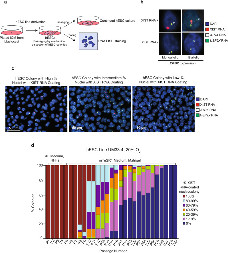
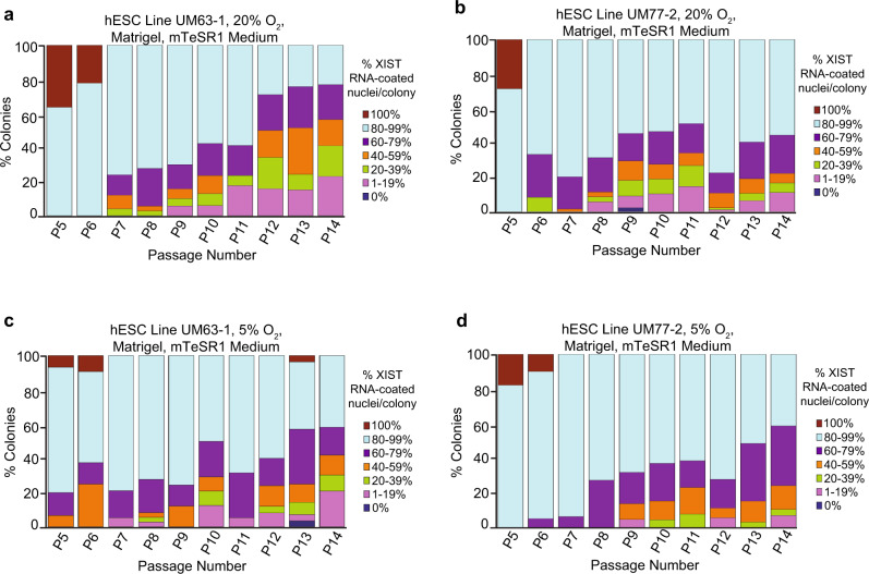
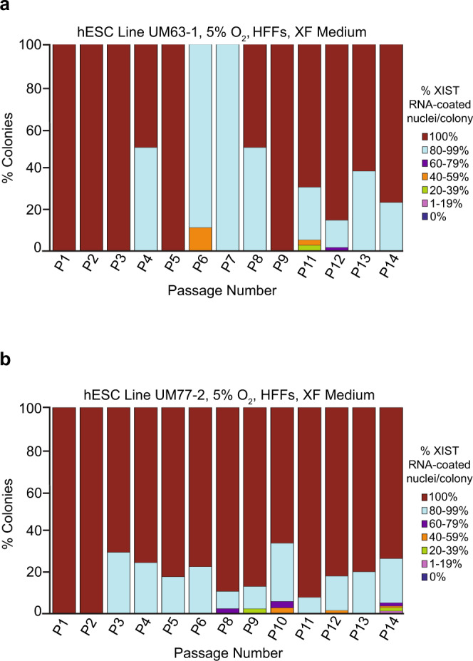
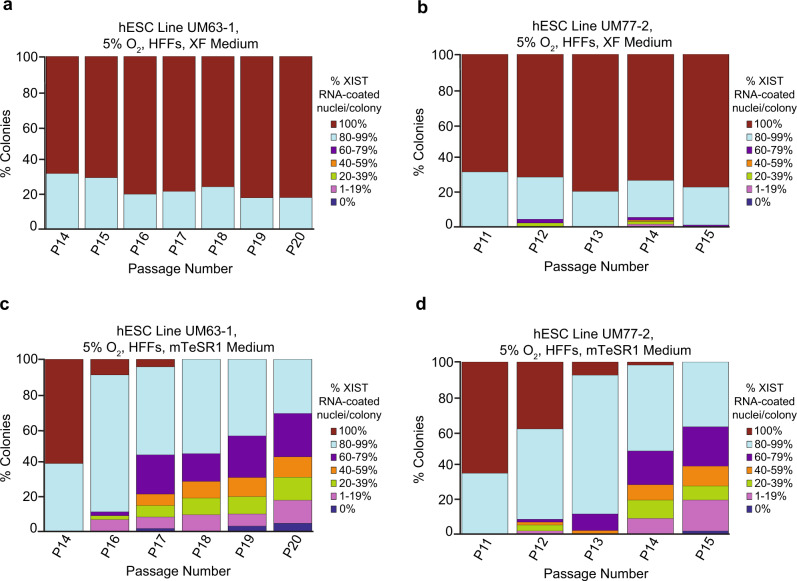
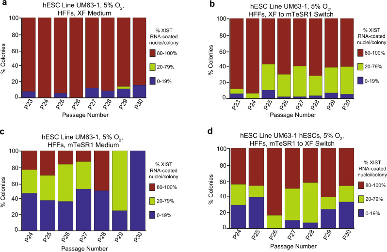
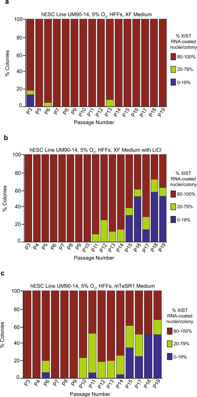
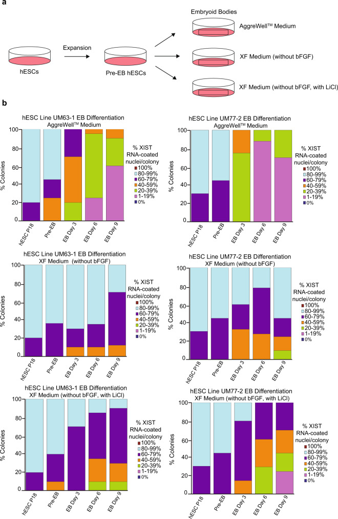
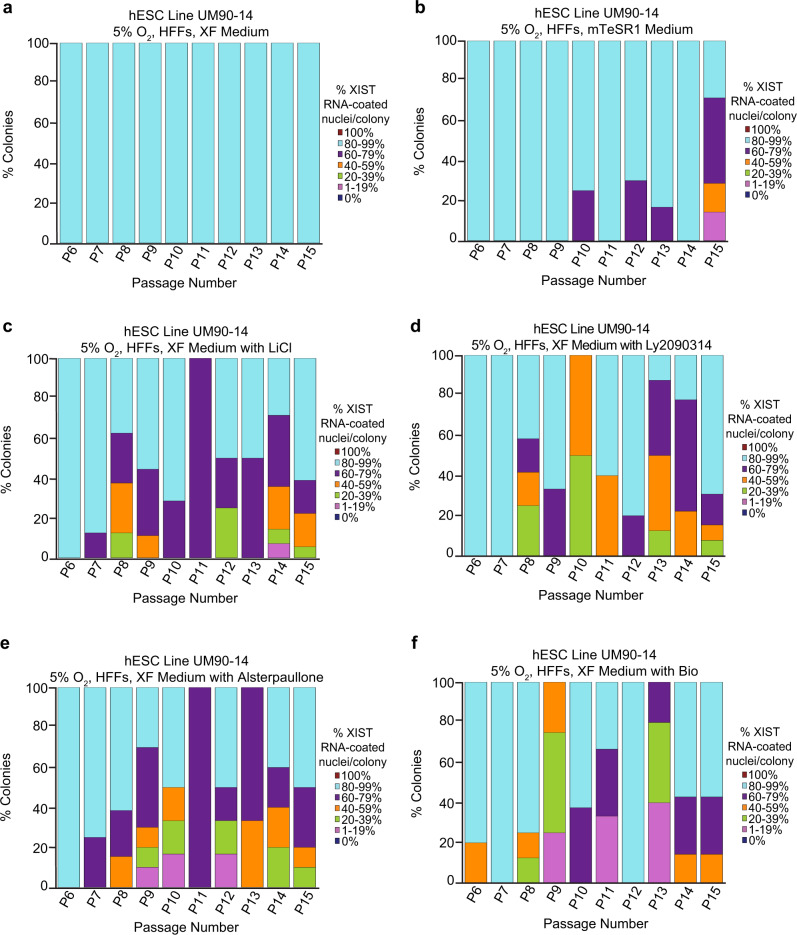
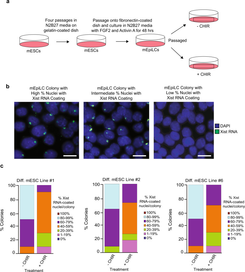
X-chromosome inactivation is a paradigm of epigenetic transcriptional regulation. Female human embryonic stem cells (hESCs) often undergo erosion of X-inactivation upon prolonged culture. Here, we investigate the sources of X-inactivation instability by deriving new primed pluripotent hESC lines. We find that culture media composition dramatically influenced the expression of XIST lncRNA, a key regulator of X-inactivation. hESCs cultured in a defined xenofree medium stably maintained XIST RNA expression and coating, whereas hESCs cultured in the widely used mTeSR1 medium lost XIST RNA expression. We pinpointed lithium chloride in mTeSR1 as a cause of XIST RNA loss. The addition of lithium chloride or inhibitors of GSK-3 proteins that are targeted by lithium to the defined hESC culture medium impeded XIST RNA expression. GSK-3 inhibition in differentiating female mouse embryonic stem cells and epiblast stem cells also resulted in a loss of XIST RNA expression. Together, these data may reconcile observed variations in X-inactivation in hESCs and inform the faithful culture of pluripotent stem cells.


Conflict of interest statement

The authors declare no competing interests.

Figures

Fig. 1
Fig. 1. Loss of XIST RNA coating upon prolonged passaging of female hESCs.
a Schematic depicting the derivation, culture, passaging, and RNA FISH staining of hESCs in this study. b Representative nuclei stained to detect XIST RNA (red), RNAs from X-linked genes ATRX (white) and USP9X (green), and the nucleus with DAPI (blue). Top, representative nuclei with XIST RNA coating. Bottom, representative nuclei without XIST RNA coating. At least 100 nuclei were counted per colony for hESC RNA FISH quantification. The total number of colonies quantified at each passage range from 2–138 and are cataloged in source data. c Representative hESC colonies stained to detect XIST RNA (red), ATRX RNA (white), and USP9X RNA (green). Nuclei are stained blue with DAPI. At least 100 nuclei were counted per colony for hESC RNA FISH quantification. d Percentage of nuclei with XIST RNA coats per colony in hESC line UM33-4 derived in XenoFree (XF) medium on human fibroblast feeders (HFFs) and cultured subsequently on Matrigel under atmospheric oxygen levels (20%). The percentage of nuclei with XIST RNA coats in individual hESC colonies were stratified into 20% increments. 100% value indicates that all nuclei in a colony harbored XIST RNA coats, whereas 0% indicates that all nuclei lacked XIST RNA coats in a colony. The percentage of colonies harboring nuclei with XIST RNA coats decreased significantly with passage number (general linear model, p = 0.002). See also Supplementary Fig. 2. Source data are provided as a Source Data file.
Fig. 2
Fig. 2. XIST RNA coating in female hESCs cultured in atmospheric vs. physiological O2 concentration.
ad Percentage of nuclei with XIST RNA coats per colony of hESC lines UM63-1 (a, c) and UM77-2 (b, d) cultured in parallel under 20% (a, b) and 5% (c, d) O2 concentration on Matrigel in mTeSR1 medium. The difference in the frequency of nuclei without XIST RNA coats per colony in either cell line when cultured at physiological vs. atmospheric O2 concentration is not significant (general linear model comparison, p = 0.1). See also Supplementary Fig. 2. At least 100 nuclei were counted per colony for hESC RNA FISH quantification. The total number of colonies quantified at each passage range between 11–93 and are cataloged in source data. Source data are provided as a Source Data file.
Fig. 3
Fig. 3. Impact of culture surface on XIST RNA coating in female hESCs.
a, b Percentage of nuclei with XIST RNA coats in colonies of hESC line UM63-1 (a) and UM77-2 (b) cultured on HFFs in XF medium under 5% O2 concentration. The frequency of nuclei harboring XIST RNA coats per colony in either cell line when cultured on HFFs did not decrease significantly (general linear model, p = 0.09). At least 100 nuclei were counted per colony for hESC RNA FISH quantification. The total number of colonies quantified at each passage range between 1 and 126 and are cataloged in source data. Source data are provided as a Source Data file.
Fig. 4
Fig. 4. Impact of culture medium on XIST RNA coating in female hESCs.
ad Percentage of nuclei with XIST RNA coats per colony in hESC lines UM63-1 (a, c) and UM77-2 (b, d) cultured in parallel in XF medium (a, b) and mTeSR1 medium (c, d) on HFFs. hESCs cultured with mTeSR1 medium displayed a significant decrease in nuclei with XIST RNA coating compared to hESCs cultured in XF medium during passaging (general linear model comparison; p < 0.001). All hESCs in this experiment were cultured in 5% O2 on HFFs. The quantification data for P13-14 in b are taken from Fig. 3b. At least 100 nuclei were counted per colony for hESC RNA FISH quantification. The total number of colonies quantified at each passage range between 10 and 180 and are cataloged in source data. Source data are provided as a Source Data file.
Fig. 5
Fig. 5. Analysis of culture media switching on XIST RNA coating in female hESCs.
Percentage of nuclei with XIST RNA coats per colony of hESC line UM63-1 (a) cultured continuously in XF medium and (b) cultured initially in XF medium and subsequently switched to mTeSR1 medium. Percentage of nuclei with XIST RNA coats per colony of hESC line UM63-1 (c) continuously cultured in mTeSR1 medium and (d) cultured initially in mTeSR1 medium and then switched to XF medium. hESCs cultured initially in XF medium and subsequently switched to mTeSR1 medium displayed a significant decrease in nuclei with XIST RNA coating during passaging compared to hESCs cultured continuously in XF medium (general linear model comparison, p = 0.01). hESCs cultured continuously in mTeSR1 medium displayed a significant decrease in nuclei with XIST RNA coating during passaging compared to hESCs cultured initially in mTeSR1 medium and then switched to XF medium (general linear model comparison, p < 0.001). All hESCs in this experiment were cultured in 5% O2 on HFFs. See also Supplementary Fig. 3 and Supplementary Fig. 4. At least 100 nuclei were counted per colony for hESC RNA FISH quantification. The total number of colonies quantified at each passage range between 2 and 148 and are cataloged in source data. Source data are provided as a Source Data file.
Fig. 6
Fig. 6. LiCl in mTeSR1 Medium as a Cause of XIST RNA Loss in Female hESCs.
ac Percentage of nuclei with XIST RNA coating in colonies of hESC line UM90-14 cultured in XF medium (a), XF medium supplemented with 0.98 mM LiCl (XF with LiCl) (b), and mTeSR1 medium (c). hESCs cultured in XF medium did not display a significant decrease in nuclei with XIST RNA coats across passaging (general linear model, p = 0.2). hESCs cultured in XF medium with LiCl and mTeSR1 medium lost XIST RNA coating in a significant percentage of nuclei per colony during passaging compared to cells cultured in XF medium (general linear model, p < 0.001). All hESCs in this experiment were cultured in 5% O2 on HFFs. At least 100 nuclei were counted per colony for hESC RNA FISH quantification. The total number of colonies quantified at each passage range between 5 and 78 and are cataloged in source data. Source data are provided as a Source Data file.
Fig. 7
Fig. 7. Analysis of XIST RNA coating during differentiation of female hESCs.
a Schematic of hESC differentiation into embryoid bodies (EBs) with three different media formulations: a commercially available AggreWellTM medium; XF medium lacking bFGF; and XF medium lacking bFGF but containing 0.98 mM LiCl. b Percentage of nuclei with XIST RNA coating in EBs generated from hESC lines UM77-2 and UM63-1. EBs generated and cultured in XF medium with LiCl and AggreWellTM medium lost a significant proportion of XIST RNA coating per colony compared to EBs generated and cultured in XF medium (general linear model comparison; p < 0.001). At least 100 nuclei were counted per colony for hESC RNA FISH quantification. The total number of colonies quantified at each passage range between 10 and 17 and are cataloged in source data. Source data are provided as a Source Data file.
Fig. 8
Fig. 8. GSK-3 Inhibition and Loss of XIST RNA Coating in Female hESCs.
Percentage of nuclei with XIST RNA coating in colonies of hESC line UM90-14 cultured in XF medium (a); mTeSR1 medium (b); XF medium supplemented with 0.98 mM LiCl (XF with LiCl) (c); XF medium supplemented with 1.5 nM Ly2090314 (XF with Ly2090314) (d); XF medium supplemented with 5.0 nM Alsterpaullone (XF with Alsterpaullone) (e); and, XF medium supplemented with 5.0 nM BIO (XF with Bio) (f). hESCs cultured in mTeSR1, XF with LiCl, XF with Ly2090314, XF with Alsterpaullone, and XF with BIO media lost XIST RNA coating during passaging in a significant percentage of nuclei compared to hESCs cultured in XF medium (general linear model comparison, p < 0.001). All hESCs in this experiment were cultured in 5% O2 on HFFs. At least 100 nuclei were counted per colony for hESC RNA FISH quantification. The total number of colonies quantified at each passage range between 1 and 18 and are cataloged in source data. Source data are provided as a Source Data file.
Fig. 9
Fig. 9. GSK-3 Inhibition and Loss of Xist RNA Coating in Differentiating Female mESCs.
a Strategy for the differentiation of mESC lines into mEpiLCs and culture of mEpiLCs with and without the GSK-3 inhibitor CHIR99021 (CHIR; 3 μM). b Representative images of mEpiLCs with high, intermediate, and low percent of nuclei with Xist RNA coating (green). Nuclei are stained blue with DAPI. At least ten nuclei were counted per colony in mEpiLC RNA FISH quantification. Scale bars are ~100 microns. c Percentage of nuclei with Xist RNA coating in differentiating mEpiLCs with and without CHIR generated from three independent ESC lines. mEpiLCs cultured with CHIR lost a significant proportion of Xist RNA coating compared to mEpiLCs cultured without CHIR in all three mEpiLC replicates (general linear model comparison, p < 0.001). At least ten nuclei were counted per colony for mEpiLC RNA FISH quantification. The total number of colonies quantified at each passage range between 12 and 20 and are cataloged in source data. Source data are provided as a Source Data file.
Fig. 10
Fig. 10. GSK-3 Inhibition and Loss of Xist RNA Coating in Female mEpiSCs.
a Strategy to test the impact of GSK-3 inhibition on Xist RNA coating in mEpiSCs. b Representative RNA FISH images of colonies with high, intermediate, and low percentage of Xist RNA coating (green). Nuclei are stained blue with DAPI. At least ten nuclei were counted per colony in mEpiSC RNA FISH quantification. Scale bars are ~100 microns. c Percentage of nuclei with Xist RNA coating in three independent mEpiSC lines cultured with and without the GSK-3 inhibitor CHIR99021 (CHIR; 3 μM). All three mEpiSC lines cultured with CHIR lost a significant proportion of Xist RNA coating compared to mEpiSCs cultured without CHIR (general linear model comparison, p < 0.001). d Model of direct or indirect repression of human XIST and mouse Xist expression. Conserved mouse and human Xist/XIST sequences upstream of the Xist/XIST TSSs shown, with putative TCF binding motifs in red and surrounding conserved sequence in blue. At least ten nuclei were counted per colony for mEpiSC RNA FISH quantification. The total number of colonies quantified at each passage range between 15 and 25 and are cataloged in source data. Source data are provided as a Source Data file.

Similar articles

Cited by

References

    1. Thomson JA, et al. Embryonic stem cell lines derived from human blastocysts. Science. 1998;282:1145–1147. doi: 10.1126/science.282.5391.1145. - DOI - PubMed
    1. Rossant J, Tam PPL. New insights into early human development: lessons for stem cell derivation and differentiation. Cell Stem Cell. 2017;20:18–28. doi: 10.1016/j.stem.2016.12.004. - DOI - PubMed
    1. Nichols J, Smith A. Naive and primed pluripotent states. Cell Stem Cell. 2009;4:487–492. doi: 10.1016/j.stem.2009.05.015. - DOI - PubMed
    1. Plath K, Mlynarczyk-Evans S, Nusinow DA, Panning B. Xist RNA and the mechanism of X chromosome inactivation. Annu Rev. Genet. 2002;36:233–278. doi: 10.1146/annurev.genet.36.042902.092433. - DOI - PubMed
    1. Morey, C. A. A. P. The demoiselle of X-inactivation: 50 years old and as trendy and mesmerising as ever. PLoS Genet.7, e1002212 (2011). - PMC - PubMed

Publication types