

Impact of tissue properties on time-dependent alterations in apparent diffusion coefficient: a phantom study using oscillating-gradient spin-echo and pulsed-gradient spin-echo sequences
- PMID: 35523921
- PMCID: PMC9441423
- DOI: 10.1007/s11604-022-01281-2
Impact of tissue properties on time-dependent alterations in apparent diffusion coefficient: a phantom study using oscillating-gradient spin-echo and pulsed-gradient spin-echo sequences
Abstract
Purpose: The purpose of this study was to investigate whether the changes in apparent diffusion coefficients (ADCs) due to differences in diffusion time reflect tissue properties in actual measurements of phantoms.
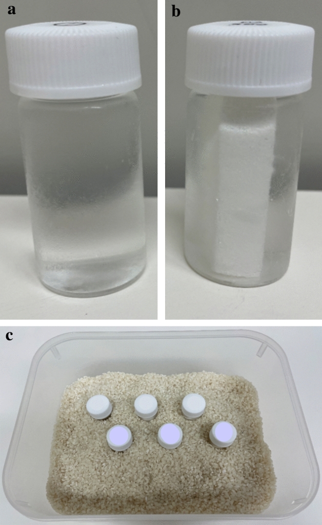
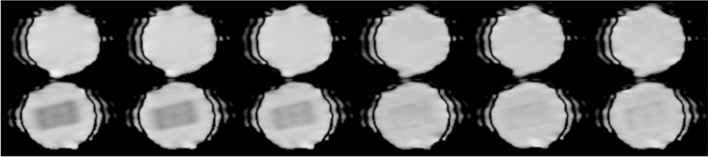
Materials and methods: Various n-alkane phantoms and sucrose/collagen phantoms with various collagen densities were set up with and without polyvinyl alcohol (PVA) foam with an average pore diameter of 300 μm. Thus, n-alkanes or sucrose/collagen represented substrate viscosity and the presence of PVA foam represented tissue structure with septum. Diffusion-weighted images with various diffusion times (7.71-60 ms) were acquired using pulsed-gradient spin-echo (PGSE) and oscillating-gradient spin-echo (OGSE) sequences. The ADCs of the phantoms with and without PVA foam were calculated.
Results: The ADCs of some of the phantoms without PVA decreased with diffusion times decreased. In the n-alkane phantoms, only C8H18 showed significantly different ADCs depending on the use of PVA foam in the OGSE sequence. On the other hand, sucrose/collagen phantoms showed significant differences according to diffusion time. The ADCs of the phantoms decreased as the molecular size of the n-alkanes or collagen density of the sucrose/collagen phantom increased. Compared to phantoms without PVA foam, the ADC of the phantoms with PVA foam decreased as the diffusion time increased.
Conclusion: Changes in ADCs due to differences in diffusion time reflect tissue properties in actual measurements of phantoms. These changes in ADCs can be used for tissue characterization in vivo.
Keywords: ADC; Diffusion time; Oscillating-gradient spin-echo; Phantom; Spatial restriction.
© 2022. The Author(s).
Conflict of interest statement
There are no other conflicts of interest to declare.
Figures


Similar articles
-
Changes in the ADC of diffusion-weighted MRI with the oscillating gradient spin-echo (OGSE) sequence due to differences in substrate viscosities.Jpn J Radiol. 2018 Jul;36(7):415-420. doi: 10.1007/s11604-018-0737-0. Epub 2018 Apr 26. Jpn J Radiol. 2018. PMID: 29700795
-
Measuring small compartment dimensions by probing diffusion dynamics via Non-uniform Oscillating-Gradient Spin-Echo (NOGSE) NMR.J Magn Reson. 2013 Dec;237:49-62. doi: 10.1016/j.jmr.2013.09.009. Epub 2013 Sep 23. J Magn Reson. 2013. PMID: 24140623
-
Validation of surface-to-volume ratio measurements derived from oscillating gradient spin echo on a clinical scanner using anisotropic fiber phantoms.NMR Biomed. 2017 May;30(5):10.1002/nbm.3708. doi: 10.1002/nbm.3708. Epub 2017 Mar 22. NMR Biomed. 2017. PMID: 28328013 Free PMC article.
-
Measuring restriction sizes using diffusion weighted magnetic resonance imaging: a review.Magn Reson Insights. 2013 May 19;6:59-64. doi: 10.4137/MRI.S11149. eCollection 2013. Magn Reson Insights. 2013. PMID: 25114548 Free PMC article. Review.
-
Characterization of tissue structure at varying length scales using temporal diffusion spectroscopy.NMR Biomed. 2010 Aug;23(7):745-56. doi: 10.1002/nbm.1531. NMR Biomed. 2010. PMID: 20677208 Free PMC article. Review.
Cited by
-
Current State of Artificial Intelligence in Clinical Applications for Head and Neck MR Imaging.Magn Reson Med Sci. 2023 Oct 1;22(4):401-414. doi: 10.2463/mrms.rev.2023-0047. Epub 2023 Aug 1. Magn Reson Med Sci. 2023. PMID: 37532584 Free PMC article. Review.
References
-
- Nagaraja N. Diffusion weighted imaging in acute ischemic stroke: a review of its interpretation pitfalls and advanced diffusion imaging application. J Neurol Sci. 2021;15:425. - PubMed
-
- Lo CP, Kao HW, Chen SY, Chu CM, Hsu CC, Chen YC, et al. Comparison of diffusion-weighted imaging and contrast-enhanced T1-weighted imaging on a single baseline MRI for demonstrating dissemination in time in multiple sclerosis. BMC Neurol. 2014;14:100. doi: 10.1186/1471-2377-14-100. - DOI - PMC - PubMed
-
- Stejskal EO, Tanner JE. Spin diffusion measurements: spin echoes in the presence of a time-dependent field gradient. J Chem Phys. 1965;42:288–292. doi: 10.1063/1.1695690. - DOI
MeSH terms
Substances
LinkOut - more resources
Full Text Sources
Miscellaneous
