. 2022 May 6;12(1):7494.
doi: 10.1038/s41598-022-11634-6.

Investigation of role of CpG methylation in some epithelial mesenchymal transition gene in a chemoresistant ovarian cancer cell line

Affiliations

Investigation of role of CpG methylation in some epithelial mesenchymal transition gene in a chemoresistant ovarian cancer cell line

Yaman Alghamian et al. Sci Rep. .

Abstract

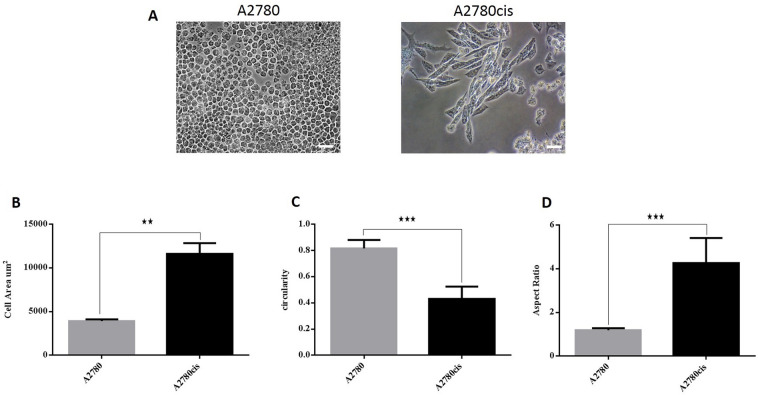
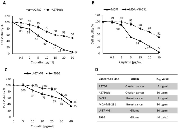
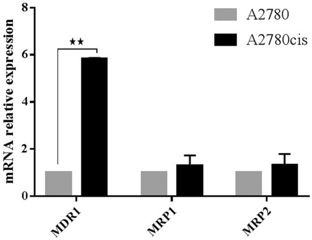
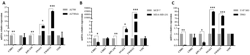
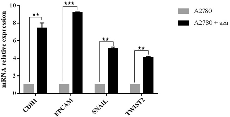
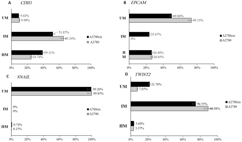
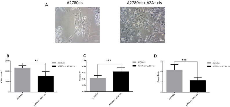
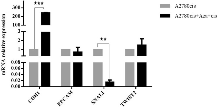
Ovarian cancer is one of the lethal gynecologic cancers. Chemoresistance is an essential reason for treatment failure and high mortality. Emerging evidence connects epithelial-mesenchymal transition (EMT) like changes and acquisition of chemoresistance in cancers. Including EMT, DNA methylation influences cellular processes. Here, EMT-like changes were investigated in cisplatin-resistant A2780 ovarian cancer cells (A2780cis), wherein role of DNA methylation in some EMT genes regulations was studied. Cell viability assay was carried out to test the sensitivity of A2780, and A2780cis human cancer cell lines to cisplatin. Differential mRNA expression of EMT markers using qPCR was conducted to investigate EMT like changes. CpG methylation role in gene expression regulation was investigated by 5-azacytidine (5-aza) treatment. DNA methylation changes in EMT genes were identified using Methylscreen assay between A2780 and A2780cis cells. In order to evaluate if DNA methylation changes are causally underlying EMT, treatment with 5-aza followed by Cisplatin was done on A2780cis cells. Accordingly, morphological changes were studied under the microscope, whereas EMT marker's gene expression changes were investigated using qPCR. In this respect, A2780cis cell line has maintained its cisplatin tolerance ability and exhibits phenotypic changes congruent with EMT. Methylscreen assay and qPCR study have revealed DNA hypermethylation in promoters of epithelial adhesion molecules CDH1 and EPCAM in A2780cis compared to the cisplatin-sensitive parental cells. These changes were concomitant with gene expression down-regulation. DNA hypomethylation associated with transcription up-regulation of the mesenchymal marker TWIST2 was observed in the resistant cells. Azacytidine treatment confirmed DNA methylation role in regulating gene expression of CDH1, EPCAM and TWIST2 genes. A2780cis cell line undergoes EMT like changes, and EMT genes are regulated by DNA methylation. To that end, a better understanding of the molecular alterations that correlate with chemoresistance may lead to therapeutic benefits such as chemosensitivity restoration.


Conflict of interest statement

The authors declare no competing interests.

Figures

Figure 1
Figure 1
Cellular shapes of A2780cis and A2780 cells. (A) A2780 cells showed small round-shaped cells and A2780cis exhibit larger, more elongated shaped cells. Scale bar = 200 µm. (B) The mean cell area of A2780 and A2780cis cells shows that A2780cis cells have a larger area compared to A2780. (C) Cell circularity confirms that A2780 cells have a higher (mean) circularity value compared to A2780cis. (D) A2780cis have an aspect ratio above 4 while the round shape of A2780 is reflected by an aspect ratio ~ 1.
Figure 2
Figure 2
Cellular shapes of MDA-MB-231 and MCF7 cells. (A) MCF7 cells showed squamous epithelial cell shape and MDA-MB-231 exhibit spindle-like morphology. Scale bar = 200 µm. (B) The mean cell area of MCF7 and MDA-MB-231 cells shows that MDA-MB-231 cells have a larger area compared to MCF7. (C) Cell circularity confirms that MCF7 cells have a higher (mean) circularity value compared to MDA-MB-231. (D) MDA-MB-231 cells have an aspect ratio above 5 while the round shape of MCF7 is reflected by an aspect ratio ~ 1.
Figure 3
Figure 3
Cellular shapes of T98G and U-87 MG cells. (A) U-87 MG cells and T98G cells showed spindle-like morphology. Scale bar = 200 µm. (B) The mean cell area of U-87 MG and T98G cells shows that T98G cells have a larger area compared to U-87 MG cells. (C) Cell circularity confirms that U-87 MG cells have a higher (mean) circularity value compared to T98G cells. (D) T98G cells have a higher aspect ratio ~ 5 than U-87 MG (aspect ratio ~ 2).
Figure 4
Figure 4
Cancer cells exhibit different resistance to cisplatin-induced cytotoxicity. (A): A2780 and A2780cis cells. (B): MCF7 and MDA-MB-231 cells. (C) U-87 MG and T98G cells. (D) Table showed IC50s of the studied cell lines.
Figure 5
Figure 5
qPCR analysis of the MDR genes in A2780cis cells. MDR1 Expression increases in the resistant variant A2780cis cells. The expression levels were normalized to parental cells A2780.
Figure 6
Figure 6
Resistant cancer cells display molecular changes consistent with partial EMT. Down-regulation in epithelial genes: CDH1 and EPCAM, and up-regulation in mesenchymal marker SNAIL and TWIST2, were assessed using qPCR, no significant expression changes in CDH2 and VIM genes were observed. (A) A2780cis compared to parental cells A2780. (B) MDA-MB-231 cisplatin resistant cells compared to MCF7 cisplatin sensitive cells. (C) T98G cisplatin resistant cells compared to U-87 MG the more sensitive cisplatin cells.
Figure 7
Figure 7
EMT marker gene expression changes after exposure to the demethylating agent 5-azacytidine. qPCR validation of gene expression showed a significant up-regulation of EMT genes in A2780 cells treated with azacytidine compared with control.
Figure 8
Figure 8
Methyscreen assay for CDH1, EPCAM, SNAIL, and TWIST2 in A2780 and A2780cis cell lines. (A) Charts display the result of CDH1 assay as a percentage of each portion of DNA. (B) Charts display the result of EPCAM assay as a percentage of each portion of DNA. (C) Charts display the result of SNAIL assay as a percentage of each portion of DNA. (D) Charts display the result of TWIST2 assay as a percentage of each portion of DNA.
Figure 9
Figure 9
A2780cis cancer cell line treated with aza and cisplatin displays morphological changes associated with partial MET. (A) A2780cis cells showed elongated shaped cells and treated cells exhibit smaller, more circular shaped cells. Scale bar = 200 µm. (B) The mean cell area of A2780cis and A2780cis treated cells shows that treatment cause a reduction in cell size. (C) Cell circularity confirms that A2780cis treated cells have a higher (mean) circularity value compared to A2780cis. (D) Treatment of A2780cis with aza and cisplatin reduces the aspect ratio to 2.
Figure 10
Figure 10
Treatment of A2780cis cells with aza and cisplatin causes molecular changes consistent with partial MET. Up-regulation in epithelial gene CDH1, and down-regulation in mesenchymal marker SNAIL, were assessed using qPCR. No significant changes in EPCAM and TWIST2 gene expression were observed in A2780cis treated cells.

Similar articles

Cited by

References

    1. Torre LA, et al. Ovarian cancer statistics, 2018. CA Cancer J. Clin. 2018;68:284–296. doi: 10.3322/caac.21456. - DOI - PMC - PubMed
    1. Siegel RL, Miller KD, Fuchs HE, Jemal A. Cancer statistics, 2021. CA Cancer J. Clin. 2021;71:7–33. doi: 10.3322/caac.21654. - DOI - PubMed
    1. Motohara T, Katabuchi H. Ovarian cancer stemness: Biological and clinical implications for metastasis and chemotherapy resistance. Cancers (Basel) 2019 doi: 10.3390/cancers11070907. - DOI - PMC - PubMed
    1. Momenimovahed Z, Tiznobaik A, Taheri S, Salehiniya H. Ovarian cancer in the world: Epidemiology and risk factors. Int. J. Womens Health. 2019;11:287–299. doi: 10.2147/IJWH.S197604. - DOI - PMC - PubMed
    1. Raja FA, Chopra N, Ledermann JA. Optimal first-line treatment in ovarian cancer. Ann. Oncol. 2012;23(Suppl 10):x118–127. doi: 10.1093/annonc/mds315. - DOI - PubMed

MeSH terms