. 2022 May 6;5(1):425.
doi: 10.1038/s42003-022-03392-y.

Laminar shear stress inhibits inflammation by activating autophagy in human aortic endothelial cells through HMGB1 nuclear translocation

Affiliations

Laminar shear stress inhibits inflammation by activating autophagy in human aortic endothelial cells through HMGB1 nuclear translocation

Qingyu Meng et al. Commun Biol. .

Abstract

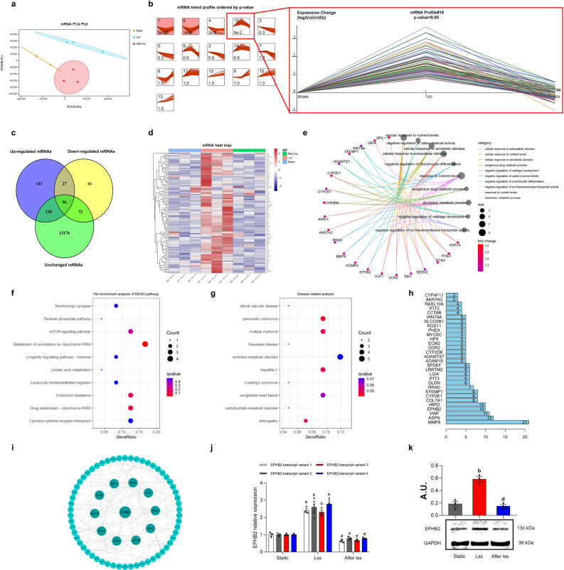
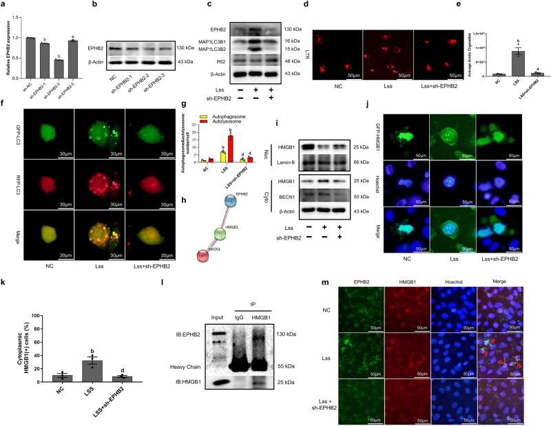
Prevention and treatment of atherosclerosis (AS) by targeting the inflammatory response in vascular endothelial cells has attracted much attention in recent years. Laminar shear stress (LSS) has well-recognized anti-AS properties, however, the exact molecular mechanism remains unclear. In this study, we found that LSS could inhibit the increased expression of intercellular adhesion molecule-1 (ICAM-1), vascular cell adhesion molecule-1 (VCAM-1), cyclooxygenase-2 (COX-2), and matrix metallopeptidase-9 (MMP-9) caused by TNF-α in an autophagy-dependent pathway in human aortic endothelial cells (HAECs) and human umbilical vein endothelial cells (HUVECs). Whole-transcriptome sequencing analysis revealed that erythropoietin-producing hepatocyte receptor B2 (EPHB2) was a key gene in response to LSS. Moreover, co-immunoprecipitation assay indicated that LSS could enhance the EPHB2-mediated nuclear translocation of high mobility group box-1 (HMGB1), which interacts with Beclin-1 (BECN1) and finally leads to autophagy. Simultaneously, we identified an LSS-sensitive long non-coding RNA (lncRNA), LOC10798635, and constructed an LSS-related LOC107986345/miR-128-3p/EPHB2 regulatory axis. Further research revealed the anti-inflammatory effect of LSS depends on autophagy activation resulting from the nuclear translocation of HMGB1 via the LOC107986345/miR-128-3p/EPHB2 axis. Our study demonstrates that LSS could regulate the expression of EPHB2 in HAECs, and the LOC107986345/miR-128-3p/EPHB2 axis plays a vital role in AS development.


Conflict of interest statement

The authors declare no competing interests.

Figures

Fig. 1
Fig. 1. LSS inhibits the inflammatory response in endothelial cells by activating autophagy.
a, c Western Blot shows the expression of MAP1LC3B2 and SQSTM1/P62 in HAECs and HUVECs. b, d Relative expression of MAP1LC3B2 and SQSTM1/P62 in HAECs and HUVECs. Cells were treated with 12 dyn/cm2 LSS for 12 h, CQ (50 μM) precedes LSS for 6 h, and e, g Western Blot shows the expression of MAP1LC3B2 in HAECs and HUVECs. f, h Relative expression of MAP1LC3B2 in HAECs and HUVECs. Cells were stimulated with TNF-α (10 ng/ml) for 12 h and then treated with LSS for another 12 h, and i, k Western Blot shows the expression of ICAM-1, VCAM-1, COX-2, MMP-9, MAP1LC3B2, and SQSTM1/P62 in HAECs and HUVECs. j, l Relative expression of ICAM-1, VCAM-1, COX-2, MMP-9, MAP1LC3B2, and SQSTM1/P62 in HAECs and HUVECs. Cells were pretreated with TNF-α (10 ng/ml) for 12 h and in the presence or absence of 3-MA (5 mM) for another 2 h, followed by LSS treatment for another 12 h, and m, o Western Blot shows the expression of ICAM-1, VCAM-1, COX-2, MMP-9, MAP1LC3B2, and SQSTM1/P62 in HAECs and HUVECs. n, p. Relative expression of ICAM-1, VCAM-1, COX-2, MMP-9, MAP1LC3B2, and SQSTM1/P62 in HAECs and HUVECs. Data are presented as mean ± SEM of three independent experiments. aP < 0.05, bP < 0.01 vs. Static or NC group; cP < 0.05, dP < 0.01 vs. LSS or TNF-α or CQ group; eP < 0.05, fP < 0.01 vs. TNF-α + LSS group. AU arbitrary units.
Fig. 2
Fig. 2. Whole-transcriptome sequencing analysis revealed that EPHB2 was a key gene in response to LSS.
a mRNA PCA analysis of HAECs in three groups. b mRNA trend profiles ordered by P value. Expression Change (log2(v(i)/v(0))). c DE mRNA Venn diagram. d DE mRNA heat map. e DE mRNA GO biological process analysis. f DE mRNA KEGG pathway analysis. g DE mRNA disease correlation analysis. h PPI histogram of DE mRNA. i PPI network diagram with EPHB2 as the core. j qRT-PCR shows the expression of EPHB2. k Western Blot shows the expression of EPHB2. Data are presented as mean ± SEM of three independent experiments. aP < 0.05, bP < 0.01 vs. Static group; cP < 0.05, dP < 0.01 vs. LSS group. AU arbitrary units.
Fig. 3
Fig. 3. LSS activates autophagy flux by enhancing the EPHB2-mediated nuclear translocation of HMGB1.
a qRT-PCR shows the expression of EPHB2. b Western Blot shows the expression of EPHB2. c Western Blot shows the expression of EPHB2, MAP1LC3B2, and SQSTM1/P62. d, e Confocal microscopy shows HAECs stained with Lyso Tracker Red, and lysosomes show red fluorescence. f, g The mRFP-GFP-LC3 adenovirus were used to observe autophagic flux under a laser confocal microscope. mRFP was used to label and track LC3. Weakening of GFP indicates fusion of lysosomes and autophagosomes to form autophagolysosomes. h PPI network functional enrichment analysis of EPHB2, HMGB1, and BECN1. i The nucleoplasmic separation experiment shows the subcellular localization of HMGB1. j, k. GFP-HMGB1 plasmid were used to observe the nuclear translocation of HMGB1 under a laser confocal microscope. l Co-immunoprecipitation assay shows the physical interaction between HMGB1 and EPHB2 in HAECs under the action of LSS, and the cell lysates were immunoprecipitated by anti-HMGB1 antibody. m. Immunofluorescence assay shows the co-localization of EPHB2 and HMGB1 in HAECs under the action of LSS. Data are presented as mean ± SEM of three independent experiments. aP < 0.05, bP < 0.01 vs. NC group; cP < 0.05, dP < 0.01 vs. LSS group.
Fig. 4
Fig. 4. Overexpression of EPHB2 can activate autophagy and exert the same anti-inflammatory effect as LSS.
a, b Western Blot shows the effect of GA (100 μM) on anti-inflammatory effect of LSS. c Confocal microscopy shows the effect of GA on LSS activated autophagy (HAECs stained with Lyso Tracker Red) and inhibited monocyte adhesion. d qRT-PCR shows the expression of EPHB2. e The nucleoplasmic separation experiment shows the subcellular localization of HMGB1 when EPHB2 was overexpressed. f Co-immunoprecipitation assay shows the physical interaction between HMGB1 and EPHB2 in HAECs when EPHB2 was overexpressed, and the cell lysates were immunoprecipitated by anti-HMGB1 antibody. g Immunofluorescence assay shows the co-localization of EPHB2 and HMGB1 in HAECs when EPHB2 was overexpressed. h, i Western Blot shows the effect of GA on anti-inflammatory effect of pri-EPHB2. j Confocal microscopy shows the effect of GA on pri-EPHB2 activated autophagy (HAECs stained with Lyso Tracker Red) and inhibited monocyte adhesion. k, l. Western Blot shows the effect of HMGB1 siRNA on anti-inflammatory effect of LSS. m, n. Western Blot shows the effect of HMGB1 siRNA on anti-inflammatory effect of pri-EPHB2. Data are presented as mean ± SEM of three independent experiments. aP < 0.05, bP < 0.01 vs. NC group; cP < 0.05, dP < 0.01 vs. TNF-α group; eP < 0.05, fP < 0.01 vs. TNF-α + LSS or pri-EPHB2 group. AU, arbitrary units.
Fig. 5
Fig. 5. Construction of the LSS-related LOC107986345/miR-128-3p/EPHB2 network.
a DE lncRNA Venn diagram. b DE lncRNA heat map. c ceRNA network regulation diagram with EPHB2 as the core. d qRT-PCR shows the expression of 17 miRNAs. e qRT-PCR shows the expression of LOC107986345 and LOC107986196. f Construction of the LOC107986345/miR-128-3p/EPHB2 and LOC107986196/miR-128-3p/EPHB2 networks. g The FISH experiment shows the subcellular localization of LOC107986345 under a laser confocal microscope. 18S and U6 were used as internal controls. Cy3 label shows LOC107986345 labeled with lncRNA FISH probe and internal control (red), and Hoechst staining shows the cell nucleus (blue). h qRT-PCR shows the subcellular localization of LOC107986345. U6 was used as the nuclear gene reference, and β-actin was used as the cytoplasmic gene reference. i CPAT predicts the coding ability of LOC107986345. j LncLocator predicts the subcellular localization of LOC107986345. k Mfold web server predicts the secondary structure of LOC107986345. l Luciferase reporter gene experiment shows the targeting relationship between LOC107986345 and miR-128-3p. m Luciferase reporter gene experiment shows the targeting relationship between EPHB2 and miR-128-3p. nq. Anti-AGO2 RIP assay shows the amounts of LOC107986345, miR-128-3p and EPHB2 in anti-AGO2 and anti-IgG immunoprecipitates. Data are presented as mean ± SEM of three independent experiments.
Fig. 6
Fig. 6. The inhibition of endothelial inflammatory by LSS depends on autophagy activation resulting from the nuclear translocation of HMGB1 via LOC107986345/miR-128-3p/EPHB2 axis.
a, b Confocal microscopy shows the effect of pri-LOC107986345, sh-miR-128-3p and pri-EPHB2 activated autophagy (HAECs stained with Lyso Tracker Red). ce Western Blot shows the expression of MAP1LC3B2. f, g Confocal microscopy shows the effect of pri-LOC107986345, sh-miR-128-3p and pri-EPHB2 inhibited monocyte adhesion. h, i Western Blot shows the anti-inflammatory effect of pri-LOC107986345, sh-miR-128-3p and pri-EPHB2. j, k Western Blot shows the effect of HMGB1 siRNA on anti-inflammatory effect of pri-LOC107986345. l, m Western Blot shows the effect of HMGB1 siRNA on anti-inflammatory effect of sh-miR-128-3p. Data are presented as mean ± SEM of three independent experiments. aP < 0.05, bP < 0.01 vs. NC group; cP < 0.05, dP < 0.01 vs. TNF-α or TNF-α + CQ group. eP < 0.05, fP < 0.01 vs. TNF-α + pri-LOC107986345 or sh-miR-128-3p group. AU, arbitrary units.

Similar articles

Cited by

References

    1. Wolf D, Ley K. Immunity and Inflammation in Atherosclerosis. Circ. Res. 2019;124:315–327. doi: 10.1161/CIRCRESAHA.118.313591. - DOI - PMC - PubMed
    1. Bäck M, Yurdagul A, Jr, Tabas I, Öörni K, Kovanen PT. Inflammation and its resolution in atherosclerosis: Mediators and therapeutic opportunities. Nat. Rev. Cardiol. 2019;16:389–406. - PMC - PubMed
    1. Arnold N, Koenig W. Atherosclerosis as an inflammatory disease—pathophysiology, clinical relevance and therapeutic implications. Dtsch. medizinische Wochenschr. (1946) 2019;144:315–321. doi: 10.1055/a-0657-1595. - DOI - PubMed
    1. Maurya, M. R. et al. Longitudinal shear stress response in human endothelial cells to atheroprone and atheroprotective conditions. Proc. Natl Acad. Sci. USA118, 10.1073/pnas.2023236118 (2021). - PMC - PubMed
    1. Baeyens N, Bandyopadhyay C, Coon BG, Yun S, Schwartz MA. Endothelial fluid shear stress sensing in vascular health and disease. J. Clin. Investig. 2016;126:821–828. doi: 10.1172/JCI83083. - DOI - PMC - PubMed

MeSH terms