. 2022 May 6;12(1):7459.
doi: 10.1038/s41598-022-10911-8.

A novel non-invasive method of measuring microcirculatory perfusion and blood velocity in infants: a pilot study

Affiliations

A novel non-invasive method of measuring microcirculatory perfusion and blood velocity in infants: a pilot study

Norani H Gangaram-Panday et al. Sci Rep. .

Abstract

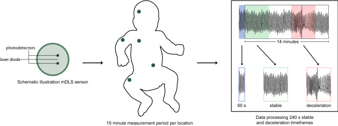
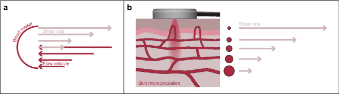
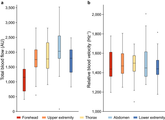
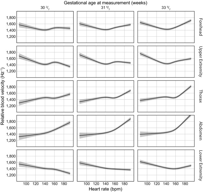
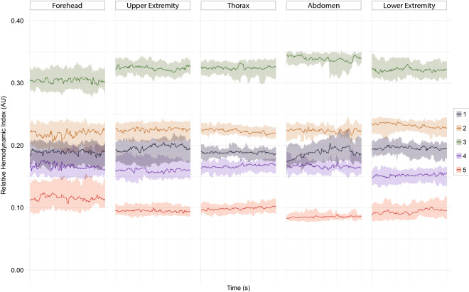
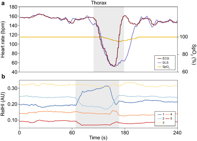
Current haemodynamic monitoring is mainly aimed at the macrocirculation. Multiple studies have demonstrated the importance of the microcirculation in relation to the patient's condition and impact of treatment strategies. However, continuous monitoring of the microcirculation is not yet possible in the neonatal field. A novel dynamic light scattering (DLS) sensor technology for continuous monitoring of the microcirculation was investigated in the neonatal population. Thirty-one haemodynamically stable infants were included. Sequential measurements at the forehead, upper extremity, thorax, abdomen and lower extremity were conducted with the DLS sensor. For analyses stable measurements were selected. The DLS parameters, total blood flow (TBF) and relative blood velocity (RBV), were compared between measurement locations. Changes in relative haemodynamic indices (relHIs), indicating the distribution of blood flow in the microcirculatory blood vessels, were associated with heart rate decelerations. Measurements performed at the forehead had significantly lower TBF levels, compared to measurements at other locations. Early changes in relHIs around a heart rate deceleration were recorded a median (IQR) of 22.0 (13.5-27.0) s before the onset. Measurement of the currently unavailable parameters TBF, RBV and relHIs is possible with DLS technology. Validation of the DLS technology is needed for clinical implementation.


Conflict of interest statement

IF is CEO of Elfi-Tech Ltd. The other authors declare that they have no competing interests.

Figures

Figure 1
Figure 1
Schematic representation of the DLS sensor and the study protocol. s seconds.
Figure 2
Figure 2
(a) The concept of vascular flow velocity and shear rates, showing high flow velocity at the centre of the blood vessel and the lowest flow velocity at the blood vessel wall. An inverse relation is shown for shear rate. (b) Schematic representation of a DLS sensor on the skin and the inverse relation between blood vessel diameter and the measured shear rate.
Figure 3
Figure 3
Boxplots of DLS parameters perfusion (total blood flow) (a) and relative blood velocity (b) per measurement location. Significance was found only between perfusion measurements on the forehead and other measurement sites. AU arbitrary unit.
Figure 4
Figure 4
Effect plots illustrating the estimate and 95% confidence intervals of the relation between relative blood velocity and heart rate, gestational age at measurement and measurement locations. The nonlinear relation of heart rate and interactions between gestational age at measurement (25th, 50th and 75th percentiles) and location are presented. Bpm beats per minute.
Figure 5
Figure 5
Plots of the medians and interquartile ranges of the relative haemodynamic indices at all measurement locations, illustrating the smallest vessels (1), small vessels (2), medium-sized vessels (3), large vessels (4) and largest vessels (5) in the microcirculation. AU arbitrary unit, s seconds.
Figure 6
Figure 6
An example of a 240 s time interval of a bradycardic event. (a) ECG heart rate, DLS heart rate and SpO2 measurements are shown. The delay in the recovery of the DLS heart rate, was caused by movement of the infant introduced by the nurses. (b) Relative haemodynamic indices are presented; smallest vessels (1), small vessels (2), medium-sized vessels (3), large vessels (4) and largest vessels (5). RelHI relative haemodynamic index, bpm beats per minute, s seconds, ECG electrocardiography, DLS dynamic light scattering, SpO2 oxygen saturation measured with pulse oximetry.

Similar articles

Cited by

References

    1. Bennett VA, Vidouris A, Cecconi M. Effects of fluids on the macro- and microcirculations. Crit. Care. 2018;22:74. doi: 10.1186/s13054-018-1993-1. - DOI - PMC - PubMed
    1. De Backer D, et al. Microcirculatory alterations in patients with severe sepsis: Impact of time of assessment and relationship with outcome. Crit. Care Med. 2013;41:791–799. doi: 10.1097/CCM.0b013e3182742e8b. - DOI - PubMed
    1. Edul VS, et al. Quantitative assessment of the microcirculation in healthy volunteers and in patients with septic shock. Crit. Care Med. 2012;40:1443–1448. doi: 10.1097/CCM.0b013e31823dae59. - DOI - PubMed
    1. Ince C. Hemodynamic coherence and the rationale for monitoring the microcirculation. Crit. Care. 2015;19(Suppl 3):S8. doi: 10.1186/cc14726. - DOI - PMC - PubMed
    1. Kuiper JW, Tibboel D, Ince C. The vulnerable microcirculation in the critically ill pediatric patient. Crit. Care. 2016;20:352. doi: 10.1186/s13054-016-1496-x. - DOI - PMC - PubMed