

Base of tongue metastasis of cutaneous malignant melanoma with rhabdoid and neuroendocrine features: Report of a rare case and review of the literature
- PMID: 35524032
- PMCID: PMC9729478
- DOI: 10.1007/s12105-022-01437-6
Base of tongue metastasis of cutaneous malignant melanoma with rhabdoid and neuroendocrine features: Report of a rare case and review of the literature
Abstract
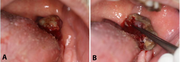
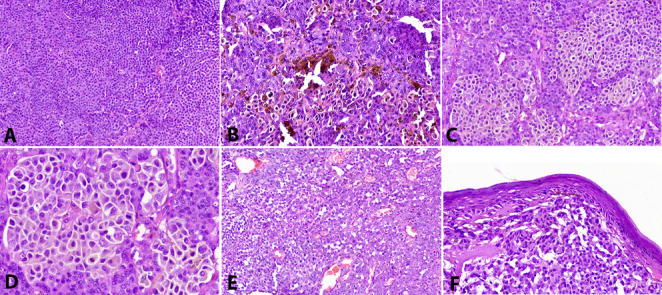
Metastatic malignant melanoma (MM) represents a highly aggressive cancer associated with overall poor prognosis. Various anatomic sites can be affected, including the oral cavity and the oropharynx. It may mimic other entities by assuming a variety of clinical appearances and exhibiting a plethora of microscopic variations. Herein, we present a case of a 63-year-old male with a MM metastasizing to the base of tongue, which developed 5 years after the original diagnosis and treatment of cutaneous MM of the chest and heralded its relapse; subsequently, neurological symptoms developed as a result of metastasis to the brain. Diagnostic challenges were encountered, as the tongue lesion clinically masqueraded as a pedunculated reactive lesion and microscopically displayed unusual rhabdoid and neuroendocrine features. Tumor cells expressed S-100, HMB-45, Melan-A, and SOX-10, while most cells with rhabdoid morphology were also positive for myogenin and Myo-D1. Chromogranin and synaptophysin positivity was further noticed in a subset of cells, suggestive of focal neuroendocrine differentiation. Molecular investigation revealed mutations for the BRAF V600E gene. Divergent differentiation of tumor cells may cause diagnostic pitfalls necessitating thorough immunohistochemical analysis. The presence of rhabdoid features and neuroendocrine differentiation are very uncommon, while their co-existence is extremely rare. Better characterization of such microscopic variations in MMs with evaluation of their potential biologic significance is warranted.
Keywords: Base of tongue; Malignant melanoma; Metastasis; Neuroendocrine differentiation; Rhabdoid features.
© 2022. The Author(s), under exclusive licence to Springer Science+Business Media, LLC, part of Springer Nature.
Conflict of interest statement
All authors declare that they have no conflict of interest to disclose. The authors have no conflicts of interest to declare that are relevant to the content of this article.
Figures


References
-
- Banerjee SS, Eyden B. Divergent differentiation in malignant melanomas: a review. Histopathology. 2008;52:119–29. - PubMed
Publication types
MeSH terms
LinkOut - more resources
Full Text Sources
Medical
Research Materials
Miscellaneous
