. 2022 May;25(5):659-674.
doi: 10.1038/s41593-022-01068-8. Epub 2022 May 6.

Trans-Seq maps a selective mammalian retinotectal synapse instructed by Nephronectin

Affiliations

Trans-Seq maps a selective mammalian retinotectal synapse instructed by Nephronectin

Nicole Y Tsai et al. Nat Neurosci. 2022 May.

Abstract

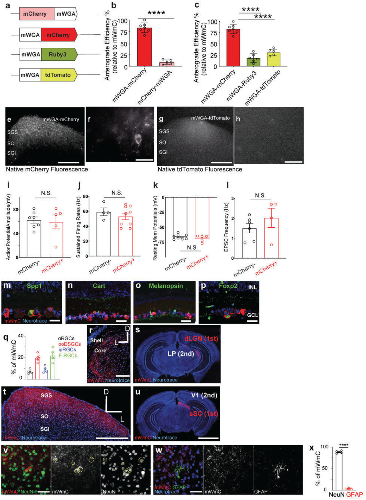
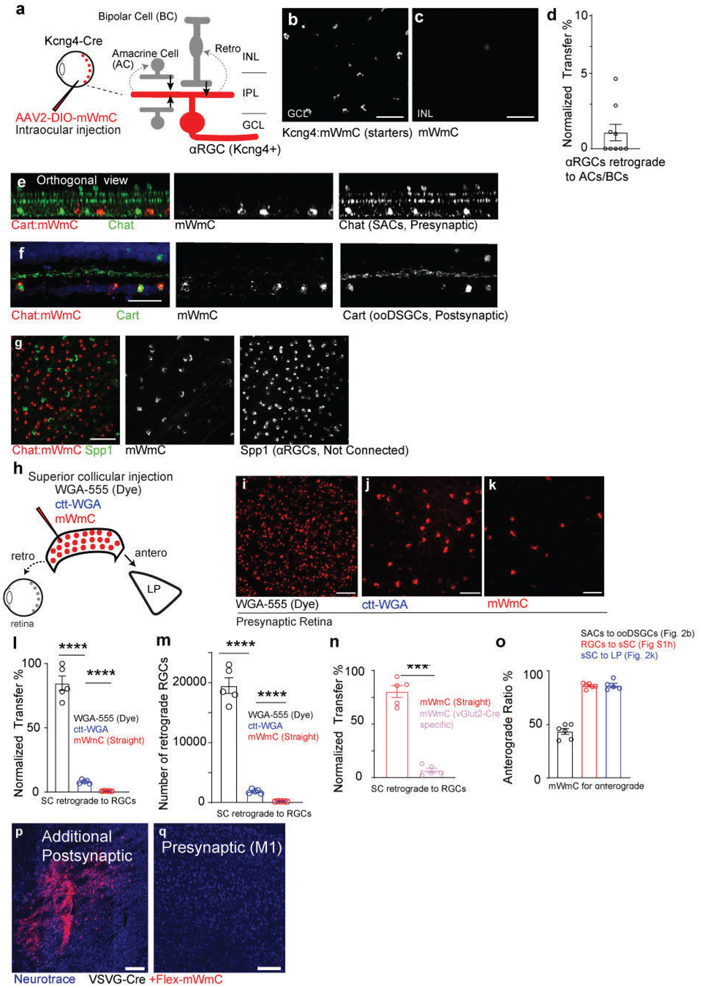
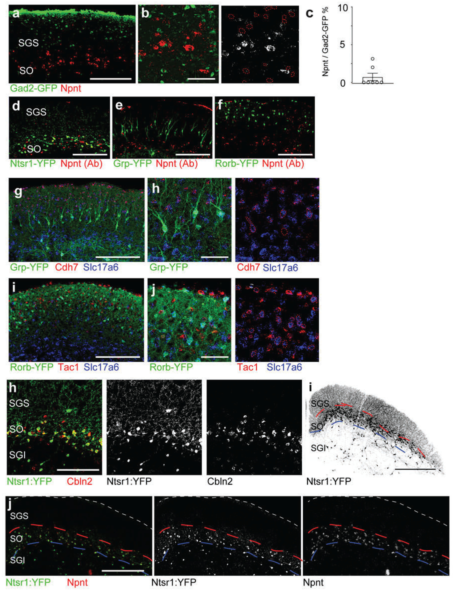
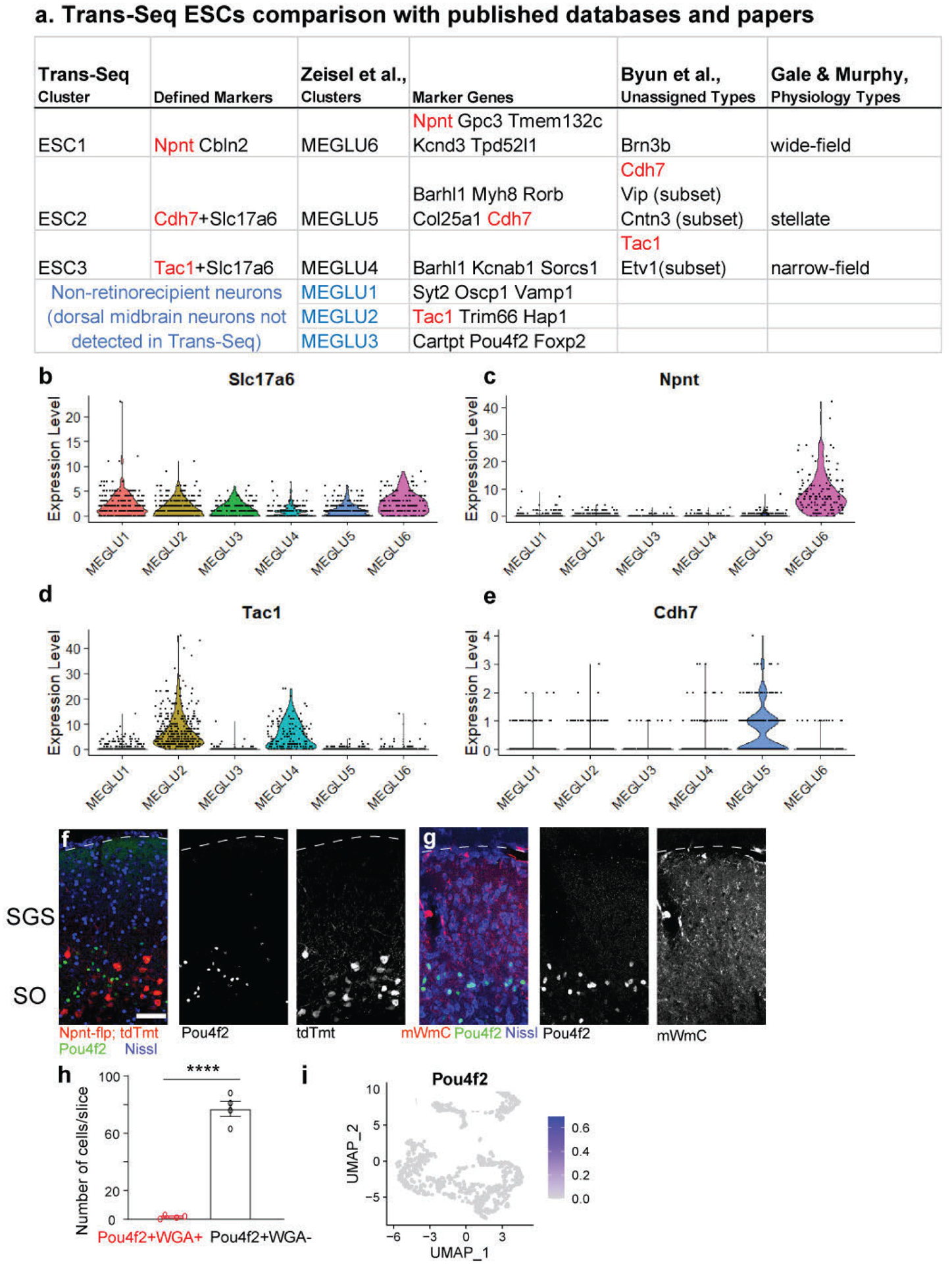
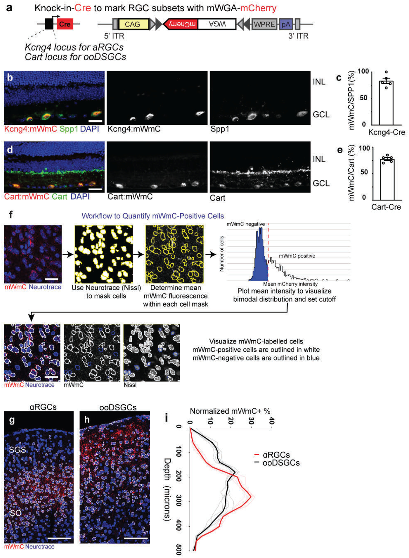
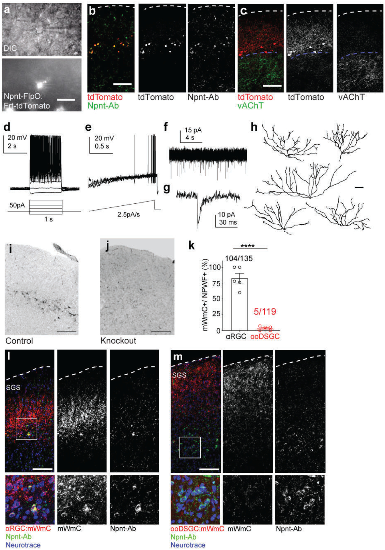
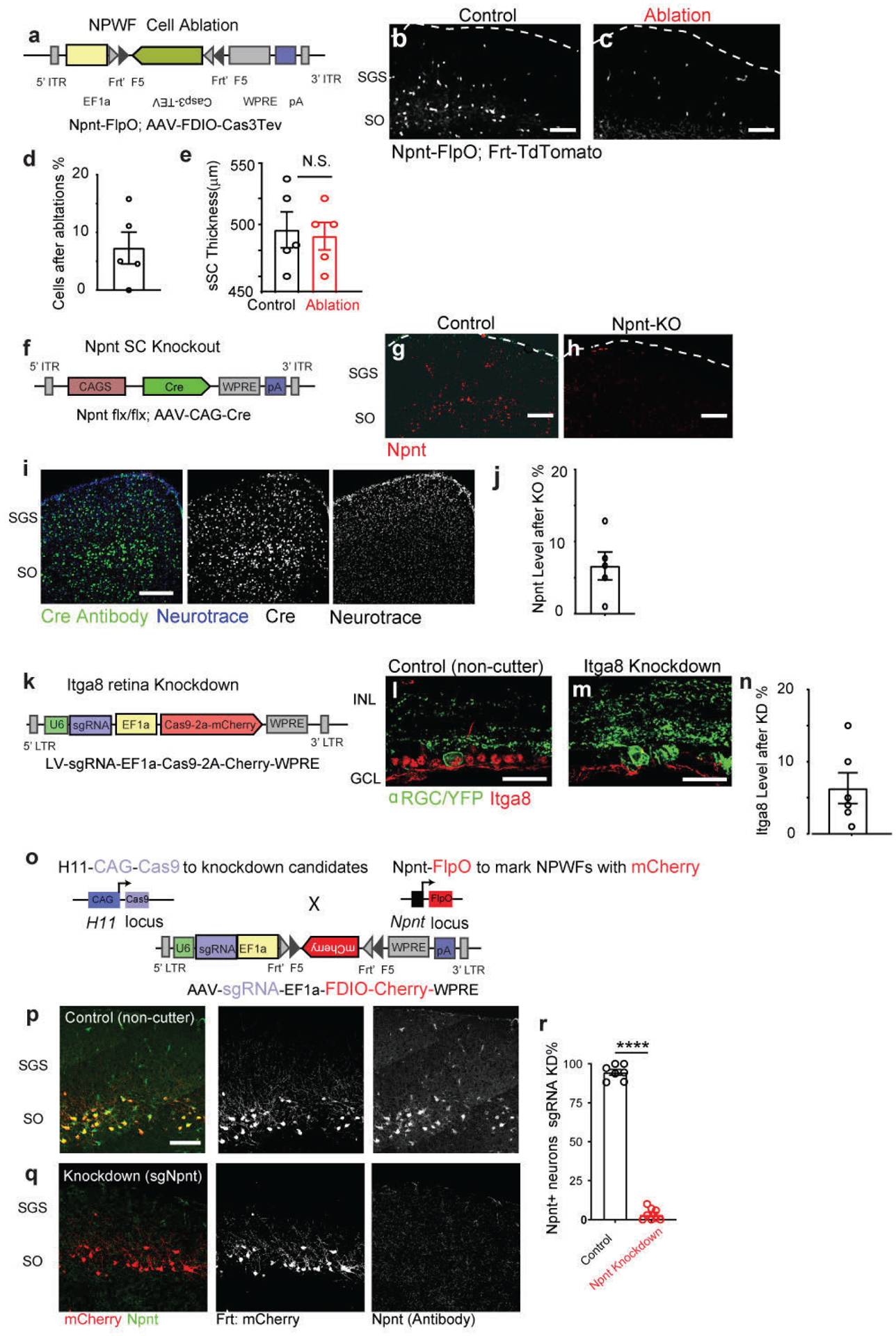
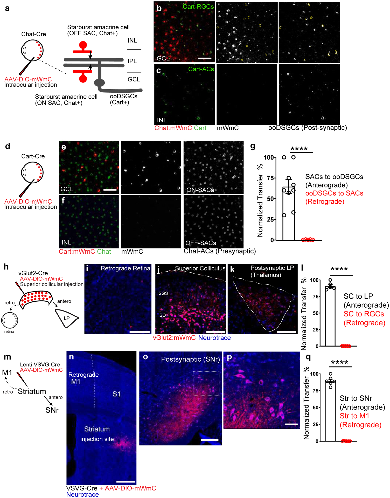
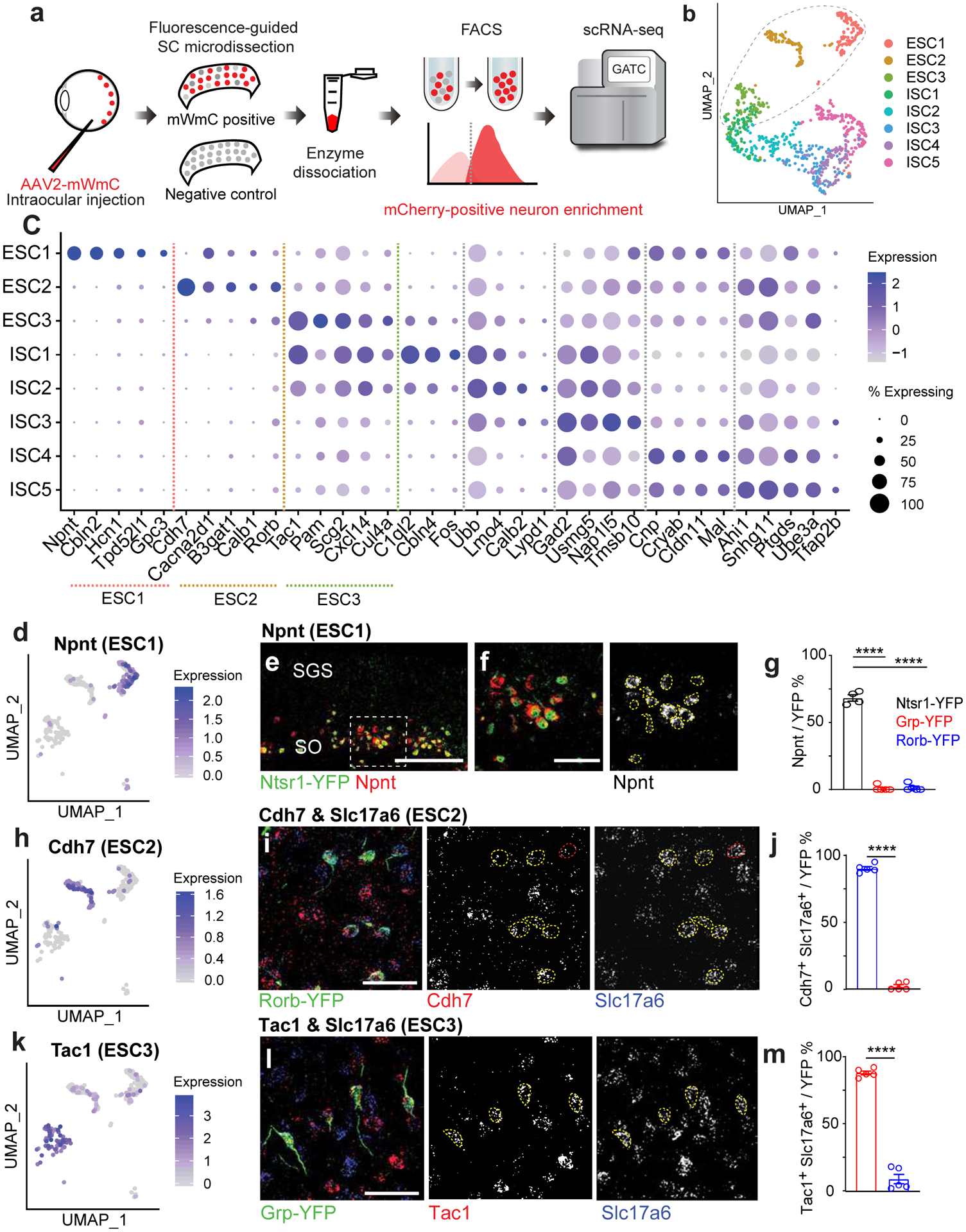
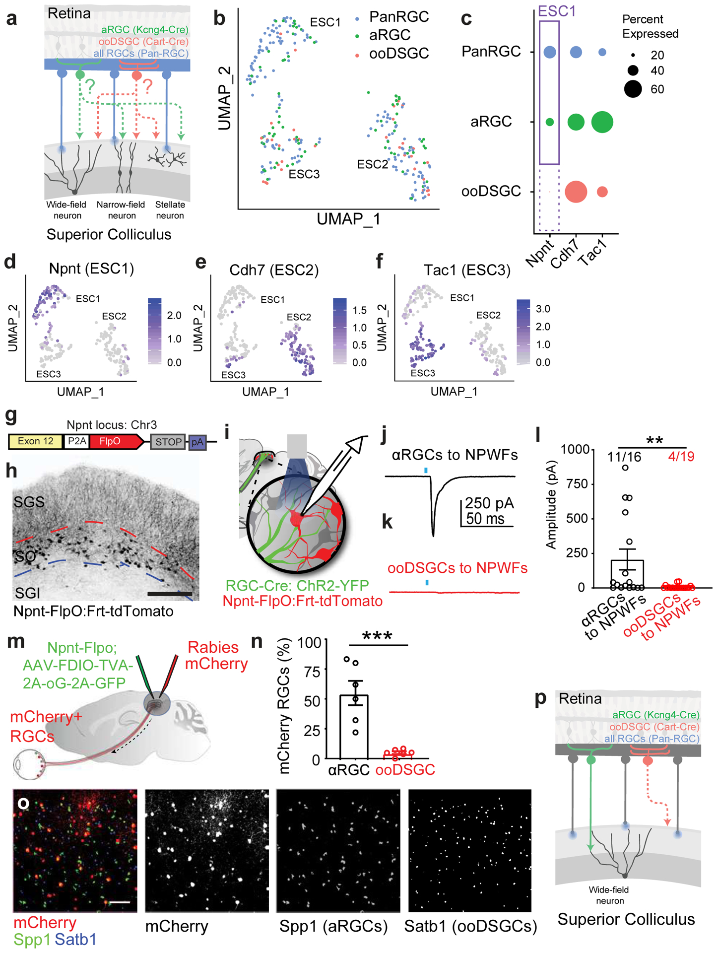
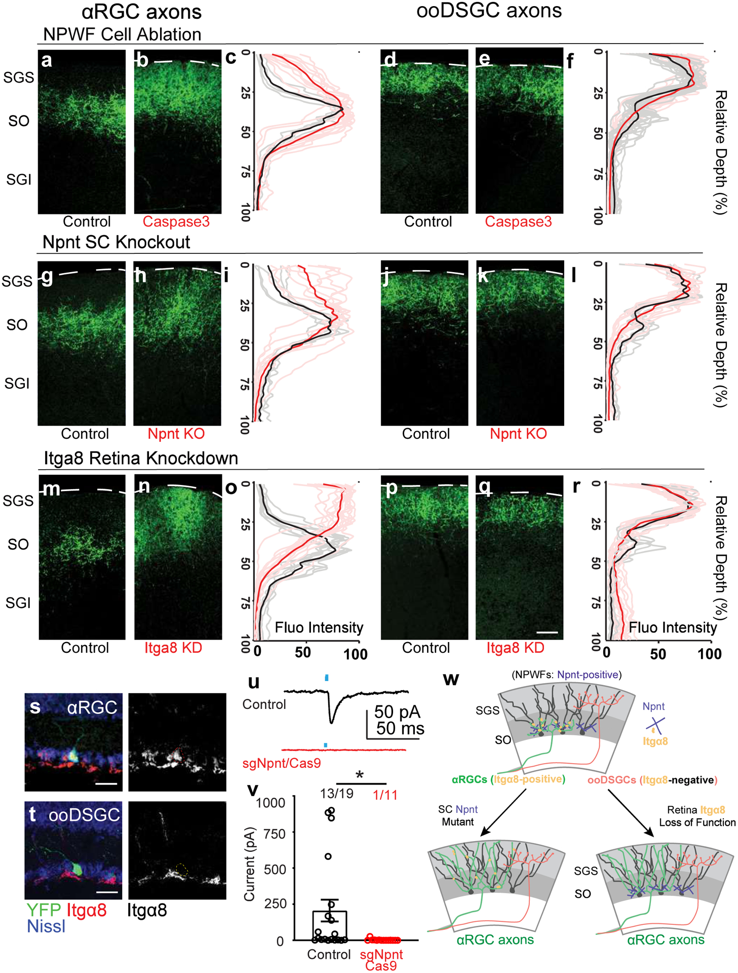
The mouse visual system serves as an accessible model to understand mammalian circuit wiring. Despite rich knowledge in retinal circuits, the long-range connectivity map from distinct retinal ganglion cell (RGC) types to diverse brain neuron types remains unknown. In this study, we developed an integrated approach, called Trans-Seq, to map RGCs to superior collicular (SC) circuits. Trans-Seq combines a fluorescent anterograde trans-synaptic tracer, consisting of codon-optimized wheat germ agglutinin fused to mCherry, with single-cell RNA sequencing. We used Trans-Seq to classify SC neuron types innervated by genetically defined RGC types and predicted a neuronal pair from αRGCs to Nephronectin-positive wide-field neurons (NPWFs). We validated this connection using genetic labeling, electrophysiology and retrograde tracing. We then used transcriptomic data from Trans-Seq to identify Nephronectin as a determinant for selective synaptic choice from αRGC to NPWFs via binding to Integrin α8β1. The Trans-Seq approach can be broadly applied for post-synaptic circuit discovery from genetically defined pre-synaptic neurons.


Conflict of interest statement

Competing interests: Authors declare that they have no competing interests.

Figures

Extended Data Fig. 1
Extended Data Fig. 1. Multiple configurations of mWGA-based tracer were examined for anterograde synaptic transfer efficiency in the retinotectal synapses.
The numbers of fluorescence-positive neurons in the SGS from different mWGA-RFP configurations were counted. The percentages were normalized to the highest number as (100%) set by the mWGA-mCherry configuration, which possessed the highest efficiency. The anterograde transfer number was quantified by counting the number of RFP-positive neurons in acute SC slices on the contralateral side (right SC) without fluorescence amplification through immunostaining. We focused on the contralateral (right) SC, as 90% of RGCs project contralaterally. We examined RGC coverage across the retina to ensure there was no retinotopy bias in SC analysis. mWGA-fusion protein configurations were compared under consistent conditions by delivering AAV2 expressing the fusion proteins into the left eyes (Fig. 1a). We used standard AAV Serotype-2 followed by 4 weeks post-injection (wpi) sampling, a commonly established window to examine RGC axonal projections and carry out the optogenetic measurement. a, Several WGA-fusion proteins were compared side-by-side using an AAV-mediated in vivo screen for highly efficient anterograde transfer from the retina to the brain. b, Comparisons between N-terminal (mCherry-mWGA) and C-terminal fusion (mWGA-mCherry) transfer efficiencies were quantified. c, Transfer efficiencies of different RFP C-terminal fusions, including mWGA-mCherry, mWGA-Ruby3, mWGA-tdTmt, were quantified, n=4 animals for each condition. Statistics for b, two-sided Student’s t-test, ****, P<0.0001; c, one-way ANOVA test. ****, P<0.0001. e-f, (e) Additional samples showing live red-fluorescent labeling of the contralateral SC after acute brain slice preparation 4 weeks post-injection (wpi), indicating transsynaptic transfer onto the recipient neurons enriched in stratum griseum superficiale (SGS) and stratum opticum (SO), but not in stratum griseum intermedium (SGI). f, Magnified view of inset from e showing individual neurons labeled with bright red fluorescent protein from RGC anterograde transfer without signal amplification through immunostaining. The ability to detect native fluorescence is unique to mWGA-mCherry (mWmC) but absent in other mWGA-RFP configurations, such as mWGA-TdTomato shown here (g, h). Scale bars: (e, g, 5 mm; f, h, 50μm). i-l, Intrinsic electrophysiological properties of mCherry-positive recipient neurons (red, n=5 animals) and neighboring mCherry-negative neurons (black, n=4 animals) are similar as showed in Fig. 1m. i, Action potential amplitudes, j, sustained firing rates, k, resting membrane potentials, and l, EPSC frequencies are shown. These intrinsic properties were unperturbed by mWmC-transfer. N.S. not significant. two-sided Student’s t-test. m-p, Retinal vertical sections to show high mWmC coverage across major RGC types, including (m) Spp1 for αRGCs, (n) Cart for ooDSGCs, (o) Melanopsin for ipRGCs, and (p) Foxp2 for F-RGCs. Scale bar: 20 μm. INL: inner nuclear layer; GCL: ganglion cell layer. The percentages of each RGC subclasses were quantified in q, representing a similar fraction of RGC subclasses among all RGCs, n=5 animals. f-i, Intraocular injections of mWmC lead to efficient monosynaptic transfer to connected neurons in multiple retino-recipient areas, including SC (t,u) and LGN (r, s). By contrast, the secondary relay neurons in V1 (u) or those in the lateral posterior nucleus of the thalamus (LP) (s) do not show mWmC transfer. Scale bars: (r, t, 2mm; s, u, 20mm). v, Immunostaining for RFP (mWmC) indicates high efficiency of anterograde transfer onto the recipient neurons in stratum griseum superficiale (SGS) and stratum opticum (SO). mWmC-positive cells are largely NeuN-positive. Dotted-yellow circles indicate mCherry and NeuN double-positive neurons from RGC anterograde transsynaptic transfer. w, mWmC-positive cells are largely GFAP-negative, which were quantified in x. n=5 animals for each condition. ****, p<0.0001, two-sided Student’s t-test. Scale bars: (v, w, 50μm). All data in this figure are presented as mean ± SEM.
Extended Data Fig. 2
Extended Data Fig. 2. Comparison of different versions of WGA tracer for their anterograde and retrograde transfer capacities in the retinotectal synapses.
a-d, a, a schematic drawing of the injection within the retinal circuit. b, Retinal wholemount view to show mWmC coverage from RGC starter neurons (Kcng4-Cre for αRGC subclass neurons) in the ganglion cell layer (GCL, b), with limited retrograde transfer to the inner plexiform layer (INL, c). Scale bar: 50μm, with the retrograde transfer ratios quantified in (d). n=9 biologically independent samples. e-g, Orthogonal view (e) of RGC labeling with mWmC (red) without any retrograde transfer onto SACs (green, Chat). Intraocular injections of mWmC into Chat-Cre led to labeling of SACs (red) for anterograde and monosynaptic tracing onto ooDSGCs (Cart-positive, green; mWmC, red double-positive) in (f). Scale bar: 50μm (e, f). Additionally, Chat-mWmC labeling does not lead transfer mWmC to SPP1-positive neurons (αRGCs, g), indicating the specificity of anterograde transsynaptic transfer as opposed to trans-cellular transfer within a local area. Scale bar: 50μm(g) 0% Spp1-positive, n=4 animals. h, Injections of WGA-protein conjugated dye, AAV-expressing ctt-WGA cDNA, and AAV-expressing mWmC into SC lead to efficient anterograde transfer to LP. However, they displayed different levels of retrograde labeling, including bright axonal uptake and faint retrograde transfer. mWmC (j) displayed with very limited retrograde spread onto the retina. This is a significant improvement in limiting retrograde transfer compared to WGA-Alexa555 dye (i) or AAV-expressing ctt-WGA (k). n=4 animals in each condition. Scale bars: (i, j, k, 50 μm). l, Retrograde transfer ratios were normalized across three configurations of the WGA, using the highest numbers from WGA-555 as 100%. n=5 biologically independent samples. ****, p<0.0001, two-sided Student’s t-test. m, Average numbers of retinal ganglion cells per retina were quantified and compared across the three conditions. n=5 biologically independent samples. ****, p<0.0001, two-sided Student’s t-test. n. Additionally, restricting the starter neurons genetically in SC vGlut2-Cre cells further eliminated the axonal retrograde uptake (compared to global mWmC expression). n=5 biologically independent samples. ****, p<0.0001, two-sided Student’s t-test. o, Retrograde transfer ratios were normalized across two conditions, using the highest numbers from straight mWmC expression as 100%. n=5 biologically independent samples. ****, p<0.0001, two-sided Student’s t-test. p, GPe, another downstream target of the striatum injection (Fig. 2m, n) was traced here; q, Magnified view of M1 in Fig. 3n from the striatum retrograde tracing showed no retrograde spread from the Striatum. Scale bar p-q, 100μm. All data in this figure are presented as mean ± SEM.
Extended Data Fig. 3
Extended Data Fig. 3. The Trans-Seq platform’s workflow ensures quality control and generates a bioinformatic prediction for neuronal clustering.
To improve adult cell isolation and survival throughout the Trans-Seq protocol, we adopted well-controlled physiological conditions to preserve adult neurons and optimized slice preparation and dissociation conditions. a, Freshly prepared brain slice for contralateral SC dissociations at 4 weeks post-injection (wpi). Bright red fluorescence without immunostaining indicates a high-efficiency anterograde transsynaptic transfer from the retina onto contralateral SC recipient neurons. Fluorescence-activated cell sorting (FACS) enriched mCherry-positive neurons and excluded dead cells, debris, and doublets. n=4 animals each preparation. b-h, FACS gating parameters to isolate mWmC+ neurons from freshly prepared SC slices reflecting the bimodal distribution of the mCherry signals enriched by mWmC tracer, feasible for FACS. b, First, cell debris was excluded using forward scatter area (FSC-A) vs. side scatter area (SSC-A) profiles. Second, cell doublets were excluded using double gating composed from c forward scatter height (FSC-W) vs. forward scatter area (FSC-A) and d side scatter height (SSC-W) vs. side scatter area (SSC-A) profiles. e, Third, gating for DAPI-negative live cells was subsequently performed, followed by f the last gating for mCherry-positive cells. g, Sample gatings for both DAPI and mCherry were performed by establishing gating parameters on unstained control SC suspensions and backgating isolated cells. h, mCherry-gating results from mWmC-positive samples, summarizing the fraction of cells passing each of the FACS parameters listed above b-g. 25000–50000 mWmC-positive cells were collected each preparation. Using the scRNA-Seq data of the connected neurons from Pan-RGC tracing, we obtained eight neuronal clusters, including three excitatory SC clusters (ESC1–3) and five inhibitory SC clusters (ISC1–5). i, UMAP plot showing the alignment of all three replicates of Pan-RGC Tran-Seq experiments, showing the even distribution of all neuronal clusters across experimental procedures. j, Contribution of each preparation from i to individual neuronal clusters to show the batch effects across three experiments. These results included all three ESCs and five ISCs, indicating no neuronal clusters were biased across the triplicates. (Red, 1st; Green, 2nd; Blue, 3rd preparation). k-m, QC plots of the sorted mCherry-positive neurons for each neuronal cluster, including ESCs and ISCs, examining the number of genes expressed k, counts of transcripts l, and the mitochondria counts m.
Extended Data Fig. 4
Extended Data Fig. 4. Data analysis of the Pan-RGC Trans-Seq data.
a-d, UMAP plots of Pan-RGC Trans-Seq data show major neuronal markers, including (a) Snap25 and (b) Syt1 as pan-neuronal markers, (c) Slc17a6 (vGluT2) for excitatory neurons including three ESCs, and (d) Gad2 for inhibitory neurons including five ISCs. These markers defined neuronal populations and separated the clusters among three ESCs (Slc17a6-positive, c) and five ISCs (Gad2-positive, d). e-g, We queried previously identified marker genes expressed in SC neuronal subsets (f),. Additionally, several cell-cell adhesion molecules, such as Type II Cdhs (g) and neuropeptides and their putative receptors (e), appeared as good molecular markers for neuronal subsets in the mammalian nervous system,. Notably, when we queried the gene expression of Ntsr1 in the Trans-Seq dataset, we found no Ntsr1 expression in any SC neuron cluster (e). Dot-plots as bioinformatics predictions of (e) Neuropeptides and receptors, and (g) Cdhs for SC subset expression.
Extended Data Fig. 5
Extended Data Fig. 5. Additional histology data to validate Trans-Seq data in the SC.
a-c, Validation of Trans-Seq marker gene expression from ESC1 using RNA-Scope in situ hybridization. Npnt (red) is not found in inhibitory neurons marked by Gad2-GFP (a). b, Higher magnification of the boxed area in a. Scale bars: (a, 250μm; b, 50μm), quantified in (c). n=4 animals, per genotype. Data in this figure are presented as mean ± SEM. d-f, Immunohistochemistry using Npnt antibody onto SC excitatory-marking lines. d, Npnt antibody (red) co-localizes with Ntsr1-GN209-YFP (green) for wide-field excitatory neurons. e, f, By contrast, the Npnt antibody (red) does not co-localize with the Grp-KH288-YFP line (green) for narrow-field neurons (e) or the Rorb-YFP line (green) for stellate cells (f). Scale bars: (d-f, 250μm). g-h, Validation of Trans-Seq marker gene expression from ESC2 using RNA-Scope in situ hybridization. (g) Cdh7 (red)/Slc17a6 (blue) double in situs do not label neurons in the Grp-KH288-YFP transgenic line for narrow-field excitatory neurons at higher magnification (h), n= 4 animals. i-j, Validation of Trans-Seq marker gene expression from ESC3 using RNA-Scope in situ hybridization. (i) Tac1 (red)/Slc17a6 (blue) double in situs do not label neurons in the Rorb-YFP knock-in line primarily for stellate excitatory neurons, at higher magnification (j), n=4 animals. Red dotted circles in (b, h, j) indicate the absence of in situ signals with GFP staining. Scale bars (h-j, 50μm). h, Validation of Trans-Seq top marker gene expression from ESC1 using RNA-Scope for in situ hybridization shows that Cbln2 RNA probes (red) are enriched in the Ntsr1-GN209-YFP transgenic line labeling wide-field excitatory neurons (green). Scale bar: 200 μm. n=3 animals. i, Neuronal distribution of Ntsr1-GN209-YFP transgenic line, including wide-field neurons present in SO and a significant fraction of neurons in the SGI and other deep SC layers. The wide distribution of YFP-positive neurons beyond SO indicates that Ntsr1-GN209-YFP cannot be used to manipulate SO neurons within the SC selectively. Red and blue dotted lines mark the borders of the three zones (SGS, SO, and SGI) within the superficial SC. j, Only wide-field neurons in the SO are Npnt-positive (between red and blue dotted lines), but not the rest of Ntsr1-YFP positive neurons in the SC’s SGI and deep layers. These data further suggest that Npnt is a faithful molecular marker for these neurons within SO. n=3 animals. Scale bars: (i, 2mm; j, 500μm).
Extended Data Fig. 6
Extended Data Fig. 6. Alignment of ESC1–3 clusters from Trans-Seq to a public database of whole-brain excitatory neuron clustering.
a, A summary of the three excitatory neuron clusters (ESCs) characterized in Trans-Seq. ESC1–3 were first correlated to a published bioinformatics database for all excitatory neuronal clusters throughout the brain, indicating that ESC1–3 are characterized as retinorecipient neurons among six excitatory neuron clusters (MEGLU1–6) annotated from the dorsal midbrain. These six clusters were classified as putative SC excitatory clusters among all 11 collicular clusters. ESC1–3 were compared to an immunohistochemical study that identified laminar markers but did not link to cell types. ESC1–3 were matched to cell types defined by morphology and physiology. b-e, Violin plots of candidate marker genes (b, Slc17a6, pan-excitatory neurons), (c, Npnt, ESC1), (d, Tac1, ESC3), and (e, Cdh7, ESC2) among MEGLU1–6. f, Immunohistochemistry of Pou4f2 (green, antibodies) showed no overlap with ESC1 neurons (Npnt-TdTomato, red). Pou4f2 was revealed as an excitatory neuron subset marker for MEGLU3 from a public scRNA-Seq database. A defined Pou4f2-positive population of neurons resides in the SGI. g, Pou4f2-positive SGI neurons did not uptake mWmC following 4wpi of mWmC infection of the RGCs, corroborating the findings from Trans-Seq in defining specific clusters of retino-recipient neurons. Scale bar: 100 μm. h. Quantifications of cell number for Pou4f2-positive and mWmC-positive neuron (red), versus all Pou4f2-positive neurons (black), n=3 animals, ****, p<0.0001, two-sided Student’s t-test. Data in this figure are presented as mean ± SEM. i. A UMAP Plot shows the absence of the MEGLU3 marker (Pou4f2) within Trans-Seq data (Fig. 5).
Extended Data Fig. 7
Extended Data Fig. 7. Cre-dependent mMmC for RGC type-specific circuit mapping.
a, Genetic design of AAV vector for Cre-dependent mWmC (AAV2-CAG-DIO-mWmC-WPRE) expression to achieve neuronal subtype-specific anterograde transsynaptic tracing. b-e, mWmC infection can achieve efficient and restricted expression in RGC starter neuron subclasses: Kcng4-Cre is a driver for αRGC subtypes (Spp1-positive, b); and Cart-Cre is a driver for all ooDSGC subtypes (Cart-positive, d). Notably, these Cre-dependent mWmC expressions are restricted to the starter neurons without retrograde spread into the inner retina (i.e., no bipolar cells and amacrine cells uptake mWmC), scale bars: 20μm. The specificities of the Cre-drivers were quantified in c (Kcng4-Cre) and e (Cart-Cre), respectively. n=5 animals, per genotype. Data in this figure are presented as mean ± SEM. f, mWmC demonstrated a bimodal distribution of red fluorescence in recipient neurons. Thresholding for the high fluorescence intensity population allowed neurons across tissue slices to be classified as transferred or un-transferred. g-i, RGC-subclass specific mWmC anterograde transfer properties can be quantified in the postsynaptic neurons. Anterograde transsynaptic tracing from different starter retinal ganglion cell (RGC) types was compared (g, αRGCs; h, ooDSGCs as Fig. 6) and quantified using this workflow. This quantification (i) demonstrated the differential distribution of SC neurons receiving mWmC transfer from RGC starter cells, supporting the electrophysiology and genetic data from Fig. 5. n=5 animals, per genotype Scale bar: 100μm. (Black line is mean intensity curve, gray lines are each example. Red line is mean intensity curve, light red lines are each example).
Extended Data Fig. 8
Extended Data Fig. 8. Characterizations of Nephronectin-positive-wide-field neurons (NPWFs) and their differential inputs from αRGCs versus ooDSGCs.
a, Sample DIC and fluorescent microscope images showing the targeted postsynaptic Npnt-FlpO-TdTomato neurons for connectivity tests. At least 30 times each experiment was repeated independently with similar results. Scale bar: 20 μm. b, The majority of the labeled Npnt-FlpO-TdTomato neurons are positive for Npnt antibody immunostaining (96.5±2.5% double-positive neurons among all TdTomato-positive neurons in b, n= 3 animals). The dotted line marks the pial surface. c, Npnt-FlpO-TdTomatao neurons have their somata positioning right above the top boundary of the SGI (dotted lines) marked by the vAChT. Scale bars: (b and c, 100 μm). d-g, Current-clamp recordings of NPWFs in response to (d) step-stimulation, 50pA per step for 1second, 10 steps per sweep, and (e) ramp-stimulation, 2.5pA/s for 20 seconds per trail, 3 trails per sweep. f, Voltage-clamp recordings of spontaneous EPSCs. g, A sample spontaneous ESPC trace from f. h, Single-cell morphologies of NPWFs (ESC1) were reconstructed after physiological recording and intracellular dye filling showing the dendritic complexity. Scale bar: 100 μm. i, j, SC-specific Npnt expression using immunohistochemistry (i) was absent in the SC-specific conditional knockout (j). Scale bar: 200μm. k-m, Sample immunohistochemistry images showing differential mWmC transfer from distinct RGC starter neurons onto retinorecipient neurons in the SC (mWmC, mCherry signal in red). k, Percentages of Npnt-positive wide-field neurons (NPWFs, ESC1) within connected neurons of the αRGCs and ooDSGCs. Data in this figure are presented as mean ± SEM. NPWFs were enriched in αRGC tracing datasets (77.0±8.2% double-positive neurons among all Npnt-positive cells in l, n=5 animals) but largely absent in ooDSGC tracing datasets (4.5±2.1% double-positive cells among all Npnt-positive neurons in m, n=5 animals). ****, p<0.0001, two-sided Student’s t-test. l, αRGC ouputomes labeled with Kcng4:mWmC. ESC1 neurons were immunostained for Npnt. m, ooDSGC tracing datasets in SC labeled with Cart:mWmC. ESC1 neurons stained with Npnt. Bottom panels are magnified view of inset from l and m labeled by solid square line.
Extended Data Fig. 9
Extended Data Fig. 9. Physiological and histological characterization of the selective RGC synapses onto NPWF neurons (ESC1).
a, Sample EPSCs from NPWF neurons driven by αRGCs with bath-applied TTX+4-AP (blue trace) and after bath perfusion of CNQX+APV (red trace). Blue dots indicate 2ms of the blue LED light. b, Quantification of the average amplitudes before and after adding pharmacological blockers in αRGC-NPWF connected pairs. Current amplitudes before adding any drugs are shown in black dots and bars; blue dots and bars show current amplitude after adding TTX+4-AP; red dots and bars show current amplitude after adding CNQX+APV. n=8 animals, N.S., no significance, *, p<0.05, two-sided Student’s t-test. The dots are amplitudes of individual cells, and the bar is the average of all cells. c, Among all the ooDSGCs and NPWF, connected pairs, only four cells showed a very small evoked current (black dots and bars). These small currents were blocked by bath-applied TTX and 4-AP (blue dots and bars), suggesting that the evoked currents are not monosynaptic inputs from ooDSGCs. The dots are amplitudes of individual cells, and the bar is the average of the three cells. n=4 cells, *, p<0.05, two-sided Student’s t-test. d, Sample images of NPWFs in the SC showing high efficiency of Flp-dependent EGFP-2a-TVA-2a-oG (green) and RdGV: Rabies-Cherry (red) overlap. Scale bar: 100 μm. ej, Vertical sections of contralateral retinas following rabies tracing indicating αRGC subtypes (e and h) OFF-transient αRGCs, (f and i) OFF-sustained αRGCs, and (g and j) On-sustained αRGCs, were retrogradely labeled. Scale bar: 50μm. (eg),vAChT staining (green) shows RGC dendritic sublaminations within the retina; (hj), SPP1 staining (green) to confirm cell identities as αRGCs. k, Quantifications of different RGC subclasses among mCherry+ neurons including αRGCs, ooDSGCs, and other RGC types. l, Magnified view of Fig. 6o to show retrogradely labeled RGCs (red) highly overlap with αRGCs (SPP1-positive, green) in filled arrowheads, but not ooDSGCs (Satb1-positive, blue) in open arrowheads, as quantified in Fig. 6n. n=6 animals, ***, p<0.005, two-sided Student’s t-test. Scale bar: 100 μm. m, n, Sample images with axons of m, αRGCs, and n, ooDSGCs labeled by YFP (green) in relative position to the dendrites of NPWF neurons (Npnt-RFPs). n=4 animals each genotype, Scale bar: 100μm. All data in this figure are presented as mean ± SEM.
Extended Data Fig. 10
Extended Data Fig. 10. Validation of genetic and molecular reagents for Npnt mutant analysis.
a, The AAV vector design for FlpO-dependent Caspase3-TEV expression, modified from (Addgene #45580). b, c, Confirmation of successfully eliminating ESC1s using AAV-fDIO-Caspase3 in Npnt-FlpO by comparing control b and Cas3-TEV injections c. The dotted line marks the pial surface. Scale bar: 100μm. d, Quantifications of Cas3-deletion of NPWFs efficiencies, compared to control side. N=5 animals. e, Quantifications of the superficial SC thickness subject to NPWF eliminations. n=5 animals, per genotype, N.S., no significance, two-sided Student’s t-test. f, The AAV vector design for Cre overexpression. g-h, Confirmation of SC-specific knockout of Npnt using Npnt f/f conditional mutants by comparing controls (g) and mutants (h), n=3 animals. Scale bar: 100μm. No obvious cell-autonomous migration or morphological changes of labeled wide-field neurons were observed after Npnt knockout. i. Sample images showing comprehensive Cre coverage across different sublaminae of SC subject to neonatal AAV injections. Anti-Cre staining was displayed here in the green channel. j, Quantifications of AAV-Cre mediated Npnt knockout efficiency, compared to the control side. n=5 animals. k, The lentiviral vector design for Itga8 knockout based on CRISPR-V2-system to simultaneously express sgRNA/Cas9-2a-Cherry (Addgene Plasmid #99154). l, m, Confirmation of the lentivirus-mediated Itga8 knockout in the retina (m) compared with control (l). Scale bar: 50μm. n, Quantifications of sgRNA-mediated retinal Itga8 knockdown efficacies, compared to control side. n=6 animals, Several sgRNAs were evaluated, and an efficient sgRNA targeting the fourth exon of Npnt was selected (See Methods). Cas9/sgRNA-mediated Npnt deletion was validated by immunostaining with Npnt antibody. We performed neonatal injections of AAV-encoding for sgRNA targeting Npnt or control sgRNA-non-cutter and Flp-dependent RFP expression (AAV-sgNpnt-FDIO-mCherry; or AAV-sg-Noncutter-FDIO-mCherry;) into Kcng4-YFP; Npnt-FlpO; H11Cas9 mice. o, The AAV vector is carrying sgRNAs and Frt-dependent mCherry. This vector can simultaneously knock down endogenous Npnt while labeling the NPWF neurons using Npnt-FlpO dependent RFP reporter. The Cas9 was genetically harbored in the H11-Cas9 line via a genetic cross to Npnt-FlpO. p, q, Npnt in the SC was knocked down (q) compared to sg-Non-cutter control (p). Scale bar: 100μm. The percentages of Npnt knockdown were quantified in (r), n=4 animals, per genotype, ****. p<0.0001, two-sided Student’s T-test. All data in this figure are presented as mean ± SEM.
Fig. 1.
Fig. 1.. Engineering a genetically encoded fluorescent anterograde transsynaptic tracer (mWGA-mCherry) to map postsynaptic neurons.
a, Schematic diagram of intraocular injections of anterograde transsynaptic red-fluorescent tracers to label postsynaptic retinorecipient neurons in the superior colliculus (SC). b, Optimized adeno-associated virus (AAV) vector design encoding mWGA-mCherry (mWmC). mCherry is fused at the C-terminal of a codon-optimized mWGA. c, Retina wholemount showing mWmC coverage to the ganglion cell layer (GCL). d, magnified view of inset of (c). Both images were stained with anti-RFP. Scale bars: (c, 2mm; d, 200 μm). 3 times each experiment was repeated independently with similar results. e, Live red fluorescent labeling of the contralateral SC after acute brain slice preparation, indicating transsynaptic transfer onto the recipient neurons enriched in stratum griseum superficiale (SGS) and stratum opticum (SO), but not in stratum griseum intermedium (SGI). f, Magnified view of inset from e. showing individual neurons labeled with bright red fluorescent protein from RGC anterograde transfer without signal amplification through immunostaining. Scale bars: (e, 500μm; f, 50μm). 3 times each experiment was repeated independently with similar results. g, Schematic drawing of mWmC-mediated monosynaptic anterograde transsynaptic tracing using electrophysiology recording after co-injection of AAV-mWmC (red) and AAV-ChR2-YFP (green). h, A sample image of patched mWmC-positive red fluorescent neurons under DIC camera (top) and epi-fluorescence scope (bottom). Scale bar: 25 μm. At least 10 times each experiment was repeated independently with similar results. i, Whole-cell recordings of mWmC-positive fluorescent neurons, indicating that excitatory postsynaptic currents (EPSCs) in response to a short pulse (2ms, blue line) of blue-light (475nm) excitation possess an onset latency of <5ms, suggesting mWmC+ SC neurons receive monosynaptic inputs from RGCs. j to m. Evoked currents of the mWmC+ SC neurons are glutamatergic and monosynaptic. j, Postsynaptic currents persisted in TTX (1μM) and 4-AP (100μM) (black) and were blocked by CNQX (10μM) and APV (50μM) (red). k. Average ESPC amplitudes in control conditions (black) and the presence of CNQX and APV (red). Both conditions had bath-applied (TTX+4-AP). n=20 cells ****, p<0.0001, two-sided Student’s t-test. l, Distributions of the response latency of the evoked EPSCs to the short pulse blue-light, with the average latency at 2.2±0.2ms. n=27 cells m, EPSCs measured from randomly patched mCherry-positive (red, n=5 animals) and mCherry-negative (black, n=4 animals) neurons under the same electrophysiological recording conditions and subject to the same LED stimulations. In total, 28 out 30 (93%) of mCherry-positive neurons received monosynaptic inputs from the retina. In comparison, only 1 out of 20 (5%) mCherry-negative neurons showed monosynaptic inputs. ****, p<0.0001, two-sided Student’s t-test. All data in this figure are presented as mean ± SEM.
Fig. 2.
Fig. 2.. Lysosomal enrichment of mWmC leads to stable red-fluorescent labeling of postsynaptic neurons, facilitating electrophysiology and FACS identification of postsynaptic neurons in vivo.
a-f, mWmC-mediated anterograde trans-synaptic labeling (e, f) leads to a selective enhancement of tracer signals in the lysosomes (Lamp1) of postsynaptic neurons, compared to ctt-WGA tracer (c, d) or WGA-protein conjugated with Alexa555 (a, b). Transferred mWmC signals within recipient neurons were observed to aggregate in a perisomatic manner, facilitating the identification of labeled neurons (as in Figs. 1&3). The mWmC aggregates highly overlap with lysosomes (f, marked by Lamp1), suggesting that the perisomatic structures are formed through mWmC enrichment within the lysosomes of postsynaptic neurons (Green, Lamp1, lysosomal marker; Red, mWmC fluorescence). 4 times each experiment was repeated independently with similar results. Scale bars: (a, c, e, 20μm; b, d, e, 5μm). g, The percentages of WGA-protein-conjugates, ctt-WGA, and mWmC overlapping with Lamp1 were quantified, showing the unique properties of mWmC in live. ****, p<0.0001, two-sided Student’s t-test. n=7 biologically independent samples. Data in this figure are presented as mean ± SEM.
Fig. 3.
Fig. 3.. Anterograde but not retrograde transfer of mWmC in retinal and brain circuits.
a-c, (a) Intraocular injections of mWmC into Chat-Cre led to (b) expression of mWmC within Starburst Amacrine Cells (SACs, red) within the GCL as starter cells. ooDSGCs (Cart-positive, green), which receive direct input from SACs, were labeled by mWmC through anterograde transfer (mWmC double-positive cells labeled with yellow-dotted circles). No signal was detected in the INL, indicating that bipolar cells were not labeled from SACs retrogradely (c), 8 times each experiment was repeated independently with similar results. d-f, (d) Intraocular injections of mWmC into Cart-Cre led to (e) Efficient transduction of ooDSGCs as starter cells for anterograde tracing into the brain (Fig. 5). No retrograde spread was seen into SACs (Chat-positive) or bipolar cells in either the GCL or INL (f), 5 times each experiment was repeated independently with similar results. Scale bars: b, c, e, f, 50μm. g, Quantifications of the anterograde transfer ratio from SACs to ooDSGCs (black) n=9 biologically independent samples, versus retrograde transfer ratio from ooDSGCs to SACs (red), n=5 biologically independent samples. ****, p<0.0001, two-sided Student’s t-test. Within each retina region, 43±6% of the Cart-positive RGCs received mWmC transfer from SACs. h-l, (h) Introduction of mWmC into excitatory SC neurons (vGlut2-positive) for anterograde and retrograde transfer tests. (j) Efficient start neurons at the SC demonstrated very few retrograde spread back to the retina (i), but specific and efficient anterograde transfer onto the LP of the thalamus (k). Scale bars: (i, 50μm, j, 200μm, k, 100μm). l. Quantifications of the anterograde transfer ratio from SC to LP (black) versus retrograde transfer ratio from SC to RGCs (red), n=5 animals. ****, p<0.0001, two-sided Student’s t-test. Within each SC starting region, 81±6% of the NeuN-positive SC neurons were infected with mWmC; within the LP recipient neuron regions, 86±4% received mWmC transfer from the SC. m-q, (m) Introduction of Cre-dependent mWmC with VSVG-lentivirus expressing Cre into the dorsal lateral striatal neurons for anterograde and retrograde transfer tests. (n) Efficient start neurons at the striatum demonstrated no retrograde transfer back to M1 on the same brain slice. In contrast, mWmC displayed specific anterograde transfer onto the Substantia Nigra (o, SNr) as zoomed-in (p). Notably, axons are also filled by mWmC, in addition to somata filling. Scale bars: (n, 500μm, o, 100μm, p, 50μm). q. Quantifications of the anterograde transfer ratio from the striatum to SNr (black) versus retrograde transfer ratio from the Striatum to M1 (red), mWmC-positive neurons, n=6 animals ****, p<0.0001, two-sided Student’s t-test. Within the SNr recipient neuron regions, 89±2% received mWmC transfer from the Striatum. All data in this figure are presented as mean ± SEM.
Fig. 4.
Fig. 4.. Utilizing mWGA-mCherry (mWmC) for downstream neuron discovery and circuit mapping in various brain regions.
a-g. Anterograde transsynaptically labeled neurons (red) in vM1 projection areas. Presynaptic starter neuron axons are labeled by the YFP signal (green). a, The injection site in the vibrissal primary motor cortex (vM1). b, The dorsal striatum with a magnified view c. d, The thalamus, and S1. e, Magnified view of the boxed area in d. (AM: anterior medial, VAL: ventral anterior-lateral, and VM: ventral medial thalamic nuclei). f, The intermediate reticular (IRt) and ventral medullary reticular (MdV) nuclei in the brainstem. g, Magnified view of the boxed area in f. Spinal trigeminal nucleus caudalis, SpVc; dorsal medullary reticular nucleus dorsal, MdD; Hypoglossal motor nucleus, 12N. Scale bars: (a, d, 1mm; b, e, f, 200μm; g, 100μm; c, 20μm), n=4 animals. h-n, Anterograde transsynaptic tracing from barrel (somatosensory) cortex (S1, h) via stereotaxic injections into the designated cortical areas as starters cells. We used a YFP marker that labels the axons of starter neurons in addition to mWmC to separate presynaptic axons from postsynaptic mWmC-positive cells. From S1 (h), we detected mWmC-transfer signals in known recipient areas, including contralateral S1, M1, thalamus, dorsal lateral ventral posterior medial (VPM), and principal trigeminal sensory nucleus (PrV) as transsynaptically labeled neurons (red) in S1 projection areas. Presynaptic starter axons are labeled by the YFP signal (green). h, The injection site in the barrel field of the primary somatosensory cortex (S1). i, Contralateral S1. Magnified view of the boxed area in h. j, M1. k, The thalamus. l, Dorsal lateral ventral posterior medial (VPM). Magnified view of the boxed area in k. m, The principal trigeminal sensory nucleus (PrV). n, Magnified view of the boxed area in m. Scale bars: (h, 1mm; k, 500μm; i, j, m, 200μm; l, 100μm; n, 20μm), 5 times each experiment was repeated independently with similar results.. Abbreviations: Posterior (PO), ventral lateral (VL), and ventral posterior lateral (VPL) nuclei of the thalamus. o to p, Monosynaptic connectivity test from the motor cortex (M1) to the striatum using mWmC-mediated anterograde transsynaptic transfer. The recordings were all done in ACSF with bath applied TTX (1μM) and 4-AP (100μM). o, An average EPSC from a sample mWmC-positive neuron responds to a 2ms light pulse (blue dot). p, All 32 cells showed EPSCs, and such responses were drastically reduced by CNQX (10μM) and APV (50μM) (N= 2 animals, 32 neurons). ***, p<0.005, two-sided Student’s t-test. Data in this figure are presented as mean ± SEM.
Fig. 5.
Fig. 5.. Trans-Seq integrates mWGA-mCherry-mediated anterograde transsynaptic tracing and scRNA-Seq to categorize the RGC connected neurons in the SC.
a, To generate a molecular atlas for all RGC-connected SC neurons, the following workflow was used to dissociate the SC and isolate mCherry-positive recipient neurons by FACS. The workflow starts with the injection of mWmC into the left retinas of mice, followed by right SC slicing, fluorescence-based microdissection, enzyme dissociation, and FACS for red fluorescence from mWmC. The scRNA-Seq library preparation and subsequent data analysis using the standard 10XGenomics platform and Seurat Packages. b, scRNA-Seq libraries were generated using the 10XGenomics platform. From three replicates, we recovered scRNA-Seq data from 898 adult SC recipient neurons, identified through neuronal markers, thereby generating a molecularly defined connectivity map from pan-RGC anterograde tracing. UMAP plots of the Trans-Seq data from all RGCs (Pan-RGC) show three excitatory neuron clusters (ESCs) and five inhibitory neuron clusters (ISCs). Among all retinorecipient SC neurons subtypes, there were 307 excitatory neurons (within the dotted circle) and 591 inhibitory neurons. c, Dot plot of top marker genes. ESC1-ESC3 are represented on the left of the plot for top feature genes, while ISC1–5 are represented on the right of the plot for top feature genes. (d, h, k) UMAP plots for primary marker genes for each ESC, d, Npnt for ESC1, h, Cdh7 for ESC2, and k, Tac1 for ESC3. e, f, Validation of Trans-Seq marker gene expression from ESC1 using RNA-Scope in situ hybridization. e, Npnt (red) is enriched in Ntsr1-GN209-YFP (green) transgenic for wide-field excitatory neurons as quantified in g. f, Higher magnification of boxed area highlighted in e. n=4 animals, Scale bars: (e, 250μm; f, 50μm). i, Validation of Trans-Seq marker gene expression from ESC2 using RNA-Scope in situ hybridization. Cdh7 (red) and Slc17a6 (blue) double-positive neurons are enriched in the Rorb-YFP line primarily for SC stellate cells as quantified in j, n=5 animals. l, Validation of Trans-Seq marker gene expression from ESC3 using RNA-Scope in situ hybridization. Tac1 (red) and Slc17a6 (blue) double-positive neurons are enriched in the Grp-KH288-YFP transgenic line for narrow-field excitatory neurons as quantified in m. n=5 animals, Scale bars: (i, l, 50μm). Dotted yellow circles (f, i, l) indicate overlapped in situ signals with GFP staining, and red circles in i indicate in situ signals non-overlapping with GFP staining. Quantification plots ****, p<0.0001, two-sided Student’s t-test. (g, j, m). All data in this figure are presented as mean ± SEM.
Fig. 6.
Fig. 6.. Etv1-positive neurons in the SO do not uptake mWmC from the retina and do not receive retinal monosynaptic inputs.
a, in situ hybridization of Etv1 (red, RNA-probe) showed no overlap with ESC1 neurons (green), even though the somata of both populations reside in the same sublamina (SO) at the SC. Scale bars: 100 μm. b, Genetically labeled Etv1-positive neurons (Etv1-CreER; LSL-YFP) were detected as a subset of neurons within the SO, above the vAChT-positive bands marking the SGI (red). Scale bar: 100 μm. The dotted line marks the pial surface. c Etv1-positive neurons were not labeled by mWmC following retinal injections of AAV2-WmC. In contrast, neighboring cells within the SO were labeled by mWmC. Red dotted circles mark cells that were not labeled by mWmC. Scale bar: 20um. d-e, Whole-cell recordings showed Etv1-positive SC neurons do not receive retinal inputs (e), while the ESC1s (Ntsr1-GN209-YFP) receive direct retinal inputs (d). The stimulation paradigms were established in Fig. 1. The blue lines indicate the onset of blue light to active ChR2.Postsynaptic currents persisted in TTX (1μM) and 4-AP (100μM). F, Average ESPC amplitudes in ESC1s (black) and Etv1-positive neurons (red). n= 5animals, ****, p<0.0001, two-sided Student’s t-test. Data in this figure are presented as mean ± SEM.
Fig. 7.
Fig. 7.. Comparative analysis of the Trans-Seq data predicted a selective synapse from αRGCs, but not ooDSGCs to Npnt-positive Wide-field neurons (NPWFs), confirmed experimentally.
a, Schematic drawing for a comparative Trans-Seq between the downstream SC neuron types of αRGCs (green) and ooDSGCs (red), in addition to the existing pan-RGC (blue). Tracing dataset, with three replicates for each. The goal is to identify selective retinotectal circuits from RGC types to SC neuron types. RGC subclass-specific Cre-drivers include Kcng4-Cre for αRGCs and Cart-Cre for ooDSGCs. b, UMAP plots generated after aligning the pan-RGC (blue), αRGC (green), and ooDSGC (red) tracing datasets and clustering 268 excitatory neurons into the three ESCs established in the pan-RGC mapping (Fig. 4). Notably, very few red dots from ooDSGC tracing are present in ESC1, indicating limited ooDSGCs innervation of ESC1s (NPWFs); By contrast, the αRGC tracing dataset (green) contain significant ESC1 [ESC1, 67 blue, 18 green, 2 red; ESC2, 62 blue, 29 green, 18 red; ESC3 32 blue, 25 green, 15 red). c, Dot-plot of three ESCs showing differential gene expression of validated marker genes among three different tracing datasets from pan-RGCs (blue), αRGCs (green), and ooDSGCs (red). Confirmed marker genes were established in Fig. 3, including Npnt for ESC1, Cdh7 for ESC2, and Tac1 for ESC3. The sizes of the dots encode the percentages of cells expressing each marker gene within each RGC tracing dataset. The presence of ESC1 (Npnt+) in αRGC tracing datasets (solid-line frame), but the absence of ESC1 (Npnt+) in ooDSGC tracing datasets (dotted-line frame), indicate that ESC1s receive selective inputs from αRGCs but not ooDSGCs. d-f, UMAP plots validating the same set of ESC markers in the combined tracing datasets of pan-RGCs, αRGCs, and ooDSGCs. d, Enriched Npnt expression in ESC1 (NPWF neurons), e, Cdh7 in ESC2, and f, Tac1 in ESC3. The normalized log expression of each gene is presented here. g, Design of Npnt-FlpO targeting vector to mark and manipulate Nephronectin-positive wide-field neurons (NPWFs) using the endogenous Npnt locus on mouse Chr3. h, Npnt-FlpO; Frt-Td-Tomato specifically labels ESC1 as a unique neuronal population in the SO but not in SGI. Scale bar: 250 μm. i, Schematic drawing of the binary genetic strategy to examine selective connectivity from specific RGC-subclasses (RGC-Cre; AAV-FLEX-ChR2-YFP, green) to NPWF neurons (Npnt-FlpO; Frt-TdTomato, red), using optogenetics-mediated electrophysiology. Kcng4-Cre; Npnt-Flp and Cart-Cre; Npnt-Flp crosses were compared. j, Average of five trials of evoked EPSCs recorded from one NPWF neuron, driven by ChR2-YFP expressed in αRGC (Kcng4-Cre; AAV-DIO-ChR2-YFP). The blue dot indicates the 2-ms blue LED stimulation, followed by a monosynaptic evoked current, n=7animals. k, Sample EPSC trace recorded from NPWF neurons, driven by ooDSGCs (Cart-Cre; AAV-DIO-ChR2-YFP), n=4animals. l, Average EPSC amplitudes were quantified. Percentages of connectivity were compared. Significantly higher ChR2-mediated synaptic currents onto Npnt-positive ESC1 neurons were detected from αRGCs (black). In contrast, very small currents can be detected in a few ooDSGCs (red), which can be further blocked by TTX (1μM) and 4-AP (100μM)(Extended Data Fig. 13c). **, p<0.001, two-sided Student’s t-test. m, Schematic drawing of Npnt-FlpO-dependent retrograde tracing from NPWFs to the retina at neuronal type resolution, using two viral components (Flp-dependent EGFP-2a-TVA-2a-oG, green; and RdGV-mCherry, red) that infect SC neurons in green and red, and retrogradely labeled RGCs in red only. n, Percentage of αRGCs (Spp1, black) and ooDSGCs (Satb1, red) RGCs among all mCherry-positive RGCs were quantified, n= 6 animals. ***, p<0.005, two-sided Student’s t-test. o, Retina wholemount images showing that retrogradely labeled RGCs (mCherry-positive, red) highly overlap with αRGCs (SPP1-positive, green), but not ooDSGCs (Satb1-positive, blue). Scale bar: 100 μm. p, Schematic drawing for the finding based on Trans-Seq prediction and experimental validations shows a selective retinotectal synapse from αRGCs (green), but not ooDSGCs (red) to NPWFs in the SC. All data in this figure are presented as mean ± SEM.
Fig. 8.
Fig. 8.. Npnt instructs the selective retinotectal circuit wiring from αRGCs to NPWFs.
a, b, Sample images of αRGC axons (Kcng4-YFP) in the SC, subject to NPWF neuron elimination mediated by (Npnt-FlpO; AAV-FDIO-Caspase3-Tev) b and control a. c, Normalized GFP axon fluorescence intensity of line scans drawn perpendicular to the SC of control (black line is mean intensity curve, gray lines are each example) and after NPWF elimination (red line is mean intensity curve, light red lines are each example). The dotted line marks the pial surface. d, e, Sample images of ooDSGC axons in the SC, subject to NPWF neuron elimination mediated by (Npnt-FlpO; AAV-FDIO-Caspase3-Tev) e and control d. f, Quantification as in c for ooDSGC axons. (n >=20 axons per animal, n=4 animals, per genotype). g to l, Sample images of αRGC axons in the SC subject to SC-specific Npnt knockout mediated by AAV-Cre injection, leading to axonal sublamination deficits h and control g. i as in c, Quantification shows the ectopic sublaminar distribution of αRGC axons in the upper SC, phenocopying NPWF-elimination c. j, k, Sample images of ooDSGC axons in the SGS, subject to SC-specific Npnt knockout mediated by AAV-Cre injection, k and control j. l, Quantification as in c showing that ooDSGC axons are not perturbed subject to Npnt knockout. (n >=20 axons per animal, n=4 animals, per genotype). m to r, sgRNA/Cas9 targeting Itga8 was delivered at embryonic (E) day 14.5 through in utero intraocular injections. m, n, Sample images of αRGC axons in the SC, subject to retina-specific Itga8 knockout mediated by sgRNA-Itga8/Cas9, leading to axonal sublamination deficits n and control m. o, Quantification shows the ectopic sublaminar distribution of αRGC axons in the upper SGS, phenocopying Npnt mutants h. Sample images of ooDSGC axons in the SC, subject to retina-specific Itga8 knockout mediated by sgRNA-Itga8/Cas9 q and control p. r, Quantification shows that ooDSGC axons are not perturbed subject to Itga8 knockout. (n>=20 axons per animal, n=4 animals, per genotype). Scale bars: (a-q, 50 μm). s, t, Sample images show selectively enriched expression of Itga8 in αRGCs (s, 74.6±8.3%, n=5 animals) but very limited if any expression in ooDSGCs (t, 3.4±1.2%, N= 5 animals). Scale bar: 25 μm. u, v AAV-sgNpnt-FDIO-mCherry was injected into the right SC hemisphere (Kcng4-Cre; Npnt-FlpO; H11Cas9) mice, followed by injections of AAV-DIO-ChR2-YFP to induce ChR2-YFP expression in αRGC cells. Kcng4-Cre; Npnt-FlpO; H11Cas9 mice injected with a non-cutter sgRNA-mCherry construct in the SC served as controls. mCherry+ NPWFs were targeted for whole-cell patch-clamp recordings. u, Sample evoked ESPCs traces showing the loss of specific connectivity subject to Npnt-loss-of-function (red), compared to controls (black). Blue dots mark the onsets of blue-light stimulations. v, The average ESPC amplitudes and percent connectivity from αRGCs to NPWFs were assessed by ChR2 activation in αRGCs. Both amplitudes and percentages connectivity decrease after Npnt-knockout (red, n=8 animals), compared to controls (black, n=8 animals). *, p<0.05, two-sided Student’s t-test. w, Model summarizing how Npnt-Integrin interactions guide RGC axons and specify the specific retinotectal synapse. Npnt acts as an anchor for axons from α8 integrin-positive αRGCs to laminate within the lower SGS and forms synapses onto NPWFs. ooDSGCs, which do not express Itgα8, do not bind Npnt and do not synapse onto NPWFs. Data in this figure are presented as mean ± SEM.

References

    1. Yemini E et al. NeuroPAL: A Multicolor Atlas for Whole-Brain Neuronal Identification in C. elegans. Cell, doi: 10.1016/j.cell.2020.12.012 (2020). - DOI - PMC - PubMed
    1. He M & Huang ZJ Genetic approaches to access cell types in mammalian nervous systems. Curr Opin Neurobiol 50, 109–118, doi: 10.1016/j.conb.2018.02.003 (2018). - DOI - PMC - PubMed
    1. Sanes JR & Zipursky SL Synaptic Specificity, Recognition Molecules, and Assembly of Neural Circuits. Cell 181, 1434–1435, doi: 10.1016/j.cell.2020.05.046 (2020). - DOI - PubMed
    1. Siegert S et al. Genetic address book for retinal cell types. Nature neuroscience 12, 1197–1204, doi: 10.1038/nn.2370 (2009). - DOI - PubMed
    1. Yonehara K et al. The first stage of cardinal direction selectivity is localized to the dendrites of retinal ganglion cells. Neuron 79, 1078–1085, doi: 10.1016/j.neuron.2013.08.005 (2013). - DOI - PubMed

Publication types

MeSH terms