Case Reports
. 2022 May 6;22(1):205.
doi: 10.1186/s12886-022-02409-8.

Combined therapy guided by multimodal imaging of fifteen retinal capillary hemangioblastomas in a monocular Von Hippel- Lindau syndrome case report

Affiliations
Case Reports

Combined therapy guided by multimodal imaging of fifteen retinal capillary hemangioblastomas in a monocular Von Hippel- Lindau syndrome case report

Ju Guo et al. BMC Ophthalmol. .

Abstract

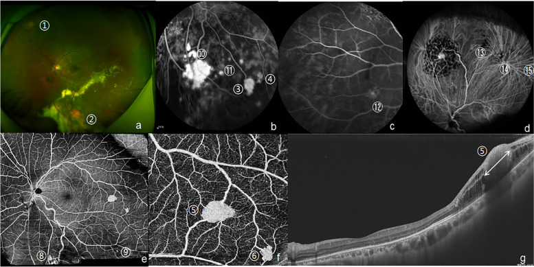
Background: To report the multimodal imaging and treatment of fifteen retinal capillary hemangioblastomas (RCHs) associated with Von Hippel-Lindau syndrome in a monocular patient during a long-term following-up, which supply high-resolution exquisite SS-OCTA images (VG200; SVision Imaging, Ltd., Luoyang, China) and management experience about multiple RCHs.

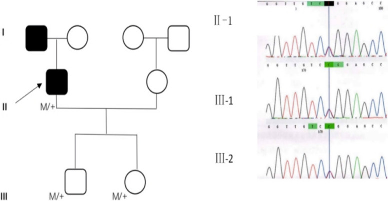
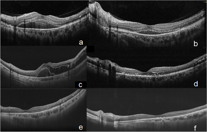
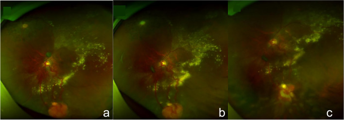
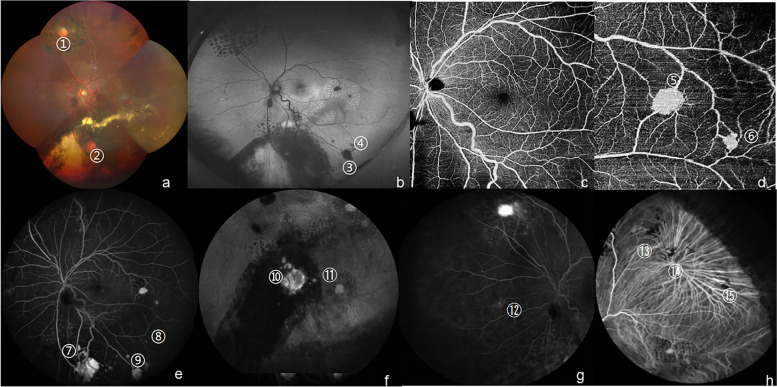
Case presentation: A 34-year-old monocular male patient complained decreased visual acuity (20/100) without pain and redness in the left eye five years ago. Von Hippel-Lindau syndrome were diagnosed with genetic testing. He, his son and daughter all carried a heterozygosity missense variant c.499C > T (p. Arg167Trp) in the Hg19 gene, a VHL gene located in Chr3:10,191,506. Fifteen RCHs were identified by the application of multimodal imaging, which including fundus photo, fundus autofluorescence (FAF), B-scan ultrasonography (US), fluorescein fundus angiography (FFA), indocyanine green angiography (ICGA) and swept-source optical coherence tomography angiography (SS-OCTA). Transscleral cryotherapy and laser photocoagulation were performed to destroy the largest RCH with the size of 4 PD in diameter. Laser photocoagulation was conducted to seal the middle or tiny RCHs (< 1.5 PD) and their nourishing vessels. The retinal edema and exudative macular detachment were successfully relieved by intraocular injection of bevacizumab for 5 times. The RCHs in the left eye responded well to these treatments and best corrected visual acuity was 20/25 for three years. Three-month recall visits were recommended for him.

Conclusion: For multiple retinal capillary hemangioblastomas in monocular patients, precise combined therapy guided by multimodal imaging has a profound impact on the management of new and recurrent RCHs.

Keywords: Case report; Multiple retinal capillary hemangioblastomas; Precise combined therapy; Swept-source optical coherence tomography angiography; Von Hippel-Lindau syndrome.


Conflict of interest statement

The authors declare that they have no competing interests.

Figures

Fig. 1
Fig. 1
Pedigree chart and Sanger sequencing results of VHL gene variants using blood samples. The black square represents the patients who were clinically diagnosed with VHL. M/ + means heterozygous variants. The Sanger sequencing results show heterozygous missense variants in the VHL gene of patients II-1, III-1, and III-2
Fig. 2
Fig. 2
The first multimodal image evaluation of eleven RCHs in a Von Hippel-lindau syndrome patient on June 6th, 2017. a. Fundus photo identified huge RCHs (No.1, 2, 7 RCHs). b. FFA visualized a newly developed small RCH in temporal retina (No.3 RCH). c-e. FFA sketched the contours of several small peripheral RCHs (No.4–11 RCHs). f. B-scan ultrasonography displayed the largest oblate lesion with the size of 6 mm* 6 mm*2 mm was located in the inferior nasal retina (No.2 RCH). F. SS-OCT (single line scan mode) showed an elevated lesion wrapped much exudation and the largest RCH with the size of 2.15*2.60*1.35 mm, arrow represent the height (1.35 mm). (No.2 RCH). RCH retinal capillary hemangioblastomas, FFA fluorescein fundus angiography, SS-OCT swept-source optical coherence tomography
Fig. 3
Fig. 3
OCT (single line scan mode) image of exudation in the macular region during five-year following up. a.SD-OCT showed macular detachment with exudative subretinal fluid(SRF)at his first visit on May 19th, 2017. b. SD-OCT exhibited reduced SRF and exudation after twice anti-VEGF therapy and laser photocoagulation on September 10th, 2017. c. SS-OCT revealed recurrent exudative macular detachment complicated with macular cystoid edema on August 17th, 2018, one year after the combined therapy. d. SD-OCT visualized SRF and macular edema were completely absorbed with much exudation left after anti-VEGF therapy for four times on December 10th, 2018. ef. Nine months and two years after the cryotherapy (August 10th, 2019, November 6th, 2020), the macular region was still in good structure by the observations of SS-OCT Spectral-domain optical coherence tomography, SD-OCT Swept- source optical coherence tomography, SS-OCT subretinal fluid, SRF subretinal fluid
Fig. 4
Fig. 4
Fundus photo of rapidly enlarged RCHs and the therapeutic effect of cryotherapy. a. Fundus photo (taken in August 17th, 2018) showed an orange-red ellipse RCH with the diameter of approximately 3 PD was located in the nasal inferior retina, nourishing by two twisted and engorged vessels. b. Fundus photo (taken in December 10th, 2018) showed the RCH located in the nasal inferior retina was rapidly enlarged to 4 PD in diameter and its nourishing vessels dilated with much exudation. c. One month after cryotherapy, the RCH in the nasal inferior retina appeared to be pale and atrophied accompanied by reduced retinal exudation based on the fundus photo taken in January 28th, 2019. RCH retinal capillary hemangioblastomas, PD papillary diameter
Fig. 5
Fig. 5
The second multimodal imaging of eleven RCHs in a Von Hippel-lindau syndrome patient on November 6th, 2020. a. Fundus photo showed a stationary white round-like RCH (1.5 PD in diameter) was in nasal superior retina (No.1 RCH) and an active orange round-like RCH (1 PD in diameter) along with diffuse exudation located in nasal inferior retina (No.2 RCH)b. FFA distinguished several middle RCHs from exudations (No.3–4, 10–11 RCH). c-d. FFA and ICGA showed four shrunk RCHs, which were small in previous evaluation, and now were almost traceless. e–f. SS-OCTA images at 15 × 12 mm2 and 6 × 6 mm2 sections with the center located on the fovea represented the angiography of the SCP, which respectively revealed a good structure in macular region and displayed small and tiny RCHs and their nourishing vessels clearly in posterior pole (No. 5–6, 8–9 RCH). g. SS-OCT (single line scan mode) showed a solid small hemangioma with the size of 1.35*0.84*0.62 mm, which was tiny in previous evaluation and overlooked in the laser treatment. (No.5 RCH). Arrow in figure g represent the length of No. 5 RCH, namely 1.35 mm. RCH retinal capillary hemangioblastomas, FFA fluorescein fundus angiography, SS-OCT(A) swept- source optical coherence tomography (angiography), ICGA indocyanine green angiography, PD papillary diameter, mm: millimeter, SCP superficial retinal capillary plexus (the layer between internal limiting membrane and 15 μm below inner plexiform layer)
Fig. 6
Fig. 6
The third multimodal image evaluation of eleven RCHs in a Von Hippel-Lindau syndrome patient three month after the last laser treatment. a. Fundus photo identified huge RCHs and yellow-white exudation around RCHs (No.1–2 RCHs). b. FAF revealed the middle RCHs from dilated vessels (No.3 RCH). c-d. SS-OCTA images at 12 × 12 mm2 and 6 × 6 mm2 sections with the center located on the fovea showed the angiography of the SCP (c, d), which respectively showed the good structure in macular region and visualized No.5–6 RCHs begun to shrunk with decreased retinal blood flow density around the tumors after laser treatment. e. FFA visualized several small peripheral RCHs (No 7–9 RCHs). f. ICGA distinguished small RCHs from fluorescent leakage points (No.10–11). g-h. FFA and ICGA showed four traceless RCHs, which were atrophied by combined therapy. RCH retinal capillary hemangioblastomas, FAF fundus autofluorescence, FFA fluorescein fundus angiography, ICGA indocyanine green angiography, SS-OCTA swept- source optical coherence tomography angiography, SCP superficial retinal capillary plexus (the layer between internal limiting membrane and 15 μm below inner plexiform layer)

Similar articles

Cited by

References

    1. Haddad NM, Cavallerano JD, Silva PS. Von hippel-lindau disease: a genetic and clinical review. Semin Ophthalmol. 2013;28(5–6):377–386. doi: 10.3109/08820538.2013.825281. - DOI - PubMed
    1. Minervini G, Quaglia F, Tabaro F, Tosatto SCE. Insights into the molecular features of the von Hippel-Lindau-like protein. Amino Acids. 2019;51(10–12):1461–1474. doi: 10.1007/s00726-019-02781-8. - DOI - PubMed
    1. Niemela M, Lemeta S, Sainio M, Rauma S, Pukkala E, Kere J, Bohling T, Laatikainen L, Jaaskelainen J, Summanen P. Hemangioblastomas of the retina: impact of von Hippel-Lindau disease. Invest Ophthalmol Vis Sci. 2000;41(7):1909–1915. - PubMed
    1. Chun LY, Massamba N, Silas MR, Blair MP, Hariprasad SM, Skondra D. Use of optical coherence tomography angiography in the diagnosis of small retina lesions in Von Hippel-Lindau disease. Eye (Lond) 2020;34(12):2345–2346. doi: 10.1038/s41433-020-0792-5. - DOI - PMC - PubMed
    1. Reich M, Glatz A, Boehringer D, Evers C, Daniel M, Bucher F, Ludwig F, Nuessle S, Lagreze WA, Maloca PM, et al. Comparison of Current Optical Coherence Tomography Angiography Methods in Imaging Retinal Hemangioblastomas. Transl Vis Sci Technol. 2020;9(8):12. doi: 10.1167/tvst.9.8.12. - DOI - PMC - PubMed

Publication types

MeSH terms