. 2022 May 6;12(1):10.
doi: 10.1186/s13395-022-00292-x.

Growth differentiation factor 11 induces skeletal muscle atrophy via a STAT3-dependent mechanism in pulmonary arterial hypertension

Affiliations

Growth differentiation factor 11 induces skeletal muscle atrophy via a STAT3-dependent mechanism in pulmonary arterial hypertension

Guiling Xiang et al. Skelet Muscle. .

Abstract

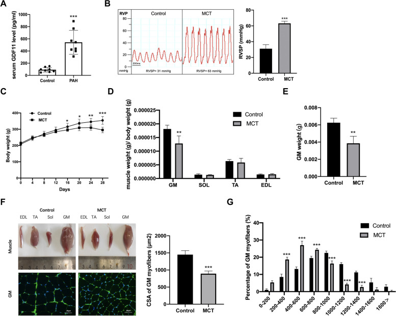
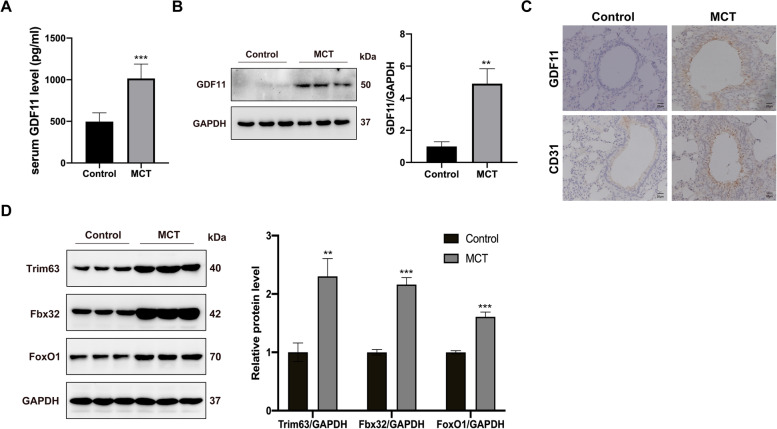
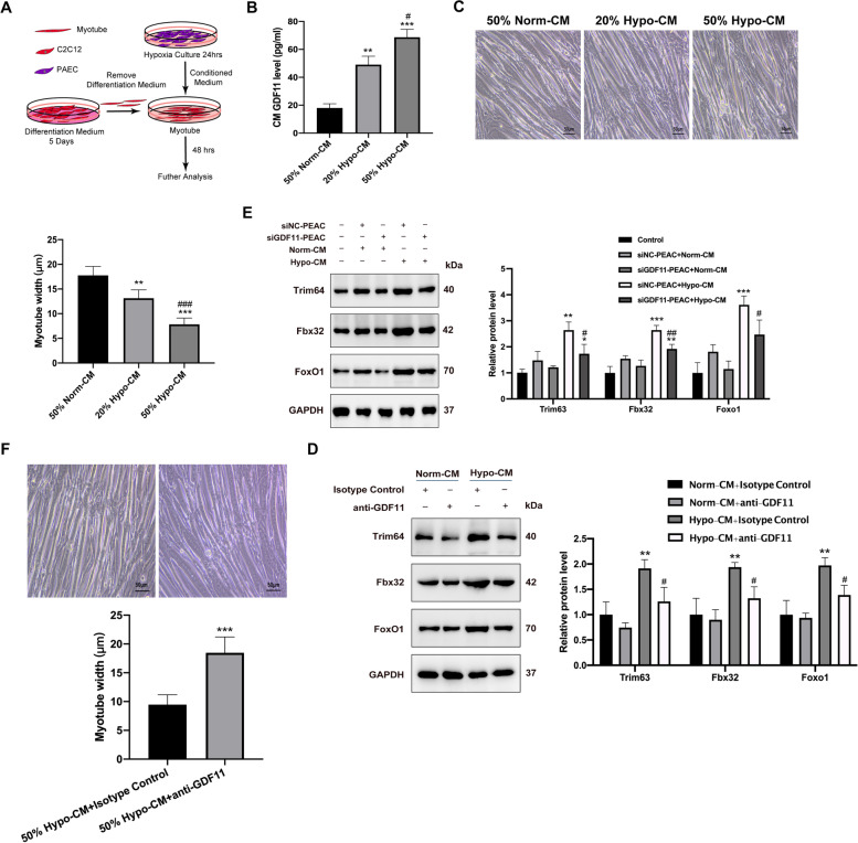
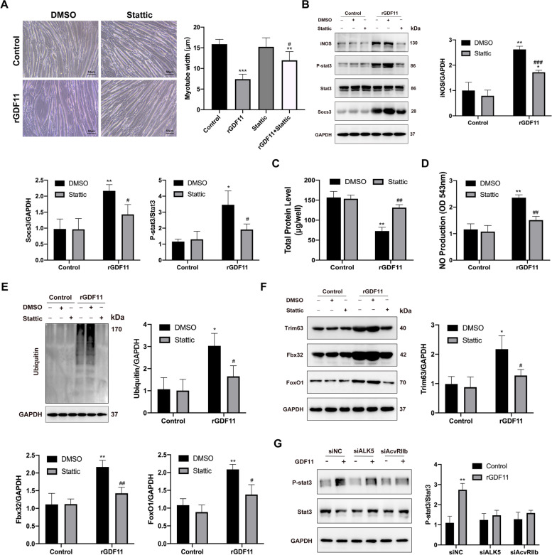
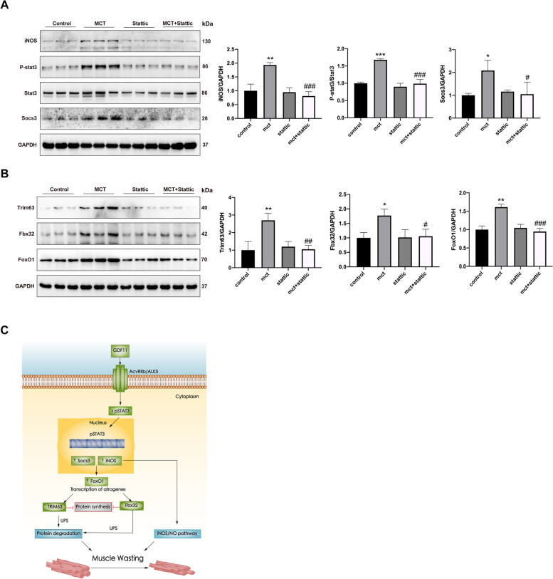
Skeletal muscle wasting is a clinically remarkable phenotypic feature of pulmonary arterial hypertension (PAH) that increases the risk of mortality. Growth differentiation factor 11 (GDF11), centrally involved in PAH pathogenesis, has an inhibitory effect on skeletal muscle growth in other conditions. However, whether GDF11 is involved in the pathogenesis of skeletal muscle wasting in PAH remains unknown. We showed that serum GDF11 levels in patients were increased following PAH. Skeletal muscle wasting in the MCT-treated PAH model is accompanied by an increase in circulating GDF11 levels and local catabolic markers (Fbx32, Trim63, Foxo1, and protease activity). In vitro GDF11 activated phosphorylation of STAT3. Antagonizing STAT3, with Stattic, in vitro and in vivo, could partially reverse proteolytic pathways including STAT3/socs3 and iNOS/NO in GDF11-meditated muscle wasting. Our findings demonstrate that GDF11 contributes to muscle wasting and the inhibition of its downstream molecule STAT3 shows promise as a therapeutic intervention by which muscle atrophy may be directly prevented in PAH.

Keywords: GDF11; Pulmonary arterial hypertension; STAT3; Skeletal muscle atrophy.


Conflict of interest statement

The authors declare that they have no competing interests.

Figures

Fig. 1
Fig. 1
MCT-induced PAH caused body weight loss and muscle atrophy. A Circulating GDF11 levels in PAH patients and health control (n=8). B RVSP of rats 4 weeks after MCT-treated, C body weight, and D the weight of gastrocnemius muscle, soleus muscle, tibialis anterior, and Extensor Digitorum Longus normalized per body weight (BW). E The weight of gastrocnemius muscle. F Representative images of EDL, TA, Sol, GM, and the cross-sectional areas of approximately 250 myofibers per group were determined. Scale bar represents 25 μm. G The distribution of myofiber cross-sectional area. n = 8 rats/group. Data are shown as mean ± SEM. Versus vehicle control, *P < 0.05, **P < 0.01, ***P < 0.001
Fig. 2
Fig. 2
GDF11 levels are accumulated in serum and lung in MCT rats. A GDF11 was measured by ELISA in the serum from rats. B GDF11 expression in lung was detected by Western blot, and GAPDH served as a loading control. C Representative immunohistochemistry of lung sections showing pulmonary arteries stained for GDF11 or CD31 in rats, scale bar is 20 μm. D Western blot analysis of trim63, fbx32, and foxo1 was assessed by Western blot, and protein expression levels were quantified by densitometry. Data are shown as mean ± SEM. Versus vehicle control, *P < 0.05, **P < 0.01, ***P < 0.001, n = 8 rats/group
Fig. 3
Fig. 3
The GDF11 is involved in myotube atrophy induced by CM from PAEC. A Schematic drawing depicting the generation of CM by hypoxia-culture of PAEC, then myotube was stimulated with CM for 48 h. B Concentrations of GDF11 (pg/mL) in 50% Norm-CM, 20% or 50% Hypo-CM. C Bright-field images of C2C12-derived myotubes treated with either 50% Norm-CM, 20% or 50% Hypo-CM from PAEC, and myotube diameter for conditions represented in the panel. Scale bar is 50 μm. D Myotubes were transfected with GDF11 siRNA or NC siRNA and 50% Norm-CM or 50% Hypo-CM. Protein levels were examined by immunoblotting. E Bright-field images of myotubes treated with 50% Hypo-CM with GDF11 antibody or isotype control, and myotube diameter for conditions represented in the panel. Scale bar is 50μm. F Immunoblots of trim63, fbx32, and foxo1 using lysates from myotubes treated with 50% Norm-CM or 50% Hypo-CM with GDF11 antibody or isotype control; and protein expression levels were quantified by densitometry. Values are presented as average ± SEM. Versus vehicle control, *P < 0.05, **P < 0.01, ***P < 0.001; versus Hypo-CM control, #P < 0.05, ##P < 0.01, ###P < 0.001; n=3
Fig. 4
Fig. 4
GDF11 acts via STAT3, SOCS3, and iNOS to induce proteolysis in muscle wasting in vitro. AD NF-κB, ERK, Smad, or STAT3 dependent luciferase reporters in C2C12 myotubes treated with rGDF11 with the dose ranging from 0 to 100 ng/ml. E Representative Western blots of target proteins (iNOS, phosphorylation and total STAT3, phosphorylation and total Smad2/3, socs3) and loading control (GAPDH) from myotubes treated with rGDF11 with the dose ranging from 0 to 100 ng/ml for 48 h. F NO levels were measured in supernatant from the myotubes described in the panel. G Total protein content of rGDF11-treated myotubes. H Representative western blotting images of ubiquitin from myotubes. I Protein expression of trim63, fbx32, and foxo1 in myotubes treated with rGDF11 with the dose ranging from 0 to 100 ng/ml. GAPDH was used as an internal control. J Bright-field images of myotubes treated with rGDF11, with or without the 26S ribosome inhibitor MG-132 (10 μM) for 48 h; diameter of myotubes for conditions represented in the panel. Scale bar is 50μm. Data presented as mean ± SEM. Versus vehicle control, *P < 0.05, **P < 0.01, ***P < 0.001; versus rGDF11 control, #P < 0.05, ##P < 0.01, ###P < 0.001; n=3
Fig. 5
Fig. 5
Blocking STAT3 activation with Stattic, a STAT3 inhibitor, prevents GDF11 mediated atrophy in vitro. A Bright-field images of myotubes treated with rGDF11 (50ng/ml), with or without STAT3 inhibitor Stattic for 48 h. Scale bars = 50 μm. The fiber widths were measured and calculated (right panel). B Myotubes treated with rGDF11 then with Stattic for 48h were used for Western blot analysis with antibodies against iNOS, pY-STAT3, total STAT3, socs3, and GAPDH. C Total protein content of rGDF11-treated myotubes, with or without STAT3 inhibitor Stattic for 48 h. D NO levels were measured in supernatant from the myotubes described in the panel. E Representative western blotting images of ubiquitin from myotubes. F Protein expression of trim63, fbx32, and foxo1 in myotubes treated with rGDF11 then with Stattic for 48h. G Representative Western blots of phosphorylation and total STAT3 from myotubes treated with rGDF11, siALK5, or AcvRIIb. Data presented as mean ± SEM. Versus vehicle control, *P < 0.05, **P < 0.01, ***P < 0.001; versus rGDF11 control, #P < 0.05, ##P < 0.01, ###P < 0.001; n=3
Fig. 6
Fig. 6
STAT3 inhibition prevents muscle atrophy in the MCT rat. A SD rats were treated with MCT or saline on day 1, with or without intraperitoneal injection of Stattic daily for 2 weeks since day 14. The effects of Stattic on the main features of PAH model were examined, including B RVSP, C body weight, and D gastrocnemius muscles mass. E Cross-sections cut from the gastrocnemius muscle and stained with wheat germ agglutinin (blue). Scale bar is 25μm, n=3. F The cross-sectional areas of approximately 250 myofibers per group were determined and the distribution of myofiber cross-sectional area. Data are shown as mean ± SEM. Versus vehicle control, *P < 0.05, **P < 0.01, ***P < 0.001; versus MCT control, #P < 0.05, ##P < 0.01, ###P < 0.001; n=8 rats
Fig. 7
Fig. 7
Pathway of STAT3 inhibition in improvement muscle atrophy in the PAH model. A, B The expression of indicated proteins in gastrocnemius muscles was detected by western blot. The band intensities were quantified and total STAT3 or GAPDH was used as control. C Model depicting how STAT3 promotes GDF11-induced muscle wasting. The GDF11 binds to ACVR2B/ALK5 and then activates STAT3 via phosphorylation. Following p-STAT3 translocates to the nucleus and upregulates the expression of iNOS and socs3, leading to the activation of the ubiquitin-proteasome pathway and iNOS/NO pathway, which in turn promotes muscle wasting. Data are shown as mean ± SEM. Versus vehicle control, *P < 0.05, **P < 0.01, ***P < 0.001; versus MCT control, #P < 0.05, ##P < 0.01, ###P < 0.001; n=3 rats

Similar articles

Cited by

References

    1. McKenna SP, Doughty N, Meads DM, et al. The Cambridge Pulmonary Hypertension Outcome Review (CAMPHOR): a measure of health-related quality of life and quality of life for patients with pulmonary hypertension. Qual Life Res. 2006;15:103–115. doi: 10.1007/s11136-005-3513-4. - DOI - PubMed
    1. Meyer FJ, Lossnitzer D, Kristen AV, et al. Respiratory muscle dysfunction in idiopathic pulmonary arterial hypertension. Eur Respir J. 2005;25:125–130. doi: 10.1183/09031936.04.00095804. - DOI - PubMed
    1. Bauer R, Dehnert C, Schoene P, et al. Skeletal muscle dysfunction in patients with idiopathic pulmonary arterial hypertension. Respir Med. 2007;101:2366–2369. doi: 10.1016/j.rmed.2007.06.014. - DOI - PubMed
    1. Zafrir B. Exercise training and rehabilitation in pulmonary arterial hypertension: rationale and current data evaluation. J Cardiopulm Rehabil Prev. 2013;33:263–273. doi: 10.1097/HCR.0b013e3182a0299a. - DOI - PubMed
    1. Donaldson AV, Maddocks M, Martolini D, et al. Muscle function in COPD: a complex interplay. Int J Chron Obstruct Pulmon Dis. 2012;7:523–535. - PMC - PubMed

Publication types

MeSH terms

Substances