. 2022 May 6;22(1):157.
doi: 10.1186/s12903-022-02195-1.

Efficiency of biofilm removal by combination of water jet and cold plasma: an in-vitro study

Affiliations

Efficiency of biofilm removal by combination of water jet and cold plasma: an in-vitro study

Rutger Matthes et al. BMC Oral Health. .

Abstract

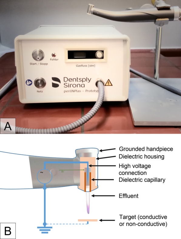
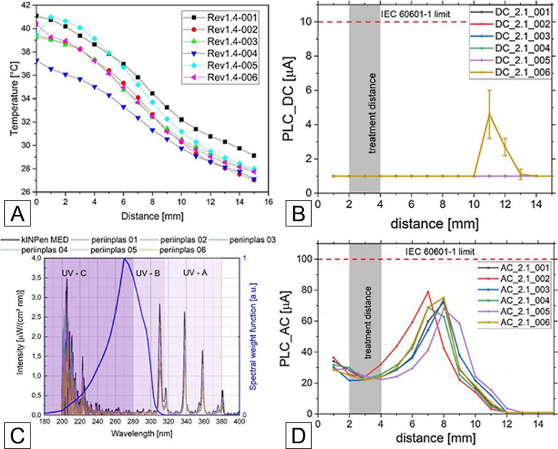
Background: Peri-implantitis therapy is a major problem in implantology. Because of challenging rough implant surface and implant geometry, microorganisms can hide and survive in implant microstructures and impede debridement. We developed a new water jet (WJ) device and a new cold atmospheric pressure plasma (CAP) device to overcome these problems and investigated aspects of efficacy in vitro and safety with the aim to create the prerequisites for a clinical pilot study with these medical devices.

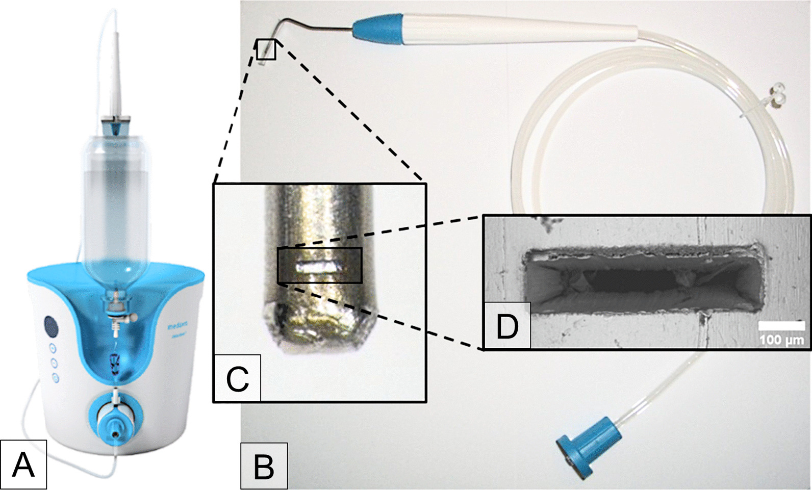
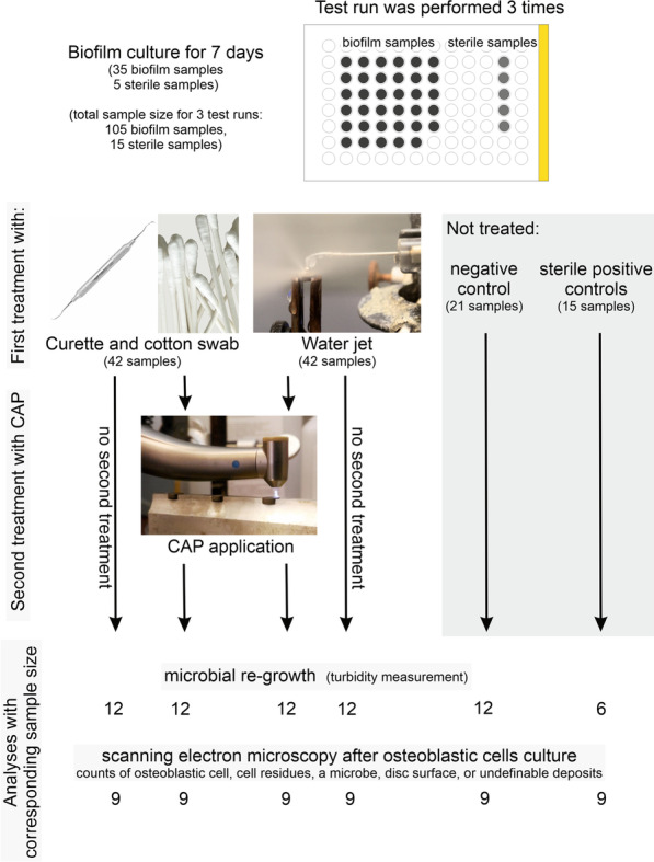
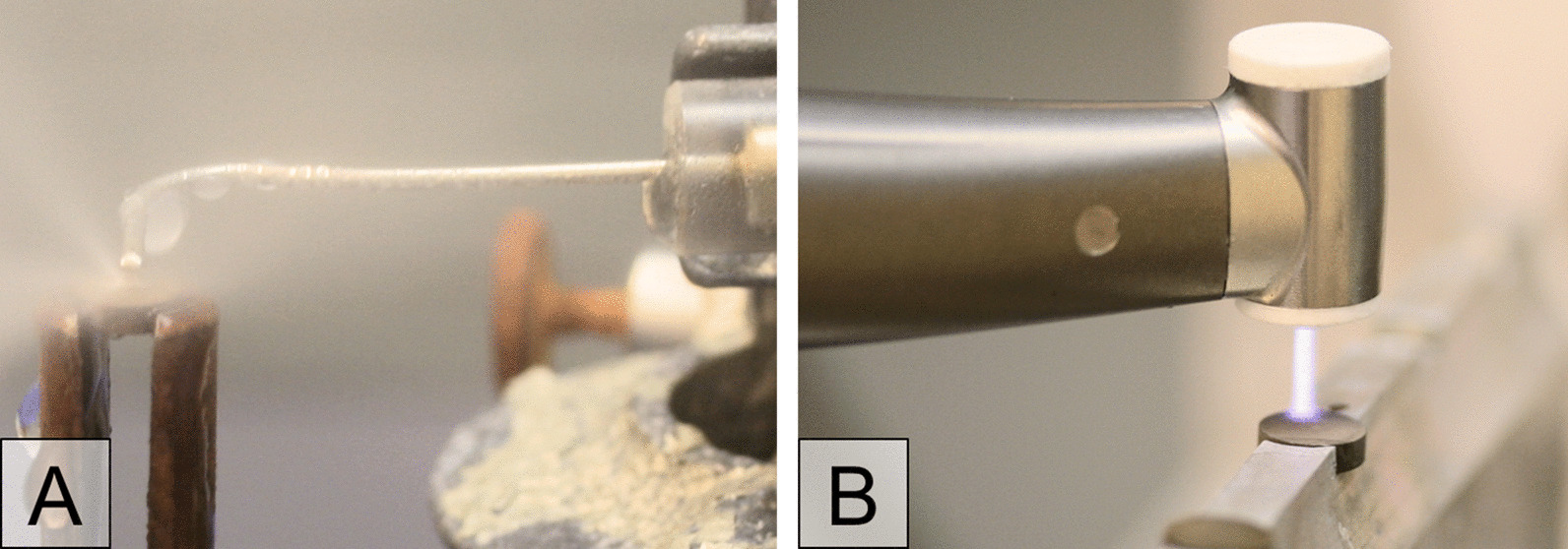
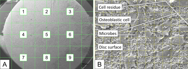
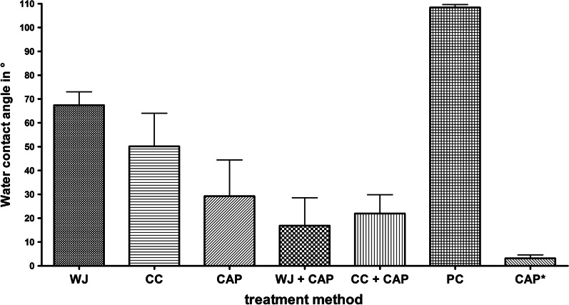
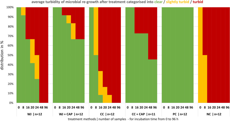
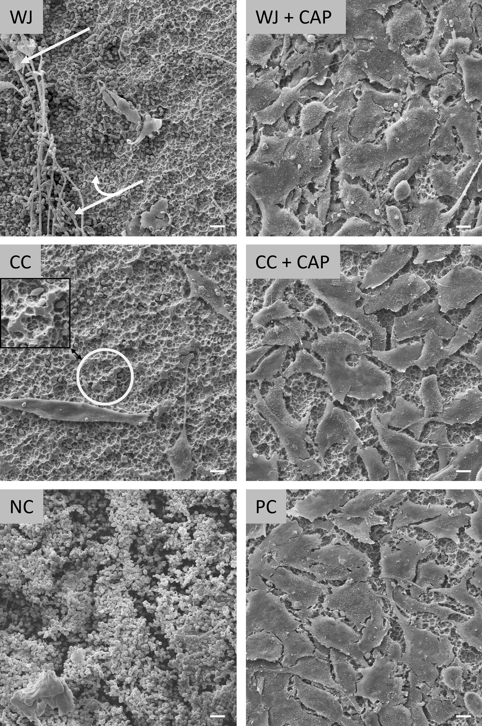
Methods: We compared the efficiency of a single treatment with a WJ or curette and cotton swab (CC) without or with adjunctive use of CAP (WJ + CAP, CC + CAP) to remove biofilm in vitro from rough titanium discs. Treatment efficacy was evaluated by measuring turbidity up to 72 h for bacterial re-growth or spreading of osteoblast-like cells (MG-63) after 5 days with scanning electron microscopy. With respect to application safety, the WJ and CAP instruments were examined according to basic regulations for medical devices.

Results: After 96 h of incubation all WJ and CC treated disks were turbid but 67% of WJ + CAP and 46% CC + CAP treated specimens were still clear. The increase in turbidity after WJ treatment was delayed by about 20 h compared to CC treatment. In combination with CAP the cell coverage significantly increased to 82% (WJ + CAP) or 72% (CC + CAP), compared to single treatment 11% (WJ) or 10% (CC).

Conclusion: The newly developed water jet device effectively removes biofilm from rough titanium surfaces in vitro and, in combination with the new CAP device, biologically acceptable surfaces allow osteoblasts to grow. WJ in combination with CAP leads to cleaner surfaces than the usage of curette and cotton swabs with or without subsequent plasma treatment. Our next step will be a clinical pilot study with these new devices to assess the clinical healing process.

Keywords: Biofilm; Cold plasma; Peri-implantitis; Titanium surface; Water jet.


Conflict of interest statement

The authors declare that no conflict of interest with all sources of institutional, private and corporate financial support for their study exists. The authors Christian Eberhard and Leo Seifert, neither of whom had any influence on the data collection and data analysis of this study, are employees of Sirona Dental Systems GmbH and have filed a patent application (WO2020182669A1).

Figures

Fig. 1
Fig. 1
The photograph shows A the debritom + as manufactured by the Swiss company Medaxis AG. Our B adapted handpiece has an elongated application tip resembling a periodontal probe. The nozzle at the end of the tip, see a photograph in the inset C, produces the micro water jet for cleaning of dental implants. The nozzle has a size of about 500 × 100 µm, as can be seen in the scanning electron micrograph D
Fig. 2
Fig. 2
A Photograph of the plasma device periINPlas with handpiece, B schematic construction inside the handpiece head where the plasma is generated
Fig. 3
Fig. 3
Flow chart of the timing (1st period biofilm culture for 7 days in the 96-well microplate), the distribution of the discs and number of samples among the test groups (curette and cotton swab, water jet and cold atmospheric pressure plasma (CAP) and among the analytical methods
Fig. 4
Fig. 4
The two photographs show A the water jet application during treatment and B the plasma device handpiece during CAP treatment on titanium specimen in special disc holders
Fig. 5
Fig. 5
Depiction of SEM analyse procedure. A) Scanning electron micrograph (× 18) with a grid to define the 9 places to scan. B Scanning electron micrograph (× 500) with grid to count the visibility of osteoblastic cells, cell residues, microbes, blank disk surface, and objects that are not definable (deposits)
Fig. 6
Fig. 6
Pictures of the oral mucosa of the mandibula of pig cadavers were treated with the Dental water jet (left column, A1–B1) and a powder jet device (right column, A2–B2). Upper panel shows the tissue before (A) and the lower panel after treatment (B) in the region of keratinised gingiva tissue (x) and non-keratinised gingiva (y). The Dental water jet was used with 5 different power settings (L1–L5). Three different areas were tested for the powder jet device. No abrasion is seen in the keratinised mucosa near the teeth (x-range), tissue abrasion and blistering are observed in the non-keratinised mucosa (y-range)
Fig. 7
Fig. 7
The graphs show the results of physical measurements of the different versions of the periINPlas devices, A temperature over distance curve, B DC part of patient leakage current (PLC) over distance, C spectral emission for all plasma devices (including kINPen MED), D AC part of PLC over distance
Fig. 8
Fig. 8
Mean values and standard deviation of water contact angles (°) of specimen treated by water jet (WJ), curette + cotton swab (CC), cold atmospheric pressure plasma (CAP), combined treatment of WJ + CAP and CC + CAP, and the untreated positive control (PC) (n = 3, time span between treatment and measurement was 2 h). Additionally, mean water contact angle data for specimen immediately measured after CAP (CAP*, time span 10 min) are shown
Fig. 9
Fig. 9
The stack diagram shows the distribution of the categorised OD values (clear, slightly turbid, and turbid) after treatment with water jet (WJ), curette + cotton swab (CC), WJ or CC combined with cold atmospheric pressure plasma CAP (WJ + CAP, CC + CAP), and the two untreated controls, the sterile positive control (PC), and biofilm covered negative control (NC). Turbidity was measured after 0, 8, 16, 20, 24, 48, and 96 h incubation time
Fig. 10
Fig. 10
Scanning electron micrographs showing examples of specimen after 5 days of osteoblastic cell cultivation for water jet (WJ), curette + cotton swab (CC) and their combinations with plasma (WJ + CAP, CC + CAP), negative control (NC) with untreated plaque-biofilm, and untreated positive control PC. The white single arrow points to destroyed osteoblastic cells, the double arrow points to microbes, and the circle marks a scratched area (see also Additional file 3). Scale bars: 10 µm

References

    1. Fiorillo L. We do not eat alone: Formation and maturation of the oral microbiota. Biology. 2020 doi: 10.3390/biology9010017. - DOI - PMC - PubMed
    1. Derks J, Schaller D, Håkansson J, Wennström JL, Tomasi C, Berglundh T. Effectiveness of implant therapy analyzed in a Swedish population prevalence of peri-implantitis. J Dent Res. 2016;95:43–49. doi: 10.1177/0022034515608832. - DOI - PubMed
    1. Heitz-Mayfield LJA, Lang NP. Comparative biology of chronic and aggressive periodontitis vs. peri-implantitis. Periodontology. 2000;2010(53):167–81. doi: 10.1111/j.1600-0757.2010.00348.x. - DOI - PubMed
    1. Rosen P, Clem D, Cochran D, Bradley SF, Renvert S, Wang H-L. Peri-implant mucositis and peri-implantitis: a current understanding of their diagnoses and clinical implications. J Periodontol. 2013;84:436–443. doi: 10.1902/jop.2013.134001. - DOI - PubMed
    1. Mahato N, Wu X, Wang L. Management of peri-implantitis: a systematic review, 2010–2015. Springerplus. 2016;5:105. doi: 10.1186/s40064-016-1735-2. - DOI - PMC - PubMed

Publication types

LinkOut - more resources