. 2022 May 20;50(9):5226-5238.
doi: 10.1093/nar/gkac315.

A programmable pAgo nuclease with RNA target preference from the psychrotolerant bacterium Mucilaginibacter paludis

Affiliations

A programmable pAgo nuclease with RNA target preference from the psychrotolerant bacterium Mucilaginibacter paludis

Wenqiang Li et al. Nucleic Acids Res. .

Abstract

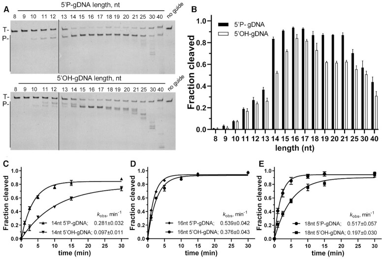
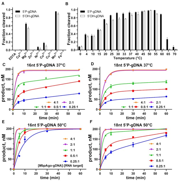
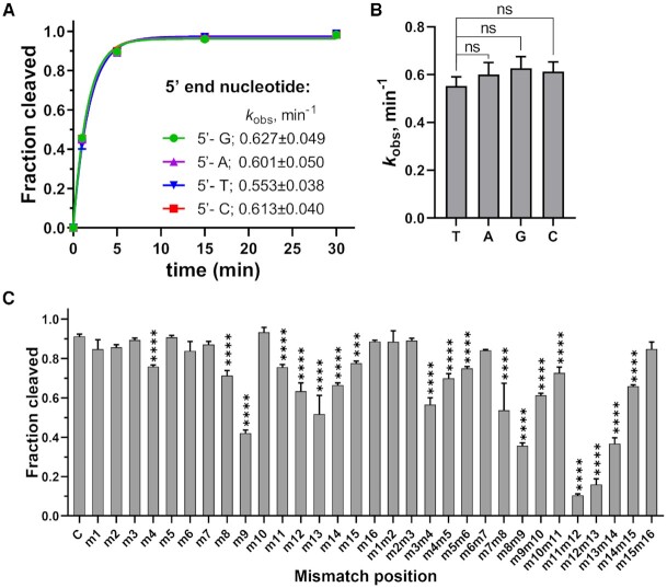
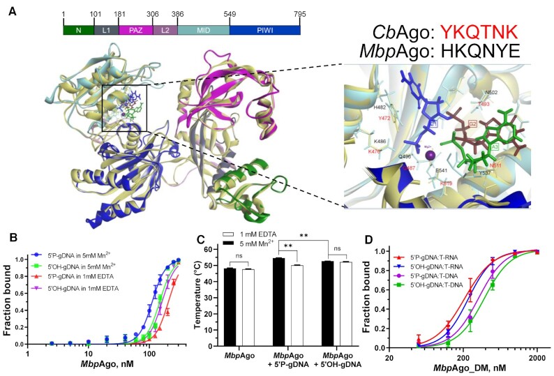
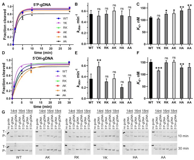
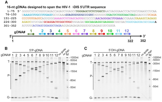
Argonaute (Ago) proteins are programmable nucleases found in eukaryotes and prokaryotes. Prokaryotic Agos (pAgos) share a high degree of structural homology with eukaryotic Agos (eAgos), and eAgos originate from pAgos. Although eAgos exclusively cleave RNA targets, most characterized pAgos cleave DNA targets. This study characterized a novel pAgo, MbpAgo, from the psychrotolerant bacterium Mucilaginibacter paludis which prefers to cleave RNA targets rather than DNA targets. Compared to previously studied Agos, MbpAgo can utilize both 5'phosphorylated(5'P) and 5'hydroxylated(5'OH) DNA guides (gDNAs) to efficiently cleave RNA targets at the canonical cleavage site if the guide is between 15 and 17 nt long. Furthermore, MbpAgo is active at a wide range of temperatures (4-65°C) and displays no obvious preference for the 5'-nucleotide of a guide. Single-nucleotide and most dinucleotide mismatches have no or little effects on cleavage efficiency, except for dinucleotide mismatches at positions 11-13 that dramatically reduce target cleavage. MbpAgo can efficiently cleave highly structured RNA targets using both 5'P and 5'OH gDNAs in the presence of Mg2+ or Mn2+. The biochemical characterization of MbpAgo paves the way for its use in RNA manipulations such as nucleic acid detection and clearance of RNA viruses.


Figures

Figure 1.
Figure 1.
MbpAgo exhibits DNA-guided RNA endonuclease activity at 37°C. (A) Maximum likelihood phylogenetic tree of characterized Ago proteins. (B) Guide and target oligonucleotides. gDNAs and RNA targets (T-RNA) were used in most experiments. The black triangle indicates the cleavage site. (C)MbpAgo exhibits DNA-guided RNA endonuclease activity. (D)MbpAgo exhibits no DNA cleavage activity. Positions of the cleavage products (P) are indicated on the left of the gels. MbpAgo, guide and target were mixed at a 4:2:1 molar ratio (800 nM MbpAgo preloaded with 400 nM guide, plus 200 nM target) and incubated for 30 min at 37°C. Catalytic dead mutant MbpAgo_DM (DM) was used as a control. Lanes M1 and M2 contain chemically synthesized 34-nt RNA and DNA corresponding to the cleavage products of T-RNA and the DNA target (T-DNA), respectively.
Figure 2.
Figure 2.
Both the length and the 5′P of the guide affect cleavage efficiency and precision. (A) Cleavage assays with 5′P-gDNA (Upper panel) and 5′OH-gDNA (Lower panel) of varying lengths. The positions of the targets (T) and cleavage products (P) are indicated on the left of the gels. Representative gels from three independent measurements are shown. (B) Quantification of cleavage efficiencies (the percentage of target cleavage). The fraction of the cleaved target for each guide length is shown. Experiments in (A) and (B) were carried out for 10 min at 37°C. (C–E) Kinetics analyses of RNA cleavage by MbpAgo with 14, 16 and 18 nt gDNAs, respectively. The kobsvalues were determined from the single-exponential fits of the data. Data are represented as the mean ± standard deviation (SD) from three independent experiments. In all experiments, MbpAgo, guide and target were mixed at a 4:2:1 molar ratio (800 nM MbpAgo preloaded with 400 nM guide, plus 200 nM target) and incubated at 37°C.
Figure 3.
Figure 3.
Characteristics of nuclease activity of MbpAgo. (A) DNA-guided RNA cleavage by MbpAgo with various divalent cations. (B) Temperature dependence of RNA cleavage by MbpAgo. Experiments in (A) and (B) were performed for 15 min at 37°C. (C and D) Quantified data of the MbpAgo-mediated 16 and 18 nt 5′P DNA-guided RNA cleavage turnover experiments using 200 nM RNA target and increasing concentrations of MbpAgo–gDNA (50–800 nM) at 37°C, respectively. (E and F) Quantified data of MbpAgo-mediated 16 and 18 nt 5′P DNA-guided RNA cleavage turnover experiments using 200 nM RNA target and increasing concentrations of MbpAgo–gDNA (50–800 nM) at 50°C, respectively. The MbpAgo–gDNA complex was prepared by mixing MbpAgo with a 1:1 molar ratio of 5′P-gDNA and incubating for 10 min at 37°C. Data were fitted using single-exponential functions if the [MbpAgo–gDNA]/RNA target ratio was > 1. Data were fitted using two-phase functions if the [MbpAgo–gDNA]/RNA target ratio was ≤ 1. Data are the mean ± SD from three independent measurements.
Figure 4.
Figure 4.
Effects of the 5′-nucleotide of the guide and guide-target mismatches on target cleavage. (A) Preferences for the 5′-nucleotide of the guide. The kobsvalues were determined from the single-exponential fits of the data. Data are the mean ± SD from three independent experiments. (B) P-values for all comparisons of kobs values from (A). nsP > 0.05, compared to the 5′-T guide using Student's t-test. (C) Effects of guide–target mismatches on RNA cleavage by MbpAgo. Data are the mean ± SD from three independent measurements. The reaction was performed with 16 nt 5′P-gDNA at 37°C for 5 min. ***P < 0.001 and ****P < 0.0001, compared to C, the control reactions with guide containing no mismatches, using Student′s t-test. In all experiments, MbpAgo, guide and target were mixed at a 4:2:1 molar ratio (800 nM MbpAgo preloaded with 400 nM guide, plus 200 nM target) and incubated at 37°C.
Figure 5.
Figure 5.
Binding analyses of guides and targets by MbpAgo. (A) (Left panel) A 3D model of the MbpAgo aligned to the structure of CbAgo in complex with a gDNA and a DNA target (with bound Mg2+ ions; PDB: 6QZK). MbpAgo domains are colored according to the colored domain architecture of MbpAgo with numbered residues and CbAgo is light yellow. The model was built using the SWISS-MODEL portal. (Right panel) Amino acid residues of the conserved MID-domain motif (shown for MbpAgo and CbAgo above the structure) and Mg2+ ions (purple) involved in interactions with the first nucleotide (blue) and the second nucleotide (deep red) of the guide are highlighted. Elements of the secondary structure and amino residues specific to MbpAgo and CbAgo are shown in cyan and light yellow, respectively. (B) Binding of 16 nt guides by MbpAgo with 5 mM Mn2+ or 1 mM EDTA. The fraction of bound guides was plotted against the protein concentration and fitted using the model of specific binding with the Hill slope. Data are represented as the mean ± SD from three independent experiments. (C) Thermostability of the MbpAgo and MbpAgo–gDNA complex with 5 mM Mn2+ or 1 mM EDTA. The melting temperature of MbpAgo and the MbpAgo-gDNA complex was measured by circular dichroism. Data are the mean ± SD from three independent experiments. P-values for all comparisons of the melting temperature were calculated using Student's t-test. nsP > 0.05 and **P < 0.01. (D) Binding of the target binding by the MbpAgo–gDNA complex with 5 mM Mn2+. The fraction of the bound target was plotted against the MbpAgo–gDNA complex concentration and fitted using the model of specific binding with the Hill slope. Data are represented as the mean ± SD from three independent experiments.
Figure 6.
Figure 6.
Cleavage analyses of MbpAgo variants. (A) Comparison of the kinetic analysis of RNA target cleavage by MbpAgo variants guided by 16 nt 5′P-gDNA. Data were fitted using single exponential functions. In all experiments, MbpAgo variants, guide and target were mixed at a 4:2:1 molar ratio (800 nM MbpAgo preloaded with 400 nM guide, plus 200 nM target) and incubated at 37°C. (B) Comparisons of the kobs values from (A). (C)Kd values of the MbpAgo variants for binding of 16 nt 5′P-gDNA. Experiments in (D–F) were performed with 16 nt 5′OH-gDNA. Data are represented as the mean ± SD from three independent experiments. nsP > 0.05, *P < 0.05, **P < 0.01 and ***P < 0.001 compared to the WT using Student's t-test. (G) Cleavage analyses of different MbpAgo variants with 14, 16 and 18 nt gDNAs, respectively. Positions of the targets (T) and cleavage products (P) are indicated on the left of the gels. Reaction time is indicated on the right of the gels. The black arrow indicates the shifted cleavage products of AK, HA and AA variants. All experiments were performed at 37°C.
Figure 7.
Figure 7.
Cleavage of highly-structured HIV-1 ΔDIS 5′-UTR RNA by the MbpAgo–gDNA complex. (A) Schematic overview of the MbpAgo-gDNA complex-mediated cleavage assay. Twelve regions (shown in different colors) were selected from the RNA target sequence, with each region targeted by a different 16 nt gDNA. (B) Analyses of the cleavage products obtained after incubation of 5′P-gDNA-MbpAgo complex with HIV-1 ΔDIS 5′-UTR RNA. (C) Analyses of the cleavage products obtained after incubation of the 5′OH-gDNA-MbpAgo complex with HIV-1 ΔDIS 5-′UTR RNA. Experiments in (B) and (C) were carried out with 5 mM Mn2+ at 37°C for 30 min. The positions of the targets (T), gDNAs (G) and cleavage products (P) are indicated on the left of the gels. M, RNA marker.

Similar articles

Cited by

References

    1. Swarts D.C., Makarova K., Wang Y., Nakanishi K., Ketting R.F., Koonin E.V., Patel D.J., van der Oost J.. The evolutionary journey of Argonaute proteins. Nat. Struct. Mol. Biol. 2014; 21:743–753. - PMC - PubMed
    1. Makarova K.S., Wolf Y.I., van der Oost J., Koonin E.V.. Prokaryotic homologs of Argonaute proteins are predicted to function as key components of a novel system of defense against mobile genetic elements. Biol. Direct. 2009; 4:29. - PMC - PubMed
    1. Swarts D.C., Jore M.M., Westra E.R., Zhu Y., Janssen J.H., Snijders A.P., Wang Y., Patel D.J., Berenguer J., Brouns S.J.J.J.et al. .. DNA-guided DNA interference by a prokaryotic Argonaute. Nature. 2014; 507:258–261. - PMC - PubMed
    1. Swarts D.C., Hegge J.W., Hinojo I., Shiimori M., Ellis M.A., Dumrongkulraksa J., Terns R.M., Terns M.P., van der Oost J.. Argonaute of the archaeon Pyrococcus furiosus is a DNA-guided nuclease that targets cognate DNA. Nucleic Acids Res. 2015; 43:5120–5129. - PMC - PubMed
    1. Lisitskaya L., Aravin A.A., Kulbachinskiy A.. RNA interference and beyond: structure and functions of prokaryotic Argonaute proteins. Nat. Commun. 2018; 9:5165. - PMC - PubMed

Supplementary concepts