Simultaneous Photodynamic Eradication of Tooth Biofilm and Tooth Whitening with an Aggregation-Induced Emission Luminogen
- PMID: 35524635
- PMCID: PMC9284169
- DOI: 10.1002/advs.202106071
Simultaneous Photodynamic Eradication of Tooth Biofilm and Tooth Whitening with an Aggregation-Induced Emission Luminogen
Abstract
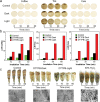
Dental caries is among the most prevalent dental diseases globally, which arises from the formation of microbial biofilm on teeth. Besides, tooth whitening represents one of the fastest-growing areas of cosmetic dentistry. It will thus be great if tooth biofilm eradication can be combined with tooth whitening. Herein, a highly efficient photodynamic dental therapy strategy is reported for tooth biofilm eradication and tooth discoloration by employing a photosensitizer (DTTPB) with aggregation-induced emission characteristics. DTTPB can efficiently inactivate S. mutans, and inhibit biofilm formation by suppressing the expression of genes associated with extracellular polymeric substance synthesis, bacterial adhesion, and superoxide reduction. Its inhibition performance can be further enhanced through combined treatment with chlorhexidine. Besides, DTTPB exhibits an excellent tooth-discoloration effect on both colored saliva-coated hydroxyapatite and clinical teeth, with short treatment time (less than 1 h), better tooth-whitening performance than 30% hydrogen peroxide, and almost no damage to the teeth. DTTPB also demonstrates excellent biocompatibility with neglectable hemolysis effect on mouse red blood cells and almost no killing effect on mammalian cells, which enables its potential applications for simultaneous tooth biofilm eradication and tooth whitening in clinical dentistry.
Keywords: Streptococcus mutans; aggregation-induced emission; biofilm; photodynamic therapy; tooth whitening.
© 2022 The Authors. Advanced Science published by Wiley-VCH GmbH.
Conflict of interest statement
The authors declare no conflict of interest.
Figures


Similar articles
-
Understanding the Basis of METH Mouth Using a Rodent Model of Methamphetamine Injection, Sugar Consumption, and Streptococcus mutans Infection.mBio. 2021 Mar 9;12(2):e03534-20. doi: 10.1128/mBio.03534-20. mBio. 2021. PMID: 33688011 Free PMC article.
-
Bifunctional defect mediated direct Z-scheme g-C3N4-x/Bi2O3-y heterostructures with enhanced piezo-photocatalytic properties for efficient tooth whitening and biofilm eradication.J Mater Chem B. 2023 Aug 2;11(30):7103-7116. doi: 10.1039/d3tb01044a. J Mater Chem B. 2023. PMID: 37417809
-
[Effects of hydrogen peroxide-containing bleaching on the growth of Streptococcus mutans biofilm on enamel disc surface].Beijing Da Xue Xue Bao Yi Xue Ban. 2014 Feb 18;46(1):30-4. Beijing Da Xue Xue Bao Yi Xue Ban. 2014. PMID: 24535343 Chinese.
-
Strategies for Streptococcus mutans biofilm dispersal through extracellular polymeric substances disruption.Mol Oral Microbiol. 2022 Feb;37(1):1-8. doi: 10.1111/omi.12355. Epub 2021 Nov 22. Mol Oral Microbiol. 2022. PMID: 34727414 Review.
-
Tooth-Bleaching: A Review of the Efficacy and Adverse Effects of Various Tooth Whitening Products.J Coll Physicians Surg Pak. 2015 Dec;25(12):891-6. J Coll Physicians Surg Pak. 2015. PMID: 26691365 Review.
Cited by
-
On-Demand Free Radical Release by Laser Irradiation for Photothermal-Thermodynamic Biofilm Inactivation and Tooth Whitening.Gels. 2023 Jul 7;9(7):554. doi: 10.3390/gels9070554. Gels. 2023. PMID: 37504433 Free PMC article.
-
Oxygen self-sufficient nanodroplet composed of fluorinated polymer for high-efficiently PDT eradicating oral biofilm.Mater Today Bio. 2024 May 15;26:101091. doi: 10.1016/j.mtbio.2024.101091. eCollection 2024 Jun. Mater Today Bio. 2024. PMID: 38800565 Free PMC article.
-
Managing Oral Health in the Context of Antimicrobial Resistance.Int J Environ Res Public Health. 2022 Dec 8;19(24):16448. doi: 10.3390/ijerph192416448. Int J Environ Res Public Health. 2022. PMID: 36554332 Free PMC article. Review.
-
A Membrane-Targeted Photosensitizer Prevents Drug Resistance and Induces Immune Response in Treating Candidiasis.Adv Sci (Weinh). 2023 Dec;10(35):e2207736. doi: 10.1002/advs.202207736. Epub 2023 Oct 24. Adv Sci (Weinh). 2023. PMID: 37875397 Free PMC article.
-
Oxidization enhances type I ROS generation of AIE-active zwitterionic photosensitizers for photodynamic killing of drug-resistant bacteria.Chem Sci. 2023 Apr 17;14(18):4863-4871. doi: 10.1039/d3sc00980g. eCollection 2023 May 10. Chem Sci. 2023. PMID: 37181775 Free PMC article.
References
-
- Peres M. A., Macpherson L. M. D., Weyant R. J., Daly B., Venturelli R., Mathur M. R., Listl S., Celeste R. K., Guarnizo‐Herreño C. C., Kearns C., Benzian H., Allison P., Watt R. G., Lancet 2019, 394, 249. - PubMed
-
- Vos T., Allen C., Arora M., Barber R. M., Brown A., Carter A., Casey D. C., Charlson F. J., Chen A. Z., Coggeshall M., Cornaby L., Dandona L., Dicker D. J., Dilegge T., Erskine H. E., Ferrari A. J., Fitzmaurice C., Fleming T., Forouzanfar M. H., Fullman N., Goldberg E. M., Graetz N., Haagsma J. A., Hay S. I., Johnson C. O., Kassebaum N. J., Kawashima T., Kemmer L., Khalil I. A., Kyu H. H., et al., Lancet 2016, 388, 1545. - PubMed
-
- Helöe L. A., Haugejorden O., Community Dent. Oral Epidemiol. 1981, 9, 294. - PubMed
-
- Marsh P. D., Microbiology 2003, 149, 279. - PubMed
Publication types
MeSH terms
Grants and funding
LinkOut - more resources
Full Text Sources
Medical
