Review
. 2023;21(5):1065-1080.
doi: 10.2174/1570159X20666220507021445.

The Role of Mitochondria-Targeting miRNAs in Intracerebral Hemorrhage

Affiliations
Review

The Role of Mitochondria-Targeting miRNAs in Intracerebral Hemorrhage

Ilgiz Gareev et al. Curr Neuropharmacol. 2023.

Abstract

Non-traumatic intracerebral hemorrhage (ICH) is the most common type of hemorrhagic stroke, most often occurring between the ages of 45 and 60. Arterial hypertension (AH) is most often the cause of ICH, followed by atherosclerosis, blood diseases, inflammatory changes in cerebral vessels, intoxication and vitamin deficiencies. Cerebral hemorrhage can occur by diapedesis or as a result of a ruptured vessel. AH is difficult to treat, requires surgery and can lead to disability or death. One of the important directions in the study of the pathogenesis of ICH is mitochondrial dysfunction and its regulation. The key role of mitochondrial dysfunction in AH and atherosclerosis, as well as in the development of brain damage after hemorrhage, has been acknowledged. MicroRNAs (miRNAs) are a class of non-coding RNAs (about 18-22 nucleotides) that regulate a variety of biological processes including cell differentiation, proliferation, apoptosis, etc., primarily through gene repression. There is growing evidence to support dysregulated miRNAs in various cardiovascular diseases, including ICH. Further, the realization of miRNAs within mitochondrial compartment has challenged the traditional knowledge of signaling pathways involved in the regulatory network of cardiovascular diseases. However, the role of miRNAs in mitochondrial dysfunction for ICH is still under-appreciated, with comparatively much lesser studies and investigations reported, than those in other cardiovascular diseases. In this review, we summarize the up-to-date findings on the published role miRNAs in mitochondrial function for ICH, and the potential use of miRNAs in clinical settings, such as potential therapeutic targets and non-invasive diagnostic/prognostic biomarker tools.

Keywords: Intracerebral hemorrhage; biomarker; miRNA; mitochondria; mitochondrial dysfunction; pathogenesis; therapeutic target.


Conflict of interest statement

The authors declare no conflict of interest, financial or otherwise.

Figures

Fig. (1)
Fig. (1)
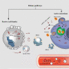
Cellular release of circulating miRNAs into biological fluids (e.g. bloodstream). In the cytoplasm, pre-miRNAs as well as mature miRNAs can be incorporated into microparticles, such as extracellular vesicles (EVs) (exosomes and microvesicles) or apoptotic bodies, and can be released from cells. In addition to being incorporated into the EVs, miRNAs are also present in the microparticle-free compartment. These miRNAs are associated with high-density lipoprotein particles (HDLPs) or RNA-binding proteins (miRNA-Argonaute 2 (Ago2) complex). In physiological conditions or diseases, miRNAs can be released passively (during apoptosis or necrosis) or actively, by secretion in the EVs, or miRNA-Ago2 complex through interaction with specific membrane channels or proteins.
Fig. (2)
Fig. (2)
Biogenesis and function miRNAs and mechanisms of mitochondrial miRNAs (mitomiRs) transport and localization to mitochondria. MitomiRs are miRNAs of nuclear origin that are associated with the mitochondrial membrane and, possibly, with their own mRNA targets (1) or localized within organelles (2), like mit-mitomiRs (mitochondrial origins). MitomiRs suppress or activate gene expression by interacting with complementary sequences of their mRNA-targets, which are nuclear (1) or mitochondrial transcripts (2). Argonaute 2 protein (Ago-2) transports mitomiRs to mitochondria. Have been put forward about three main mechanisms: (3) exchange through contact between mitochondria and P-bodies; (4) transport via PNAPASE, component; or transport across the porins of the outer mitochondrial membrane.

Similar articles

Cited by

References

    1. Sallinen H., Putaala J., Strbian D. Triggering factors in non-traumatic intracerebral hemorrhage. J. Stroke Cerebrovasc. Dis. 2020;29(8):104921. doi: 10.1016/j.jstrokecerebrovasdis.2020.104921. - DOI - PubMed
    1. Hostettler I.C., Seiffge D.J., Werring D.J. Intracerebral hemorrhage: An update on diagnosis and treatment. Expert Rev. Neurother. 2019;19(7):679–694. doi: 10.1080/14737175.2019.1623671. - DOI - PubMed
    1. Aguilar M., Freeman W.D. Spontaneous intracerebral hemorrhage. Semin. Neurol. 2010;30(5):555–564. doi: 10.1055/s-0030-1268865. - DOI - PubMed
    1. Zaidi H.A., Zabramski J.M., Safavi-Abbasi S., Preul M.C. Spontaneous intracerebral hemorrhage. World Neurosurg. 2015;84(5):1191–1192. doi: 10.1016/j.wneu.2015.06.015. - DOI - PubMed
    1. Chen W., Guo C., Feng H., Chen Y. Mitochondria: Novel mechanisms and therapeutic targets for secondary brain injury after intracerebral hemorrhage. Front. Aging Neurosci. 2021;12:615451. doi: 10.3389/fnagi.2020.615451. - DOI - PMC - PubMed