

Abnormal dynamic functional connectivity after sleep deprivation from temporal variability perspective
- PMID: 35524680
- PMCID: PMC9294309
- DOI: 10.1002/hbm.25886
Abnormal dynamic functional connectivity after sleep deprivation from temporal variability perspective
Abstract
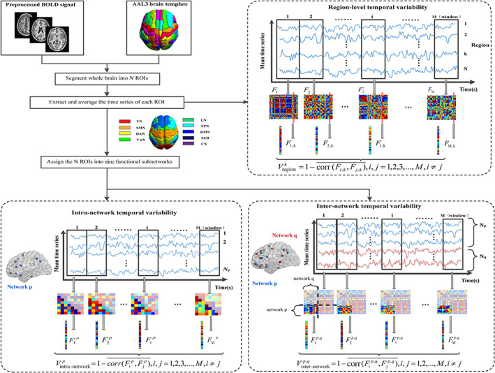
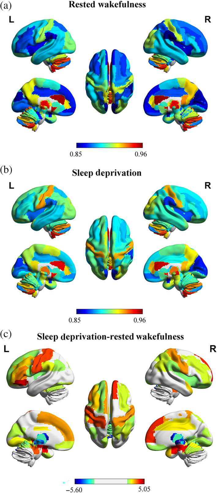
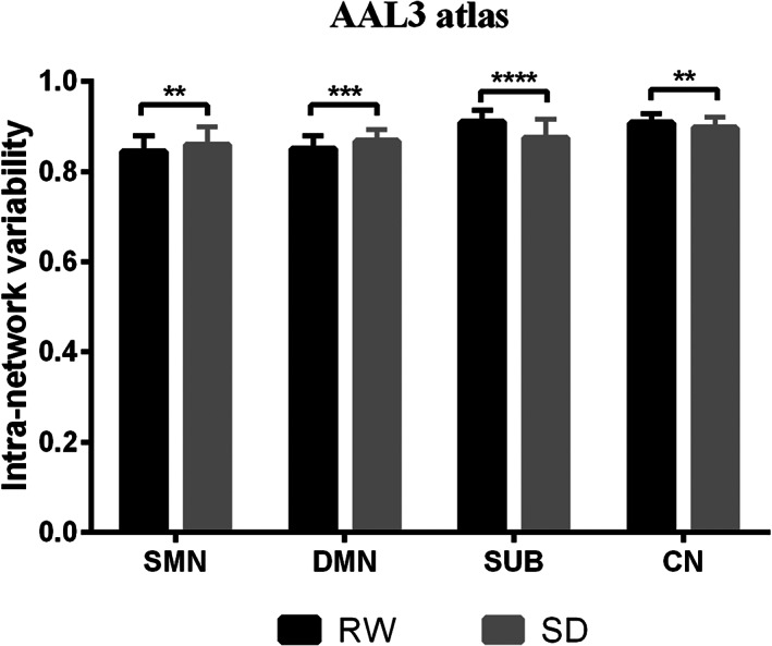
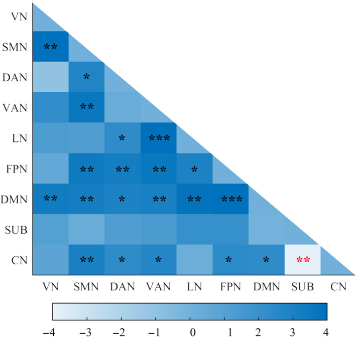
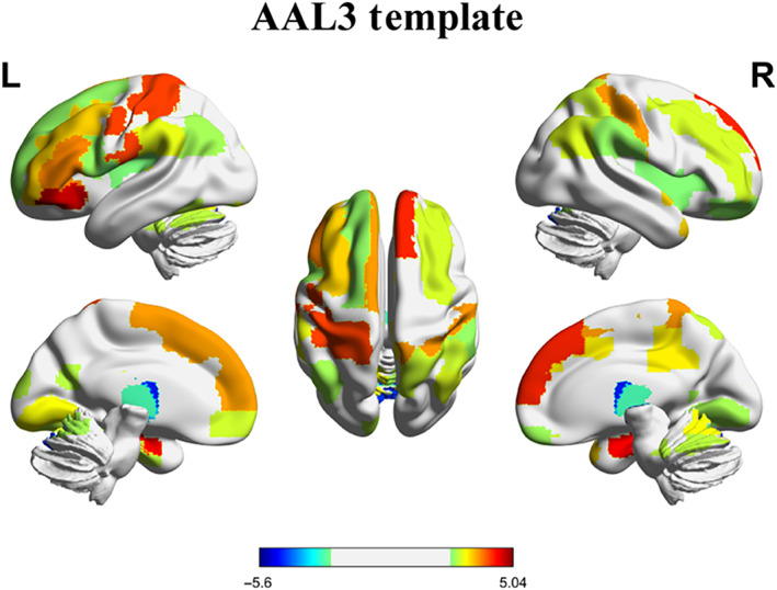
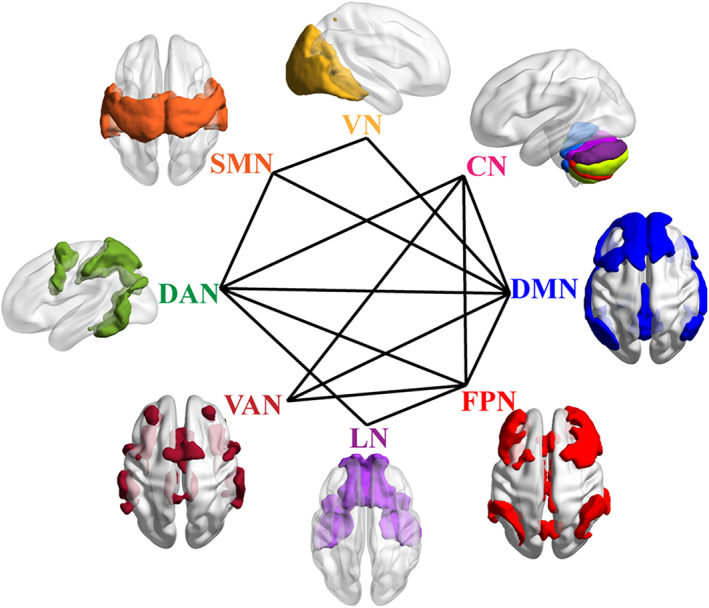
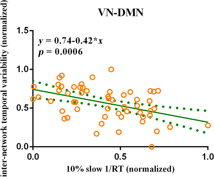
Sleep deprivation (SD) is very common in modern society and regarded as a potential causal mechanism of several clinical disorders. Previous neuroimaging studies have explored the neural mechanisms of SD using magnetic resonance imaging (MRI) from static (comparing two MRI sessions [one after SD and one after resting wakefulness]) and dynamic (using repeated MRI during one night of SD) perspectives. Recent SD researches have focused on the dynamic functional brain organization during the resting-state scan. Our present study adopted a novel metric (temporal variability), which has been successfully applied to many clinical diseases, to examine the dynamic functional connectivity after SD in 55 normal young subjects. We found that sleep-deprived subjects showed increased regional-level temporal variability in large-scale brain regions, and decreased regional-level temporal variability in several thalamus subregions. After SD, participants exhibited enhanced intra-network temporal variability in the default mode network (DMN) and increased inter-network temporal variability in numerous subnetwork pairs. Furthermore, we found that the inter-network temporal variability between visual network and DMN was negative related with the slowest 10% respond speed (β = -.42, p = 5.57 × 10⁻⁴ ) of the psychomotor vigilance test after SD following the stepwise regression analysis. In conclusion, our findings suggested that sleep-deprived subjects showed abnormal dynamic brain functional configuration, which provides new insights into the neural underpinnings of SD and contributes to our understanding of the pathophysiology of clinical disorders.
Keywords: dynamic functional connectivity; psychomotor vigilance test; resting-state functional magnetic resonance imaging; sleep deprivation; temporal variability.
© 2022 The Authors. Human Brain Mapping published by Wiley Periodicals LLC.
Conflict of interest statement
The authors declare no potential conflict of interest.
Figures
References
-
- Andersson, J. L. R. , Hutton, C. , Ashburner, J. , Turner, R. , & Friston, K. (2001). Modeling geometric deformations in EPI time series. NeuroImage, 13, 903–919. - PubMed
-
- Arslan, S. , Ktena, S. I. , Makropoulos, A. , Robinson, E. C. , Rueckert, D. , & Parisot, S. (2018). Human brain mapping: A systematic comparison of parcellation methods for the human cerebral cortex. NeuroImage, 170, 5–30. - PubMed
-
- Ashburner, J. , & Friston, K. J. (2005). Unified segmentation. NeuroImage, 26, 839–851. - PubMed
-
- Bandyopadhyay, A. , & Sigua, N. L. (2019). What is sleep deprivation? American Journal of Respiratory Critical Care Medicine, 199, P11–P12. - PubMed
Publication types
MeSH terms
LinkOut - more resources
Full Text Sources
