Review
. 2022 Jun;474(6):575-590.
doi: 10.1007/s00424-022-02695-7. Epub 2022 May 7.

Harnessing retinal phagocytes to combat pathological neovascularization in ischemic retinopathies?

Affiliations
Review

Harnessing retinal phagocytes to combat pathological neovascularization in ischemic retinopathies?

Anne Klotzsche-von Ameln et al. Pflugers Arch. 2022 Jun.

Abstract

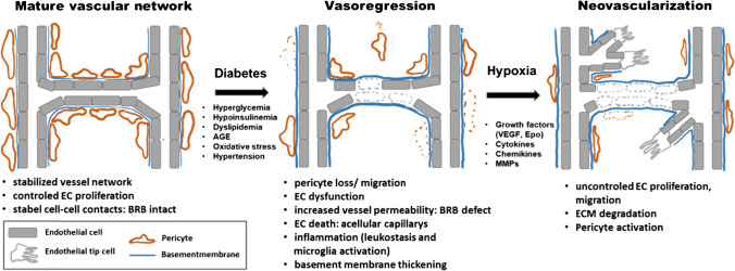
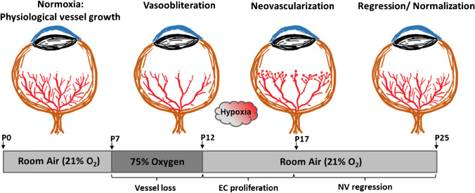
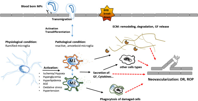
Ischemic retinopathies (IR) are vision-threatening diseases that affect a substantial amount of people across all age groups worldwide. The current treatment options of photocoagulation and anti-VEGF therapy have side effects and are occasionally unable to prevent disease progression. It is therefore worthwhile to consider other molecular targets for the development of novel treatment strategies that could be safer and more efficient. During the manifestation of IR, the retina, normally an immune privileged tissue, encounters enhanced levels of cellular stress and inflammation that attract mononuclear phagocytes (MPs) from the blood stream and activate resident MPs (microglia). Activated MPs have a multitude of effects within the retinal tissue and have the potential to both counter and exacerbate the harmful tissue microenvironment. The present review discusses the current knowledge about the role of inflammation and activated retinal MPs in the major IRs: retinopathy of prematurity and diabetic retinopathy. We focus particularly on MPs and their secreted factors and cell-cell-based interactions between MPs and endothelial cells. We conclude that activated MPs play a major role in the manifestation and progression of IRs and could therefore become a promising new target for novel pharmacological intervention strategies in these diseases.

Keywords: Inflammation; Macrophages; Microglia; Mononuclear phagocytes; Pathological angiogenesis; Retinopathy.


Conflict of interest statement

The authors declare no competing interests.

The authors declare no competing interests.

Figures

Fig. 1
Fig. 1
Schematic illustration of ROP disease manifestation. Physiological retinal vessel growth in humans starts at the beginning of the fourth month of gestational age (“natural hypoxia”) and is normally finished shortly before full-term birth. Accordingly, premature infants have incompletely vascularized retinas. After birth, loss of nutrients and physiological growth factors provided at the maternal–fetal interface together with the increased oxygen pressure (oxygen pressure of ambient air ~ 160 mmHg; plus additional oxygen supplementation) result in a persistently undervascularized (vasoobliterative phase) and later hypoxic retina. This causes excess production of VEGF and other oxygen-regulated vascular growth factors, resulting in pathological retinal neovascularization (neovascularization phase)
Fig. 2
Fig. 2
Summary of the main pathogenic events causing pathological neovascularization in the retina of patients with DR. In healthy retinal capillaries, adequate pericyte coverage supports endothelial cell survival and integrity of blood retina barrier. Long-term diabetes induces alterations and damage in several cell types resulting in progressive vasoregression. Occluded remnants of capillaries are no longer perfused, leading to tissue hypoxia and a subsequent upregulation of survival/growth factors such as VEGF. As a consequence, ischemia/hypoxia-induced, pathological neovascularization is triggered. EC endothelial cells, BRB blood retina barrier, ECM extracellular matrix, AGE advanced glycosylation end products, VEGF vascular endothelial growth factor, Epo erythropoietin
Fig. 3
Fig. 3
Schematic illustration of the mouse OIR model. Neonatal mice are kept in ambient air (21% oxygen) from birth until postnatal day 7 (P7); meanwhile, normal vascular development starts. At P7, mice are then exposed to 75% oxygen, resulting in the inhibition and regression of retinal vessel growth (vaso-obliteration). At P12, mice are returned to ambient air. The drop in oxygen pressure leads to the development of hypoxia in avascular retinal areas, triggering both normal vessel regrowth and pathological neovascularization. Pathological vessel growth reaches its maximum at P17 and spontaneously regresses thereafter until the retinal vasculature is completely normalized by P25
Fig. 4
Fig. 4
Summary of the action of MPs causing pathological neovascularization in the retina. Two immune cell populations are described to affect pathological neovascularization. These are either macrophages derived from the circulation or resident microglia. In the stressed retina, ramified microglia become activated and differentiate into an M1 or M2 phenotype, which goes along with respective changes in cell morphology, proliferation, migration, phagocytosis, and alterations in cytokine/growth factor/protease production. MP mononuclear phagocyte, EC endothelial cells, BRB blood retina barrier, ECM extracellular matrix, GF growth factor, AGE advanced glycosylation end products VEGF vascular endothelial growth factor, Epo erythropoietin

Similar articles

Cited by

References

    1. Altmann, C. and M.H.H. Schmidt, The role of microglia in diabetic retinopathy: inflammation, microvasculature defects and neurodegeneration. Int J Mol Sci, 2018. 19(1). - PMC - PubMed
    1. Antonetti DA, Klein R, Gardner TW. Diabetic retinopathy. N Engl J Med. 2012;366(13):1227–1239. - PubMed
    1. Arroba AI, et al. Modulation of microglia polarization dynamics during diabetic retinopathy in db/db mice. Biochim Biophys Acta. 2016;1862(9):1663–1674. - PubMed
    1. Baer C, et al. Reciprocal interactions between endothelial cells and macrophages in angiogenic vascular niches. Exp Cell Res. 2013;319(11):1626–1634. - PMC - PubMed
    1. Barile GR, et al. The RAGE axis in early diabetic retinopathy. Invest Ophthalmol Vis Sci. 2005;46(8):2916–2924. - PubMed

Publication types