

Headache and cognitive disturbance correlate with ganglion cell layer thickness in patients who recovered from COVID-19
- PMID: 35525105
- PMCID: PMC9040445
- DOI: 10.1016/j.clineuro.2022.107263
Headache and cognitive disturbance correlate with ganglion cell layer thickness in patients who recovered from COVID-19
Abstract
Background: Retinal abnormalities are being increasingly reported in COVID-19, in addition to the well-known symptoms of this disease accounting for the neurological involvement. In this study, we aimed to investigate whether ganglion cell layer thickness (GCLT) was different in recovered COVID-19 patients compared to controls in the subacute stage and to determine whether it correlated with COVID-19-related neurological symptoms or pneumonia.
Methods: This study involved 40 patients who had recovered from COVID-19 and 40 age- and sex-matched healthy controls. All the participants underwent ophthalmological examination, spectral domain optical coherence tomography and neurological examination. The clinical and biochemical properties of the patients were noted and their correlations with GCLT were sought.
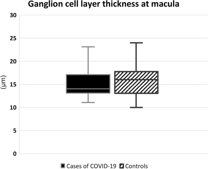
Results: The duration after COVID-19 infection was 113 ± 62 (mean ± SD) days. At this subacute stage, there was no significant difference between the GCLT measurements of the COVID-19 patients and the controls (14 ± 4.0 µm [median ± IQR] vs 16 ± 4.8 µm, respectively). When we analyzed the relationships with neurological symptoms in the patient group, we found that patients with cognitive symptoms had lower GCLT values compared to those without (13 ± 3 µm vs. 16 ± 4 µm, respectively; p = 0.002). Patients who suffered headache during the acute infection also had lower GCLT values compared to those without (14 ± 4 µm vs. 18 ± 5 µm, respectively; p = 0.015). The GCLT values did not differ significantly with respect to anosmia, ageusia, sleep disturbances, having had COVID-19 pneumonia, or smoking status. Age, duration after COVID-19, and blood levels of thyroid stimulating hormone, glucose, vitamin D and vitamin B12 were not in correlation with GCLT in our study.
Conclusion: Our findings highlight an association between GCLT values and neurological symptoms such as cognitive disturbance (brain fog) and headache in patients who had recovered after non-severe COVID-19 infection. Neuroretinal involvement by SARS-CoV2 might be linked to central neurological symptoms. The patients with lower GCLT values may benefit from close monitoring for neurological problems.
Keywords: Brain fog; COVID-19; Ganglion cell layer; Headache; Optical coherence tomography.
Copyright © 2022 Elsevier B.V. All rights reserved.
Figures
Similar articles
-
Optical coherence tomography findings in primary headache disorders: is pain duration a clinical correlate?Int J Neurosci. 2024 May 25:1-7. doi: 10.1080/00207454.2024.2358367. Online ahead of print. Int J Neurosci. 2024. PMID: 38768056
-
Evaluation of Retinal and Optic Nerve Parameters in Recovered COVID-19 Patients: Potential Neurodegenerative Impact on the Ganglion Cell Layer.Diagnostics (Basel). 2025 May 9;15(10):1195. doi: 10.3390/diagnostics15101195. Diagnostics (Basel). 2025. PMID: 40428188 Free PMC article.
-
Subacute neurological sequelae in mild COVID-19 outpatients.Tuberk Toraks. 2022 Mar;70(1):27-36. doi: 10.5578/tt.20229904. Tuberk Toraks. 2022. PMID: 35362302 English.
-
Neurological Sequelae of COVID-19.J Integr Neurosci. 2022 Apr 6;21(3):77. doi: 10.31083/j.jin2103077. J Integr Neurosci. 2022. PMID: 35633158 Review.
-
Spectrum of Neurological Manifestations in Covid-19: A Review.Neurol India. 2020 May-Jun;68(3):560-572. doi: 10.4103/0028-3886.289000. Neurol India. 2020. PMID: 32643664 Review.
Cited by
-
Prevalence and prognostic value of neurological affections in hospitalized patients with moderate to severe COVID-19 based on objective assessments.Sci Rep. 2023 Nov 10;13(1):19619. doi: 10.1038/s41598-023-46124-w. Sci Rep. 2023. PMID: 37949882 Free PMC article.
-
SARS-CoV-2 neurovascular invasion supported by Mendelian randomization.J Transl Med. 2024 Jan 24;22(1):101. doi: 10.1186/s12967-024-04907-3. J Transl Med. 2024. PMID: 38268029 Free PMC article.
-
Retinal Microvasculature Image Analysis Using Optical Coherence Tomography Angiography in Patients with Post-COVID-19 Syndrome.J Imaging. 2023 Oct 24;9(11):234. doi: 10.3390/jimaging9110234. J Imaging. 2023. PMID: 37998081 Free PMC article.
References
MeSH terms
Substances
LinkOut - more resources
Full Text Sources
Medical
Miscellaneous
