

Zinc transport via ZNT5-6 and ZNT7 is critical for cell surface glycosylphosphatidylinositol-anchored protein expression
- PMID: 35525268
- PMCID: PMC9168625
- DOI: 10.1016/j.jbc.2022.102011
Zinc transport via ZNT5-6 and ZNT7 is critical for cell surface glycosylphosphatidylinositol-anchored protein expression
Abstract
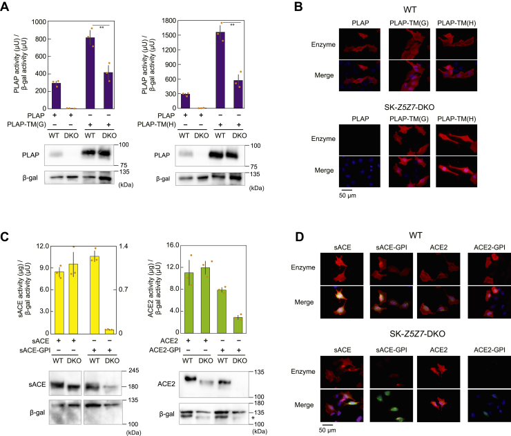
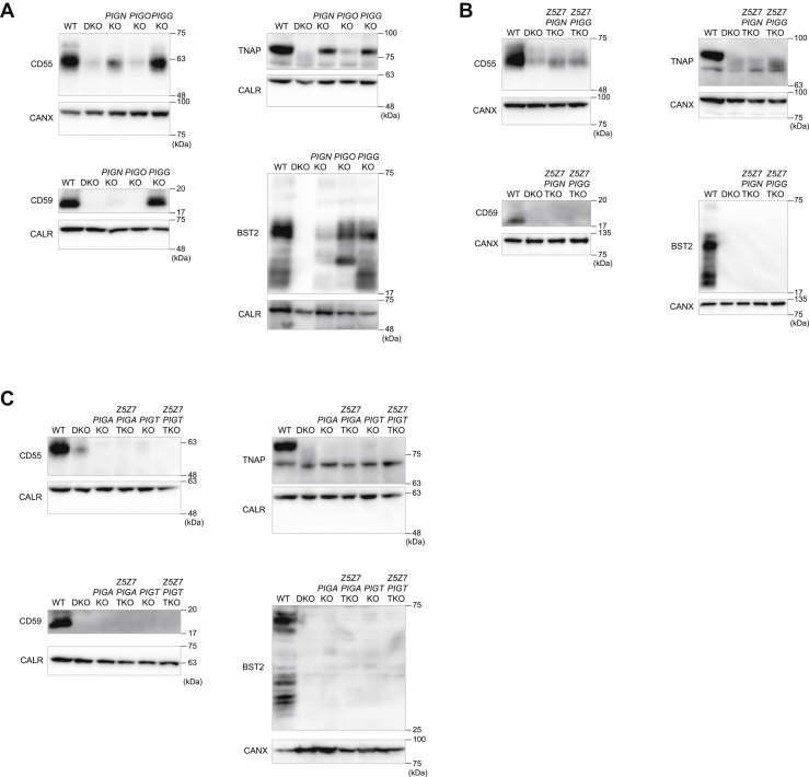
Glycosylphosphatidylinositol (GPI)-anchored proteins play crucial roles in various enzyme activities, cell signaling and adhesion, and immune responses. While the molecular mechanism underlying GPI-anchored protein biosynthesis has been well studied, the role of zinc transport in this process has not yet been elucidated. Zn transporter (ZNT) proteins mobilize cytosolic zinc to the extracellular space and to intracellular compartments. Here, we report that the early secretory pathway ZNTs (ZNT5-ZNT6 heterodimers [ZNT5-6] and ZNT7-ZNT7 homodimers [ZNT7]), which supply zinc to the lumen of the early secretory pathway compartments are essential for GPI-anchored protein expression on the cell surface. We show, using overexpression and gene disruption/re-expression strategies in cultured human cells, that loss of ZNT5-6 and ZNT7 zinc transport functions results in significant reduction in GPI-anchored protein levels similar to that in mutant cells lacking phosphatidylinositol glycan anchor biosynthesis (PIG) genes. Furthermore, medaka fish with disrupted Znt5 and Znt7 genes show touch-insensitive phenotypes similar to zebrafish Pig mutants. These findings provide a previously unappreciated insight into the regulation of GPI-anchored protein expression and protein quality control in the early secretory pathway.
Keywords: ER quality control; ZNT; cell surface; early secretory pathway; ectoenzyme; glycosylphosphatidylinositol (GPI anchor); phosphatidylinositol glycan anchor biosynthesis (PIG); transporter; zinc.
Copyright © 2022 The Authors. Published by Elsevier Inc. All rights reserved.
Conflict of interest statement
Conflict of interest The authors declare that they have no conflicts of interest with the contents of this article.
Figures


Similar articles
-
Detailed analyses of the crucial functions of Zn transporter proteins in alkaline phosphatase activation.J Biol Chem. 2020 Apr 24;295(17):5669-5684. doi: 10.1074/jbc.RA120.012610. Epub 2020 Mar 16. J Biol Chem. 2020. PMID: 32179649 Free PMC article.
-
Zinc transporters, ZnT5 and ZnT7, are required for the activation of alkaline phosphatases, zinc-requiring enzymes that are glycosylphosphatidylinositol-anchored to the cytoplasmic membrane.J Biol Chem. 2005 Jan 7;280(1):637-43. doi: 10.1074/jbc.M411247200. Epub 2004 Nov 2. J Biol Chem. 2005. PMID: 15525635
-
Two different zinc transport complexes of cation diffusion facilitator proteins localized in the secretory pathway operate to activate alkaline phosphatases in vertebrate cells.J Biol Chem. 2005 Sep 2;280(35):30956-62. doi: 10.1074/jbc.M506902200. Epub 2005 Jul 1. J Biol Chem. 2005. PMID: 15994300
-
Trafficking of glycosylphosphatidylinositol anchored proteins from the endoplasmic reticulum to the cell surface.J Lipid Res. 2016 Mar;57(3):352-60. doi: 10.1194/jlr.R062760. Epub 2015 Oct 8. J Lipid Res. 2016. PMID: 26450970 Free PMC article. Review.
-
GPI-anchor remodeling: potential functions of GPI-anchors in intracellular trafficking and membrane dynamics.Biochim Biophys Acta. 2012 Aug;1821(8):1050-8. doi: 10.1016/j.bbalip.2012.01.004. Epub 2012 Jan 11. Biochim Biophys Acta. 2012. PMID: 22265715 Review.
Cited by
-
Pigmentation and TYRP1 expression are mediated by zinc through the early secretory pathway-resident ZNT proteins.Commun Biol. 2023 Apr 18;6(1):403. doi: 10.1038/s42003-023-04640-5. Commun Biol. 2023. PMID: 37072620 Free PMC article.
-
Severe neonatal hypotonia due to SLC30A5 variant affecting function of ZnT5 zinc transporter.JIMD Rep. 2025 Jan 9;66(1):e12465. doi: 10.1002/jmd2.12465. eCollection 2025 Jan. JIMD Rep. 2025. PMID: 39790720 Free PMC article.
-
ZnT6-mediated Zn2+ redistribution: impact on mitochondrial fission and autophagy in H9c2 cells.Mol Cell Biochem. 2025 Jul;480(7):4283-4296. doi: 10.1007/s11010-025-05247-6. Epub 2025 Mar 14. Mol Cell Biochem. 2025. PMID: 40087209
-
Metalation and activation of Zn2+ enzymes via early secretory pathway-resident ZNT proteins.Biophys Rev (Melville). 2023 Dec 8;4(4):041302. doi: 10.1063/5.0176048. eCollection 2023 Dec. Biophys Rev (Melville). 2023. PMID: 38510844 Free PMC article. Review.
-
Novel SLC30A2 mutations in the pathogenesis of transient neonatal zinc deficiency.Pediatr Investig. 2023 Feb 22;7(1):6-12. doi: 10.1002/ped4.12366. eCollection 2023 Mar. Pediatr Investig. 2023. PMID: 36967740 Free PMC article.
References
-
- Liu Y.S., Fujita M. Mammalian GPI-anchor modifications and the enzymes involved. Biochem. Soc. Trans. 2020;48:1129–1138. - PubMed
Publication types
MeSH terms
Substances
LinkOut - more resources
Full Text Sources
Molecular Biology Databases
