. 2022 Jun;298(6):102009.
doi: 10.1016/j.jbc.2022.102009. Epub 2022 May 4.

A novel small-molecule selective activator of homomeric GIRK4 channels

Affiliations

A novel small-molecule selective activator of homomeric GIRK4 channels

Meng Cui et al. J Biol Chem. 2022 Jun.

Abstract

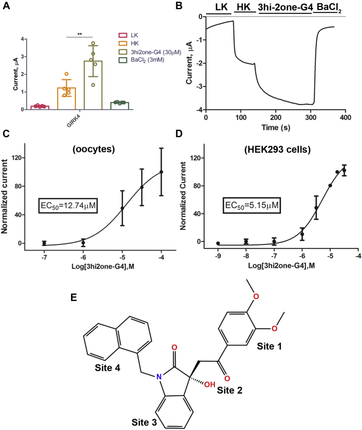
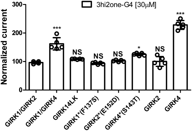
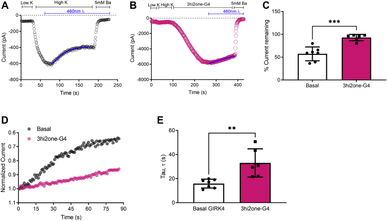
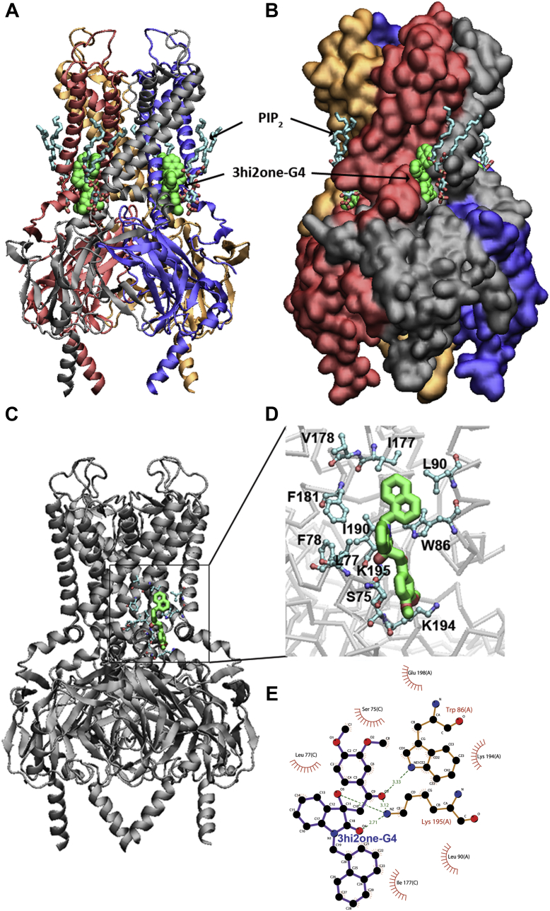
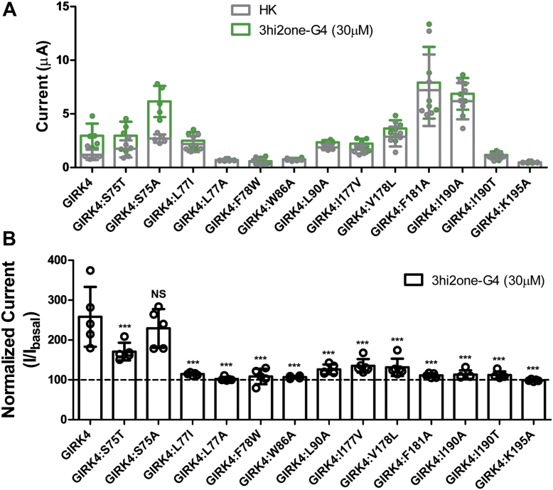
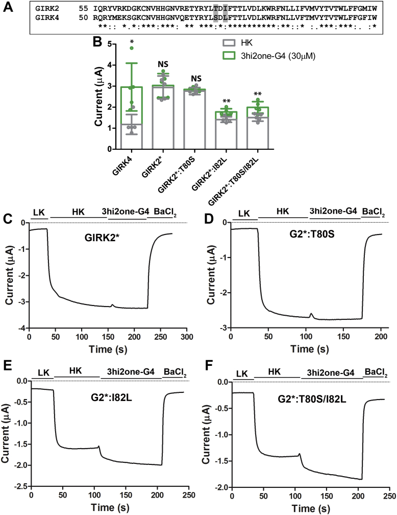
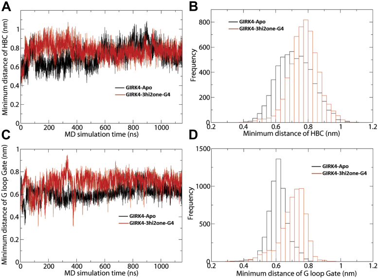
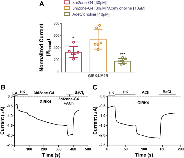
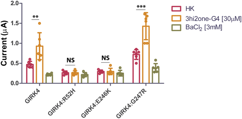
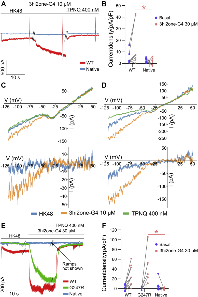
G protein-sensitive inwardly rectifying potassium (GIRK) channels are important pharmaceutical targets for neuronal, cardiac, and endocrine diseases. Although a number of GIRK channel modulators have been discovered in recent years, most lack selectivity. GIRK channels function as either homomeric (i.e., GIRK2 and GIRK4) or heteromeric (e.g., GIRK1/2, GIRK1/4, and GIRK2/3) tetramers. Activators, such as ML297, ivermectin, and GAT1508, have been shown to activate heteromeric GIRK1/2 channels better than GIRK1/4 channels with varying degrees of selectivity but not homomeric GIRK2 and GIRK4 channels. In addition, VU0529331 was discovered as the first homomeric GIRK channel activator, but it shows weak selectivity for GIRK2 over GIRK4 (or G4) homomeric channels. Here, we report the first highly selective small-molecule activator targeting GIRK4 homomeric channels, 3hi2one-G4 (3-[2-(3,4-dimethoxyphenyl)-2-oxoethyl]-3-hydroxy-1-(1-naphthylmethyl)-1,3-dihydro-2H-indol-2-one). We show that 3hi2one-G4 does not activate GIRK2, GIRK1/2, or GIRK1/4 channels. Using molecular modeling, mutagenesis, and electrophysiology, we analyzed the binding site of 3hi2one-G4 formed by the transmembrane 1, transmembrane 2, and slide helix regions of the GIRK4 channel, near the phosphatidylinositol-4,5-bisphosphate binding site, and show that it causes channel activation by strengthening channel-phosphatidylinositol-4,5-bisphosphate interactions. We also identify slide helix residue L77 in GIRK4, corresponding to residue I82 in GIRK2, as a major determinant of isoform-specific selectivity. We propose that 3hi2one-G4 could serve as a useful pharmaceutical probe in studying GIRK4 channel function and may also be pursued in drug optimization studies to tackle GIRK4-related diseases such as primary aldosteronism and late-onset obesity.

Keywords: Kir3 channels; drug–channel interaction; electrophysiology; molecular docking; molecular dynamics simulations; mutagenesis.


Conflict of interest statement

Conflict of interest The authors declare that they have no conflicts of interest with the contents of this article.

Figures

Figure 1
Figure 1
3hi2one-G4 activates GIRK4 homomeric channel.A, GIRK4 channel is activated by 3hi2one-G4 (30 μM) (TEVC). The asterisks indicate significant differences tested by unpaired Student's t test (∗∗p < 0.01) (data are mean ± SD, N = 5). B, representative traces of responses to the 3hi2one-G4 compound (30 μM) of GIRK4 channel. C, dose–response curves of the 3hi2one-G4 compound activation on the GIRK4 channel (EC50 = 12.74 μM) from TEVC recordings of Xenopus oocytes expressing GIRK4 channels (N = 5). D, dose–response curves of the 3h2one-G4 compound activation on the GIRK4 channel (EC50 = 5.15 μM) from patch-clamp recordings of HEK293 cells expressing GIRK4 channels (N > 5) (data are mean ± SD). E, the chemical structure of 3hi2one-G4. GIRK, G protein–sensitive inwardly rectifying potassium channel; HEK293, human embryonic kidney 293 cell; 3hi2one, 3-[2-(3,4-dimethoxyphenyl)-2-oxoethyl]-3-hydroxy-1-(1-naphthylmethyl)-1,3-dihydro-2H-indol-2-one; HK, high potassium solution ND96K; LK, low potassium solution ND96; TEVC, two-electrode voltage clamp.
Figure 2
Figure 2
3hi2one-G4 selectively activates GIRK4 channels. TEVC recordings of GIRK1/2, GIRK1/4, GIRK14LK, GIRK1∗(GIRK1:F137S), GIRK2∗(GIRK2:E152D), GIRK4∗(GIRK4:S143T), GIRK2, and GIRK4 channels expressed in Xenopus oocytes. The basal current (normalized) is measured in a high potassium (HK) solution ND96K. The asterisks indicate significant differences tested by one-way ANOVA Tukey's test (∗∗∗p < 0.001, ∗p < 0.05) compared with GIRK1/2 (data are mean ± SD, N ≥ 4). GIRK, G protein–sensitive inwardly rectifying potassium channel; 3hi2one, 3-[2-(3,4-dimethoxyphenyl)-2-oxoethyl]-3-hydroxy-1-(1-naphthylmethyl)-1,3-dihydro-2H-indol-2-one; TEVC, two-electrode voltage clamp.
Figure 3
Figure 3
3hi2one-G4 decreases GIRK4 current inhibition after PIP2dephosphorylation by 5-ptaseOCRL in HEK cells.A, representative plot of GIRK4 current decrease (control, black open circle). B, representative plot of 3hi2one-G4 induced GIRK4 current decrease in response to optogenetic dephosphorylation by phosphatase 5-ptaseOCRL (20 μM 3hi2one-G4, mauve open circle). C, the decrease in current (%current remaining) is reduced when GIRK4 channels are studied in the presence of 3hi2one-G4. D, 5-ptaseOCRL-mediated decrease in GIRK4 current is characterized by monoexponential fits in the presence of 5-ptaseOCRL in the presence (mauve circles) and the absence of 3hi2one-G4 (black circles). E, 3hi2one-G4 increases the τ (tau) of current decrease, following activation of 5-ptaseOCRL. Data are currents recorded from HEK293T cells using patch clamp in the whole-cell mode and are shown as means ± SD for six to seven cells per group. Statistical significance was calculated using unpaired Student's t tests using GraphPad Prism (∗∗p < 0.005; ∗∗∗p < 0.0005). GIRK, G protein–sensitive inwardly rectifying potassium channel; HEK, human embryonic kidney cell line; 3hi2one, 3-[2-(3,4-dimethoxyphenyl)-2-oxoethyl]-3-hydroxy-1-(1-naphthylmethyl)-1,3-dihydro-2H-indol-2-one; PIP2, phosphatidylinositol-4,5-bisphosphate.
Figure 4
Figure 4
Model of 3hi2one–GIRK4 channel complex and interactions between 3hi2one-G4 and GIRK4 channel.A, GIRK4 is rendered as NewCartoon; B, as molecular surface. 3hi2one molecules are rendered in van der Waals spheres colored in green, PIP2 molecules are rendered in licorice colored by atom types. C, molecular docking predicted 3hi2one-G4–GIRK4 channel complex. D, the critical interacting residues in 3hi2one-G4 binding site of GIKR4 channel. The 3hi2one-G4 is shown by sticks colored by atom types (C: green, N: blue, and O: red). E, 2D plot of the detailed interactions between 3hi2one-G4 and GIRK4 channel. Hydrogen bonds: K195 and W86; hydrophobic interactions: S75, L77, L90, I177, K194, and E198. GIRK, G protein–sensitive inwardly rectifying potassium channel; 3hi2one, 3-[2-(3,4-dimethoxyphenyl)-2-oxoethyl]-3-hydroxy-1-(1-naphthylmethyl)-1,3-dihydro-2H-indol-2-one; PIP2, phosphatidylinositol-4,5-bisphosphate.
Figure 5
Figure 5
Mutations of the predicted 3hi2one-G4 binding site on GIRK4 channel expressed in Xenopus oocytes.A, GIRK4 basal (HK, gray bars) and 3hi2one-G4 activator–induced current (green bars). B, normalized 3hi2one-G4 activator–induced current (I/Ibasal). The asterisks indicate significant differences of GIRK4 channel mutants by one-way ANOVA Tukey’s tests (∗∗∗p < 0.001) compared with GIRK4 wildtype channel (data are mean ± SD, N ≥ 4). GIRK, G protein–sensitive inwardly rectifying potassium channel; 3hi3one, 3-[2-(3,4-dimethoxyphenyl)-2-oxoethyl]-3-hydroxy-1-(1-naphthylmethyl)-1,3-dihydro-2H-indol-3-one; HK, high potassium.
Figure 6
Figure 6
3hi2one-G4 activates the GIRK2∗ channel mutants expressed in Xenopus oocytes.A, local sequence alignment between GIRK2 and GIRK4 near the slide helix region. Residues T80 and I82 in GIRK2; S75 and L77 in GIRK4 are highlighted in gray (∗: conserved, :: semiconserved, and .: similar residues). B, GIRK4, GIRK2∗, and mutant channels basal (HK, gray bars), and 3hi2one-G4 activator–induced current (green bars). The asterisks indicate significant differences between basal and 3hi2one-G4–induced currents by unpaired Student's t tests (∗∗p < 0.01, ∗p < 0.05) (data are mean ± SD, n = 5). C, representative traces of responses to the 3hi2one-G4 compound (30 μM) of GIRK2∗ channel. D, GIRK2∗:T80S. E, GIRK2∗:I82L. F, GIRK2∗:T80S/I82L. GIRK, G protein–sensitive inwardly rectifying potassium channel; HK, high potassium; 3hi2one, 3-[2-(3,4-dimethoxyphenyl)-2-oxoethyl]-3-hydroxy-1-(1-naphthylmethyl)-1,3-dihydro-2H-indol-2-one.
Figure 7
Figure 7
3hi2one-G4 facilitates the opening of HBC and G-loop gates of GIRK4 channel.A, minimum distance of HBC gate as function of time. B, histogram plot. C, minimum distance of G-loop gate as function of time. D, histogram plot. GIRK, G protein–sensitive inwardly rectifying potassium channel; HBC, helix bundle crossing; 3hi2one, 3-[2-(3,4-dimethoxyphenyl)-2-oxoethyl]-3-hydroxy-1-(1-naphthylmethyl)-1,3-dihydro-2H-indol-2-one.
Figure 8
Figure 8
3hi2one-G4 activates GIRK4 channel independent of G proteins.A, TEVC recordings of GIRK4 channel coexpressed with the M2R receptor in Xenopus oocytes. Activator (3hi2one-G4)/agonist (acetylcholine; ACh)-induced current (I/Ibasal) was normalized to HK current. The asterisks indicate significant differences tested by one-way ANOVA Tukey’s test (∗p < 0.05, ∗∗∗p < 0.001) (data are mean ± SD, N = 6). B, representative traces of responses of GIRK4 channel to the 3hi2one-G4 (30 μM)/ACh (10 μM). C, ACh (10 μM). GIRK, G protein–sensitive inwardly rectifying potassium channel; 3hi2one, 3-[2-(3,4-dimethoxyphenyl)-2-oxoethyl]-3-hydroxy-1-(1-naphthylmethyl)-1,3-dihydro-2H-indol-2-one; HK, high potassium; TEVC, two-electrode voltage clamp.
Figure 9
Figure 9
The activity of 3hi2one-G4 on disease-related GIRK4 channel mutants. TEVC recordings of GIRK4, GIRK4:R52H, GIRK4:E246K, and GIRK4:G247R channels expressed in Xenopus oocytes. The asterisks indicate significant differences tested by unpaired Student's t test (∗∗∗p < 0.001, ∗∗p < 0.01) HK basal current (data are mean ± SD, N = 6). GIRK, G protein–sensitive inwardly rectifying potassium channel; 3hi2one, 3-[2-(3,4-dimethoxyphenyl)-2-oxoethyl]-3-hydroxy-1-(1-naphthylmethyl)-1,3-dihydro-2H-indol-2-one; HK, high potassium; TEVC, two-electrode voltage clamp.
Figure 10
Figure 10
The effect of 3hi2one-G4 on native and transfected HAC15 cells.A, representative traces from native and GIRK4:WT transfected HAC15 cells. B, current density of basal and 3hi2one-G4 response in native (mean ± SD: 2.09 ± 1.94 and 3.02 ± 2.27, respectively) and GIRK4:WT transfected (mean ± SD: 4.61 ± 5.548 and 19.95 ± 23.72, respectively) HAC15 cells shows that response to 3hi2one-G4 in transfected cells was larger compared with native cells. Voltage ramps (seen as brief bidirectional deflections in the current record) have been applied in each experiment (GIRK4:WT: n = 7 and native: n = 8). C and D, representative I–V relationships from native and GIRK4:WT transfected HAC15 cells, respectively. Top, representative raw I–V curves; bottom, net GIRK I–V relationships obtained by the subtraction of I–V curves recorded in the presence of TPNQ. E, representative traces from native, GIRK4:WT, and GIRK4:G247R transfected HAC15 cells. F, current density of basal and 3hi2one-G4 response in native (6.41 ± 12.27 and −0.76 ± 2.26, respectively), GIRK4:WT (4 ± 5.48 and 26.5 ± 20.21, respectively), and GIRK4:G247R (−0.52 ± 3.44 and 44.38 ± 37.34, respectively) transfected HAC15 cells shows that the response of GIRK4:G247R was significantly larger than the native but similar to GIRK4:WT (GIRK4:WT: n = 9, GIRK4:G247R: n = 5, and native: n = 6). GIRK, G protein–sensitive inwardly rectifying potassium channel; HAC15, human adrenocortical 15 cell line; 3hi2one, 3-[2-(3,4-dimethoxyphenyl)-2-oxoethyl]-3-hydroxy-1-(1-naphthylmethyl)-1,3-dihydro-2H-indol-2-one; TPNQ, Tertiapin-Q.

References

    1. Cui M., Cantwell L., Zorn A., Logothetis D.E. Kir channel molecular physiology, pharmacology, and therapeutic implications. Handb. Exp. Pharmacol. 2021;267:277–356. - PubMed
    1. Wickman K., Nemec J., Gendler S.J., Clapham D.E. Abnormal heart rate regulation in GIRK4 knockout mice. Neuron. 1998;20:103–114. - PubMed
    1. Dobrev D., Friedrich A., Voigt N., Jost N., Wettwer E., Christ T., et al. The G protein-gated potassium current I(K,ACh) is constitutively active in patients with chronic atrial fibrillation. Circulation. 2005;112:3697–3706. - PubMed
    1. Chan K.W., Sui J.L., Vivaudou M., Logothetis D.E. Control of channel activity through a unique amino acid residue of a G protein-gated inwardly rectifying K+ channel subunit. Proc. Natl. Acad. Sci. U. S. A. 1996;93:14193–14198. - PMC - PubMed
    1. Perry C.A., Pravetoni M., Teske J.A., Aguado C., Erickson D.J., Medrano J.F., et al. Predisposition to late-onset obesity in GIRK4 knockout mice. Proc. Natl. Acad. Sci. U. S. A. 2008;105:8148–8153. - PMC - PubMed

Publication types

MeSH terms

Substances

LinkOut - more resources