. 2022 Jul;24(7):738-749.
doi: 10.1016/j.jmoldx.2022.04.003. Epub 2022 May 4.

RT-PCR/MALDI-TOF Diagnostic Target Performance Reflects Circulating SARS-CoV-2 Variant Diversity in New York City

Affiliations

RT-PCR/MALDI-TOF Diagnostic Target Performance Reflects Circulating SARS-CoV-2 Variant Diversity in New York City

Matthew M Hernandez et al. J Mol Diagn. 2022 Jul.

Abstract

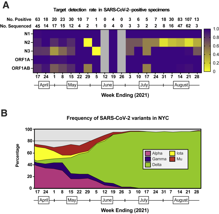
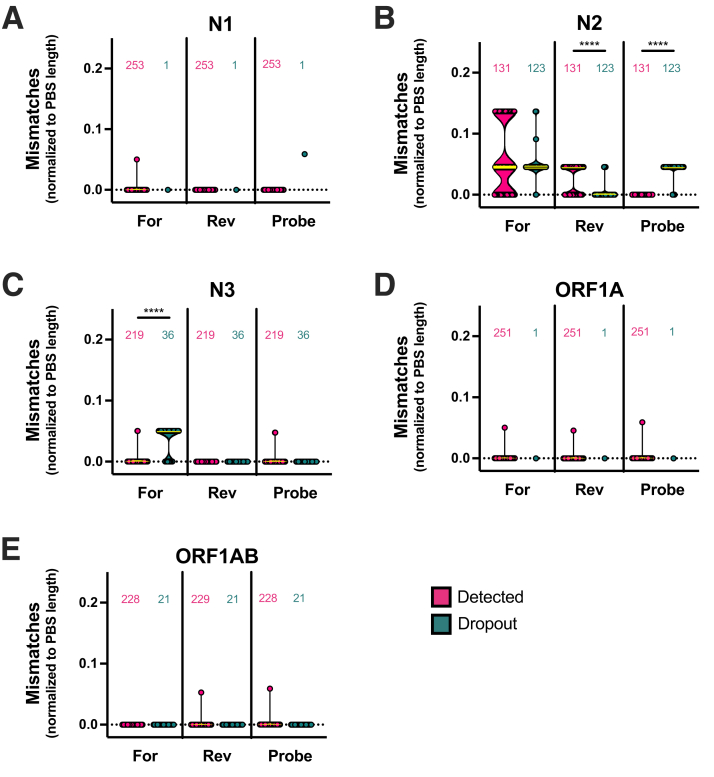
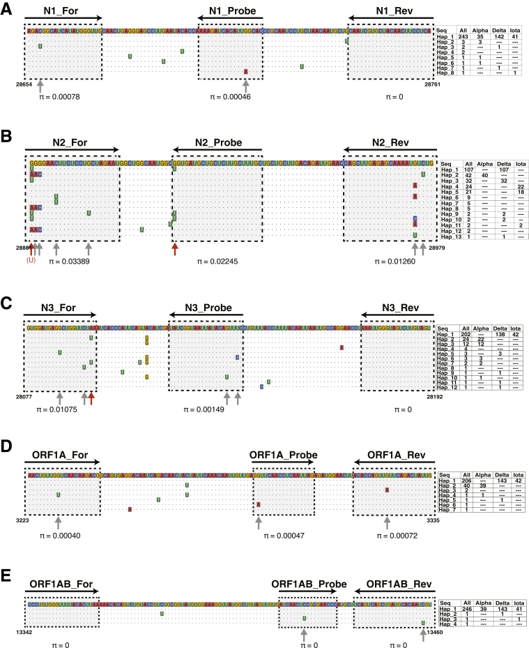
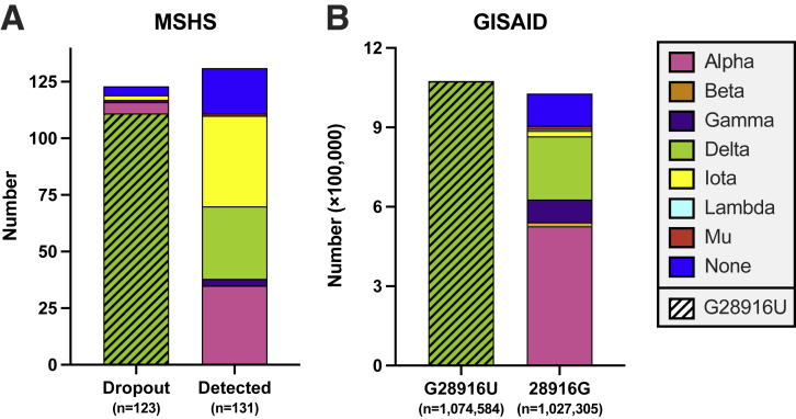
As severe acute respiratory syndrome coronavirus 2 (SARS-CoV-2) continues to circulate, multiple variants of concern have emerged. New variants pose challenges for diagnostic platforms because sequence diversity can alter primer/probe-binding sites (PBSs), causing false-negative results. The MassARRAY SARS-CoV-2 Panel (Agena Bioscience) uses RT-PCR and mass spectrometry to detect five multiplex targets across N and ORF1ab genes. Herein, we use a data set of 256 SARS-CoV-2-positive specimens collected between April 11, 2021, and August 28, 2021, to evaluate target performance with paired sequencing data. During this time frame, two targets in the N gene (N2 and N3) were subject to the greatest sequence diversity. In specimens with N3 dropout, 69% harbored the Alpha-specific A28095U polymorphism that introduces a 3'-mismatch to the N3 forward PBS and increases risk of target dropout relative to specimens with 28095A (relative risk, 20.02; 95% CI, 11.36 to 35.72; P < 0.0001). Furthermore, among specimens with N2 dropout, 90% harbored the Delta-specific G28916U polymorphism that creates a 3'-mismatch to the N2 probe PBS and increases target dropout risk (relative risk, 11.92; 95% CI, 8.17 to 14.06; P < 0.0001). These findings highlight the robust capability of MassARRAY SARS-CoV-2 Panel target results to reveal circulating virus diversity, and they underscore the power of multitarget design to capture variants of concern.


Figures

Figure 1
Figure 1
RT-PCR/matrix-assisted laser desorption-ionization time-of-flight target detection rate in severe acute respiratory syndrome coronavirus 2 (SARS-CoV-2)–positive specimens. A: Heatmap depicting the proportion of SARS-CoV-2–positive specimens that have detectable targets (N1, N2, N3, ORF1A, and ORF1AB) according to week from April 11, 2021, through August 28, 2021. The total number of SARS-CoV-2–positive specimens and the number of SARS-CoV-2–positive specimens sequenced according to pathogen surveillance are depicted above each week (column). Gray boxes indicate weeks in which no specimens were positive for SARS-CoV-2 on this platform. B: Stacked area plots depicting frequencies of SARS-CoV-2 variants reported by publicly available New York City (NYC) Department of Health surveillance data within the same time frame.
Figure 2
Figure 2
Frequencies of target primer/probe-binding site (PBS) mismatches and RT-PCR/matrix-assisted laser desorption-ionization time-of-flight target detection results. Number of mismatches normalized to the number of nucleotides in PBS (PBS length) across five diagnostic targets: N1 (A), N2 (B), N3 (C), ORF1A (D), and ORF1AB (E). Each point represents the calculated mismatches per specimen consensus genome for each target PBS. Violin plots represent the distribution as density of the points grouped according to primer/probe sequence [forward (For), reverse (Rev), and probe] and according to target detection result [detected (magenta), dropout (turquoise)]. Numbers of genomes analyzed for mismatches are depicted above each violin plot. Medians (yellow lines) are depicted, and bars above distributions represent statistical comparisons. Mann-Whitney test, ∗∗∗∗P < 0.0001.
Figure 3
Figure 3
Alignment of severe acute respiratory syndrome coronavirus 2 (SARS-CoV-2) haplotypes to RT-PCR/matrix-assisted laser desorption-ionization time-of-flight targets. Multiple sequence alignment of haplotype sequences to forward, reverse, and probe binding sites for N1 (A), N2 (B), N3 (C), ORF1A (D), and ORF1AB (E) targets. In each alignment, the reference sequence (NC_045,512.2; https://www.ncbi.nlm.nih.gov/nuccore) is annotated above, comprising color-coded nucleotides. The coordinates of the reference sequence are annotated at the bottom corners of each alignment. Each row represents a haplotype sequence that aligns to the target site. Absolute counts of each haplotype (eg, Hap_1, Hap_2, and so forth) within the data set and counts stratified according to variant lineage (eg, Alpha, Delta, Iota) are depicted to the right side of each haplotype sequence. Positions of primer/probe sequences are indicated by arrows, in which tail to arrowhead reflects 5′ to 3′ directionality. Gray boxes with black dotted borders outline primer/probe-binding sites (PBSs). Dots represent conserved nucleotides at each position and mismatched nucleotides are indicated. PBS mismatches are indicated by arrows; red arrows reflect mismatches that are significantly associated with target dropout. Notably, the association of the G28881U mismatch to the N2 forward PBS [panel B, red (U)] with target dropout is distinguished from the G28881A mismatch. Nucleotide diversity (π) of sequences at each PBS is indicated.
Figure 4
Figure 4
Delta-specific substitution interferes with N2 diagnostic target detection. A: Stacked bar plots depict the composition of severe acute respiratory syndrome coronavirus 2 variants in 254 Mount Sinai Health System (MSHS) genomes tested by RT-PCR/MALDI-TOF mass spectrometry. Bar plots reflect absolute number of genomes with N2 target dropout (left) and those with N2 target detection (right). Variant groups are color-coded, and genomes with the G28916U polymorphism are depicted by a hatched pattern. B: Stacked bar plot of publicly available sequences [Global Initiative on Sharing Avian Influenza Data (GISAID); see Materials and Methods] from the same time frame of this study depicting absolute numbers of variant genomes and presence or absence of the G28916U polymorphism. Color-coding and patterns are the same as in panel A.

Similar articles

  • A Robust, Highly Multiplexed Mass Spectrometry Assay to Identify SARS-CoV-2 Variants.
    Hernandez MM, Banu R, Shrestha P, Gonzalez-Reiche AS, van de Guchte A, Farrugia K, Sebra R; Mount Sinai PSP Study Group; Gitman MR, Nowak MD, Cordon-Cardo C, Simon V, van Bakel H, Sordillo EM, Luna N, Ramirez A, Castañeda SA, Patiño LH, Ballesteros N, Muñoz M, Ramírez JD, Paniz-Mondolfi AE. Hernandez MM, et al. Microbiol Spectr. 2022 Oct 26;10(5):e0173622. doi: 10.1128/spectrum.01736-22. Epub 2022 Sep 7. Microbiol Spectr. 2022. PMID: 36069609 Free PMC article.
  • Robust clinical detection of SARS-CoV-2 variants by RT-PCR/MALDI-TOF multitarget approach.
    Hernandez MM, Banu R, Gonzalez-Reiche AS, van de Guchte A, Khan Z, Shrestha P, Cao L, Chen F, Shi H, Hanna A, Alshammary H, Fabre S, Amoako A, Obla A, Alburquerque B, Patiño LH, Ramírez JD, Sebra R, Gitman MR, Nowak MD, Cordon-Cardo C, Schutzbank TE, Simon V, van Bakel H, Sordillo EM, Paniz-Mondolfi AE. Hernandez MM, et al. J Med Virol. 2022 Apr;94(4):1606-1616. doi: 10.1002/jmv.27510. Epub 2021 Dec 16. J Med Virol. 2022. PMID: 34877674 Free PMC article.
  • RT-PCR/MALDI-TOF mass spectrometry-based detection of SARS-CoV-2 in saliva specimens.
    Hernandez MM, Banu R, Shrestha P, Patel A, Chen F, Cao L, Fabre S, Tan J, Lopez H, Chiu N, Shifrin B, Zapolskaya I, Flores V, Lee PY, Castañeda S, Ramírez JD, Jhang J, Osorio G, Gitman MR, Nowak MD, Reich DL, Cordon-Cardo C, Sordillo EM, Paniz-Mondolfi AE. Hernandez MM, et al. J Med Virol. 2021 Sep;93(9):5481-5486. doi: 10.1002/jmv.27069. Epub 2021 May 19. J Med Virol. 2021. PMID: 33963565 Free PMC article.
  • Use of MALDI-TOF mass spectrometry for virus identification: a review.
    Do T, Guran R, Adam V, Zitka O. Do T, et al. Analyst. 2022 Jul 12;147(14):3131-3154. doi: 10.1039/d2an00431c. Analyst. 2022. PMID: 35713185 Review.
  • Universal screening for SARS-CoV-2 infection: a rapid review.
    Viswanathan M, Kahwati L, Jahn B, Giger K, Dobrescu AI, Hill C, Klerings I, Meixner J, Persad E, Teufer B, Gartlehner G. Viswanathan M, et al. Cochrane Database Syst Rev. 2020 Sep 15;9(9):CD013718. doi: 10.1002/14651858.CD013718. Cochrane Database Syst Rev. 2020. PMID: 33502003 Free PMC article.

Cited by

References

    1. Rochman N.D., Wolf Y.I., Faure G., Mutz P., Zhang F., Koonin E.V. Ongoing global and regional adaptive evolution of SARS-CoV-2. Proc Natl Acad Sci U S A. 2021;118 e2104241118. - PMC - PubMed
    1. Tao K., Tzou P.L., Nouhin J., Gupta R.K., de Oliveira T., Kosakovsky Pond S.L., Fera D., Shafer R.W. The biological and clinical significance of emerging SARS-CoV-2 variants. Nat Rev Genet. 2021;22:757–773. - PMC - PubMed
    1. Artesi M., Bontems S., Göbbels P., Franckh M., Maes P., Boreux R., Meex C., Melin P., Hayette M.-P., Bours V., Durkin K. A recurrent mutation at position 26340 of SARS-CoV-2 is associated with failure of the E gene quantitative reverse transcription-PCR utilized in a commercial dual-target diagnostic assay. J Clin Microbiol. 2020;58:e01598-20. - PMC - PubMed
    1. Rana D.R., Pokhrel N. Sequence mismatch in PCR probes may mask the COVID-19 detection in Nepal. Mol Cell Probes. 2020;53:101599. - PMC - PubMed
    1. Bal A., Destras G., Gaymard A., Stefic K., Marlet J., Eymieux S., Regue H., Semanas Q., d’Aubarede C., Billaud G., Laurent F., Gonzalez C., Mekki Y., Valette M., Bouscambert M., Gaudy-Graffin C., Lina B., Morfin F., Josset L., COVID-Diagnosis HCL Study Group Two-step strategy for the identification of SARS-CoV-2 variant of concern 202012/01 and other variants with spike deletion H69-V70, France, August to December 2020. Euro Surveill. 2021;26:2100008. - PMC - PubMed

Publication types

MeSH terms

Supplementary concepts