. 2022 Aug:111:34-48.
doi: 10.1016/j.reprotox.2022.04.011. Epub 2022 May 5.

Effects of spike protein and toxin-like peptides found in COVID-19 patients on human 3D neuronal/glial model undergoing differentiation: Possible implications for SARS-CoV-2 impact on brain development

Affiliations

Effects of spike protein and toxin-like peptides found in COVID-19 patients on human 3D neuronal/glial model undergoing differentiation: Possible implications for SARS-CoV-2 impact on brain development

Francesca Pistollato et al. Reprod Toxicol. 2022 Aug.

Abstract

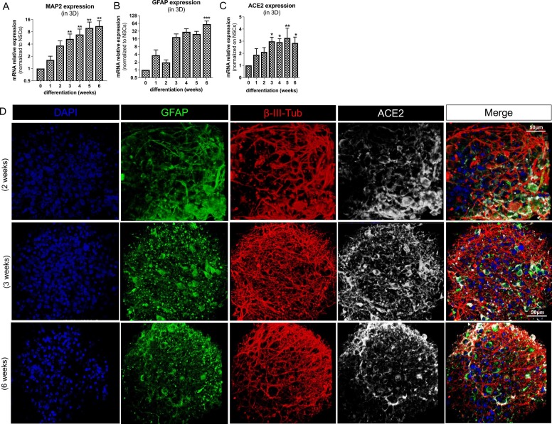
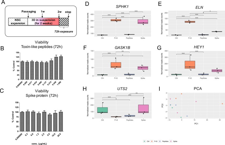
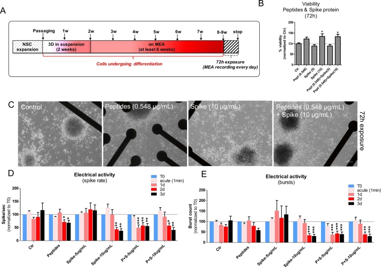
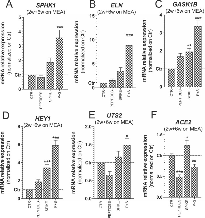
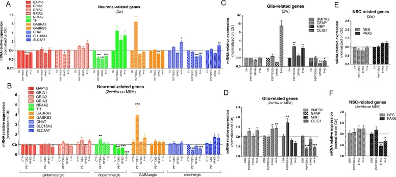
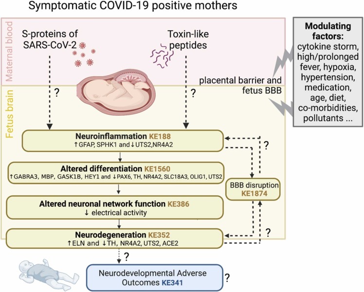
The possible neurodevelopmental consequences of SARS-CoV-2 infection are presently unknown. In utero exposure to SARS-CoV-2 has been hypothesized to affect the developing brain, possibly disrupting neurodevelopment of children. Spike protein interactors, such as ACE2, have been found expressed in the fetal brain, and could play a role in potential SARS-CoV-2 fetal brain pathogenesis. Apart from the possible direct involvement of SARS-CoV-2 or its specific viral components in the occurrence of neurological and neurodevelopmental manifestations, we recently reported the presence of toxin-like peptides in plasma, urine and fecal samples specifically from COVID-19 patients. In this study, we investigated the possible neurotoxic effects elicited upon 72-hour exposure to human relevant levels of recombinant spike protein, toxin-like peptides found in COVID-19 patients, as well as a combination of both in 3D human iPSC-derived neural stem cells differentiated for either 2 weeks (short-term) or 8 weeks (long-term, 2 weeks in suspension + 6 weeks on MEA) towards neurons/glia. Whole transcriptome and qPCR analysis revealed that spike protein and toxin-like peptides at non-cytotoxic concentrations differentially perturb the expression of SPHK1, ELN, GASK1B, HEY1, UTS2, ACE2 and some neuronal-, glia- and NSC-related genes critical during brain development. Additionally, exposure to spike protein caused a decrease of spontaneous electrical activity after two days in long-term differentiated cultures. The perturbations of these neurodevelopmental endpoints are discussed in the context of recent knowledge about the key events described in Adverse Outcome Pathways relevant to COVID-19, gathered in the context of the CIAO project (https://www.ciao-covid.net/).

Keywords: 3D neurospheres; AOP; CIAO Project; Electrical activity; RNA-Seq; Spike protein; Toxin-like peptides; brain development.


Conflict of interest statement

The authors have no conflict of interests to declare.

Figures

Fig. 1
Fig. 1
ACE2 expression in 3D neuronal/glial models undergoing differentiation. (A-C) Bar graphs showing MAP2, GFAP and ACE2 gene expression in 3D neurospheres differentiated for 0 (NSCs), 1, 2, 3, 4, 5 or 6 weeks; data were normalized to reference genes ACTB and GAPDH, and further normalized on NSCs (undifferentiated cells) (D) Representative fluorescent images (40x magnification) of 3D neurospheres differentiated for 2-, 3- and 6-weeks towards a mixed culture of neurons and glia; neurospheres were stained for GFAP (green), β-III-tubulin (red), ACE2 (white) and nuclei counterstained with DAPI (blue).
Fig. 2
Fig. 2
Effects of spike protein and toxin-like peptides on viability and whole transcriptome in 2-week old 3D neuronal/glial models after 72 h exposure. (A) NSCs were expanded, passaged and differentiated for 2 weeks as 3D neurospheres in suspension before being exposed for 72 h to different concentrations of toxin-like peptides or spike protein. (B, C) Cell viability analyses of 3D neurospheres after 72 h exposure to different concentrations of toxin-like peptides or spike protein; values were normalised to control (Ctr). (D-H) Differentially expressed genes with FDR corrected p value < 0.1; graphs show normalized reads counts for: Sphingosine kinase 1 (SPHK1) (D), Elastin (ELN) (E), Golgi associated kinase (GASK1B) (F), HEY1 (G), and Urotensin-II (UTS2) (H). (I) Principal component analysis (PCA). Data are representative of three biological replicates.
Fig. 3
Fig. 3
Effects of spike protein and toxin-like peptides on electrical activity in long-term differentiated 3D neuronal/glial models. (A) NSCs were expanded, passaged and differentiated for 2 weeks as 3D neurospheres in suspension before being transferred to 24-well MEA plates for at least 6 additional weeks (total of 8–9 weeks in differentiation). Cultures were exposed for 72 h to Spike alone (5 and 10 µg/mL), Peptides alone (0.548 µg/mL) or a combination of both, and electrical activity was recorded for 5 min before (T0), 1 min after adding compounds (acute), and after 1-, 2- and 3-day exposure. (B) Cell viability analyses of cultures after 72 h exposure as indicated in A (values were normalised to Ctr). (C) Representative phase contrast images of cell cultures on 24-well MEA plates exposed to compounds as described in A. (D, E) Bar graphs showing spike rate (i.e., number of spikes/sec) (D), and number of bursts (E) of cultures exposed to compounds as described in A (data were normalized to T0). For all analyses, mean ± S.E.M. of 4 biological replicates.
Fig. 4
Fig. 4
Effects of spike protein and toxin-like peptides on SPHK1, ELN, GASK1B, HEY1, UTS2 and ACE2 expression in long-term differentiated cultures. (A-F) Bar graphs showing expression of SPHK1, ELN, GASK1B, HEY1, UTS2 and ACE2 in long-term differentiated cultures exposed for 72 h to toxin-like peptides alone (0.548 µg/mL), spike protein alone (10 µg/mL) and a combination of both (P + S) vs Control. Data were normalized to reference genes ACTB and GAPDH, and further normalized to Ctr (mean ± S.E.M. of 3 biological replicates).
Fig. 5
Fig. 5
qPCR analyses of neuronal, glia and NSC-related genes in short and long-term differentiated cultures exposed for 72 h to peptides, spike protein and P + S. (A-F) Bar graphs showing expression of neuronal subtypes specific genes (A, B), glia-related genes (C, D), and NSC-related genes (E, F) in 2-week old neurosphere cultures (A, C, E) and 2-week old neurospheres further differentiated on MEA for 6 additional weeks (B, D, F). Both culture types were exposed at the end of differentiation period to toxin-like Peptides alone (0.548 µg/mL), Spike protein alone (10 µg/mL) and both compounds (P + S) vs Control for 72 h. Data were normalized to reference genes ACTB and GAPDH, and further normalized on Ctr (unexposed cells). For all analyses, mean ± S.E.M. of 3 biological replicates.
Fig. 6
Fig. 6
Summary of the effects possibly triggered by SARS-CoV-2 Spike protein and toxin-like Peptides, along with Key Events (KEs) describing these mechanisms, and additional modulating factors. Under each KE, the endpoints (genes and electrical activity) that have been found deregulated in this study are reported. Dashed lines and question marks indicate still unknown (not yet verified) processes

Similar articles

Cited by

References

    1. Zambrano L.D., Ellington S., Strid P., Galang R.R., Oduyebo T., Tong V.T., Woodworth K.R., Nahabedian J.F., 3rd, Azziz-Baumgartner E., Gilboa S.M., Meaney-Delman D., Pregnancy C.C.-R., Infant Linked Outcomes T. Update: characteristics of symptomatic women of reproductive age with laboratory-confirmed SARS-CoV-2 Infection by pregnancy status - United States, January 22-October 3, 2020. MMWR Morb. Mortal. Wkly. Rep. 2020;69(44)):1641–1647. - PMC - PubMed
    1. Aguilar-Valles A., Rodrigue B., Matta-Camacho E. Maternal immune activation and the development of dopaminergic neurotransmission of the offspring: relevance for schizophrenia and other psychoses. Front. Psychiat. 2020;11:852. - PMC - PubMed
    1. Figueiredo C.P., Fontes-Dantas F.L., da Poian A.T., Clarke J.R. SARS-CoV-2-associated cytokine storm during pregnancy as a possible risk factor for neuropsychiatric disorder development in post-pandemic infants. Neuropharmacology. 2021;201 - PMC - PubMed
    1. A.L. Düppers, B. Bohnhorst, E. Bültmann, T. Schulz, L. Higgins-Wood, C.S. von Kaisenberg, Severe fetal brain damage subsequent to acute maternal hypoxemic deterioration in COVID-19 Ultrasound in obstetrics & gynecology: the official journal of the International Society of Ultrasound in Obstetrics and Gynecology 58 3 2021 490 491. - PMC - PubMed
    1. G.M. Fernandes, F. Motta, L.M.P. Sasaki, P.D. Silva, Â A.M. Miranda, A.O. Carvalho, A.P.M. Gomides, A. Soares, A. Santos Jr., C.O. Alves, C.M. Gomes, C.C. Siracusa, D.A. Araújo Jr., D.L. Mendonça-Silva, J.A.L. Jesus, K.N. Costa, M.E.C. Castro, P.S. Kurizky, P.S. França, R. Tristão, Y.R. Pereira, L.C.G. Castro, A.M. Zaconeta, C.P. Albuquerque, L. Mota, Pregnancy outcomes and child development effects of SARS-CoV-2 Infection (PROUDEST Trial): Protocol for a Multicenter, Prospective Cohort Study, JMIR Research Protocols 10 4 2021 e26477. - PMC - PubMed

Substances