. 2022:667:575-610.
doi: 10.1016/bs.mie.2022.03.047. Epub 2022 Apr 18.

Methods for discovering catalytic activities for pseudokinases

Affiliations

Methods for discovering catalytic activities for pseudokinases

Miles H Black et al. Methods Enzymol. 2022.

Abstract

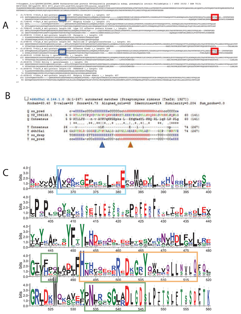
Pseudoenzymes resemble active enzymes, but lack key catalytic residues believed to be required for activity. Many pseudoenzymes appear to be inactive in conventional enzyme assays. However, an alternative explanation for their apparent lack of activity is that pseudoenzymes are being assayed for the wrong reaction. We have discovered several new protein kinase-like families which have revealed how different binding orientations of adenosine triphosphate (ATP) and active site residue migration can generate a novel reaction from a common kinase scaffold. These results have exposed the catalytic versatility of the protein kinase fold and suggest that atypical kinases and pseudokinases should be analyzed for alternative transferase activities. In this chapter, we discuss a general approach for bioinformatically identifying divergent or atypical members of an enzyme superfamily, then present an experimental approach to characterize their catalytic activity.

Keywords: Atypical kinases; Bioinformatics; Pseudokinases; Uncharacterized proteins.


Figures

Fig. 1
Fig. 1
Sequence search for novel kinases. (A) Archival FFAS results. L. pneumophila proteome compared against the PDB. Results for the SidJ protein shown, with noticeable similarity to protein kinases. Blue box—equivalent of the PKA catalytic residue, K72. Red box—equivalent of the PKA catalytic residue, D166. (B) Archival results, HHpred analysis of SidJ. Similarity to N-lobe of protein kinases. Blue triangle—equivalent of PKA K72. Brown triangle—equivalent of PKA E91. (C) Sequence logo of SidJ homologs. Blue box: K72-E91 ion pair region. Green boxes: canonical kinase active site motifs. Orange box: migrated kinase active site motifs.
Fig. 2
Fig. 2
Structure search for novel kinases. (A) TMalign results, Leishmania infantum proteome vs kinases. Known kinases marked in red. (B) As in (A), but known kinases filtered out. Data points corresponding to Leishmania E9AHR4 shown in blue. (C) Structural superposition of an AlphaFold model of Leishmania E9AHR4 (gray) vs PDB 2zv2 (CaMKK2 kinase).
Fig. 3
Fig. 3
Purification of the active SidJ-CaM complex. (A) Plasmid map of pETDuet1 SUMO-SidJ/CaM used to co-express SidJ and CaM. Black arrows indicate the coding sequences of CaM and SidJ cloned into the first and second multiple cloning sites ([MCS]), respectively, under control of a T7 promoter (T7 pro.) The map also indicates the ampicillin resistance cassette (AmpR), pUC high-copy number origin if replication (ori) and the coding sequence of the lac repressor (LacI). (B) Superdex 200 size exclusion chromatography trace of the SidJ-CaM complex purified from E. coli as described in Section 6.1.2. (C) Fractions that eluted from 67.5 to 80 mL were separated by reduced 6% SDS-PAGE and visualized with Coomassie blue, demonstrating co-elution of SidJ (the ~100kDa species) and CaM (the ~15kDa species).
Fig. 4
Fig. 4
Discovery of glutamylation of SdeA after co-expression with SidJ and CaM. (A) Plasmid maps of pETDuet1 SidJ/CaM and pSUMO-SdeA, annotated as in Fig. 3. (B) Reducing 6% SDS-PAGE analysis and Coomassie staining of SdeA isolated from E. coli following co-expression with CaM, SidJ, or the indicated mutants. Note the electrophoretic mobility shift of SdeA co-expressed with SidJ and CaM. (C) Intact mass LC/MS spectra of SdeA from lane 2 of (B). The theoretical MW of SdeA is 104,340 Da. The labeled peaks demonstrate up to five mass shifts of +129Da. D. LC/MS spectra of SdeA from lane 3 of (B), in which the D542A mutant of SidJ was co-expressed. Panels (B)–(D): Adapted from fig. 2 in Black, M. H., Osinski, A., Gradowski, M., Servage, K. A., Pawlowski, K., Tomchick, D. R., et al. (2019). Bacterial pseudokinase catalyzes protein polyglutamylation to inhibit the SidE-family ubiquitin ligases. Science, 364(6442), 787–792. doi:https://doi.org/10.1126/science.aaw7446.
Fig. 5
Fig. 5
LC/MS spectra of SidJ residues 59–851 demonstrating auto-glutamylation. SidJ was co-expressed with CaM in E. coli as described in Section 6.1.2 and analyzed by protein intact mass spectrometry.

Similar articles

Cited by

References

    1. Ashkenazy H, Abadi S, Martz E, Chay O, Mayrose I, Pupko T, et al. (2016). ConSurf 2016: An improved methodology to estimate and visualize evolutionary conservation in macromolecules. Nucleic Acids Research, 44(W1), W344–W350. 10.1093/nar/gkw408. - DOI - PMC - PubMed
    1. Baek M, DiMaio F, Anishchenko I, Dauparas J, Ovchinnikov S, Lee GR, et al. (2021). Accurate prediction of protein structures and interactions using a three-track neural network. Science, 373(6557), 871–876. 10.1126/science.abj8754. - DOI - PMC - PubMed
    1. Bhogaraju S, Bonn F, Mukherjee R, Adams M, Pfleiderer MM, Galej WP, et al. (2019). Inhibition of bacterial ubiquitin ligases by SidJ-calmodulin catalysed glutamylation. Nature, 572(7769), 382–386. 10.1038/s41586-019-1440-8. - DOI - PMC - PubMed
    1. Black MH, Osinski A, Gradowski M, Servage KA, Pawlowski K, Tomchick DR, et al. (2019). Bacterial pseudokinase catalyzes protein polyglutamylation to inhibit the SidE-family ubiquitin ligases. Science, 364(6442), 787–792. 10.1126/science.aaw7446. - DOI - PMC - PubMed
    1. Cheng H, Schaeffer RD, Liao Y, Kinch LN, Pei J, Shi S, et al. (2014). ECOD: An evolutionary classification of protein domains. PLoS Computational Biology, 10(12), e1003926. 10.1371/journal.pcbi.1003926. - DOI - PMC - PubMed

Publication types

LinkOut - more resources