. 2022 May 7;12(1):7520.
doi: 10.1038/s41598-022-11638-2.

Proteins in human body fluids contain in vivo antigen analog of the melibiose-derived glycation product: MAGE

Affiliations

Proteins in human body fluids contain in vivo antigen analog of the melibiose-derived glycation product: MAGE

Kinga Gostomska-Pampuch et al. Sci Rep. .

Abstract

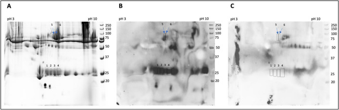
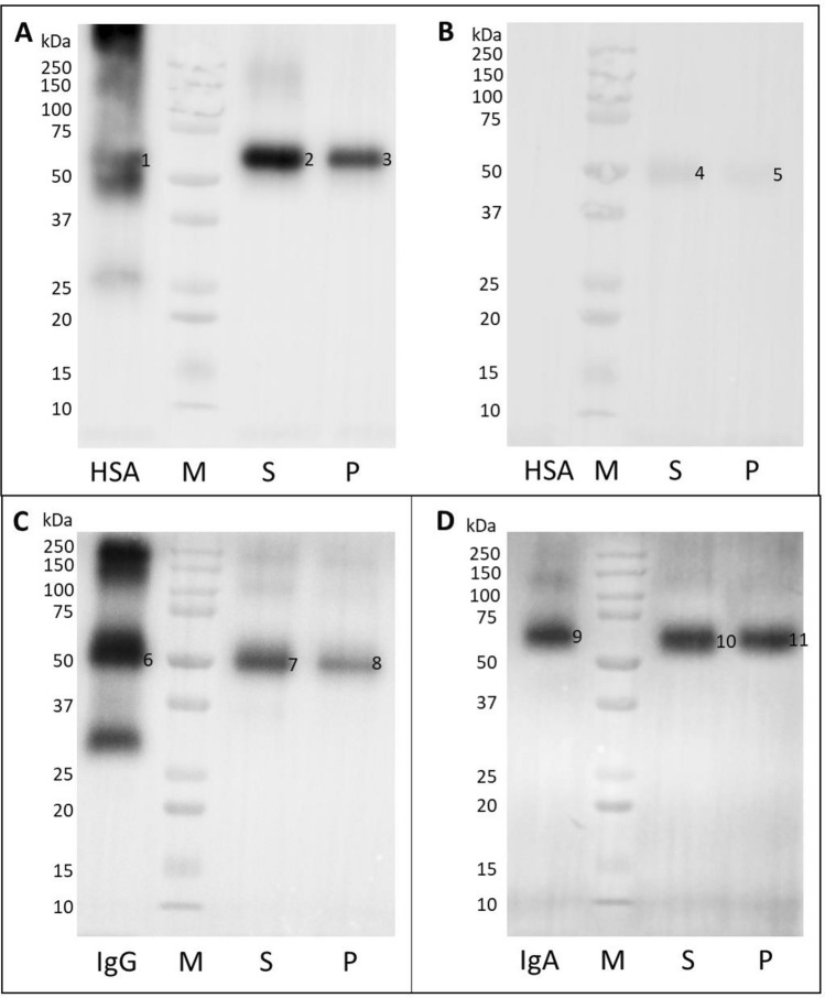
Melibiose-derived AGE (MAGE) is an advanced glycation end-product formed in vitro in anhydrous conditions on proteins and protein-free amino acids during glycation with melibiose. Our previous studies revealed the presence of MAGE antigen in the human body and tissues of several other species, including muscles, fat, extracellular matrix, and blood. MAGE is also antigenic and induces generation of anti-MAGE antibody. The aim of this paper was to identify the proteins modified by MAGE present in human body fluids, such as serum, plasma, and peritoneal fluids. The protein-bound MAGE formed in vivo has been isolated from human blood using affinity chromatography on the resin with an immobilized anti-MAGE monoclonal antibody. Using mass spectrometry and immunochemistry it has been established that MAGE epitope is present on several human blood proteins including serum albumin, IgG, and IgA. In serum of diabetic patients, mainly the albumin and IgG were modified by MAGE, while in healthy subjects IgG and IgA carried this modification, suggesting the novel AGE can impact protein structure, contribute to auto-immunogenicity, and affect function of immunoglobulins. Some proteins in peritoneal fluid from cancer patients modified with MAGE were also observed and it indicates a potential role of MAGE in cancer.


Conflict of interest statement

The authors declare no competing interests.

Figures

Figure 1
Figure 1
Identification of proteins glycated with MAGE. Protein from human serum were subjected to isoelectric focusing between pH 3–7, separated on 8% SDS-PAGE gel and stained with Coomassie Safe (A). Additional gels were subjected to WB analysis with anti-MAGE mAb followed by secondary Ab anti-mouse IgE-HRP (B) or by incubation with the secondary Ab only (C). The indicated spots 1–6 were excised from the gel. Molecular mass markers on the right side of each panel indicate mass in kDa. The original gel and blots are presented in Supplementary Fig. S4.
Figure 2
Figure 2
Extraction of blood proteins glycated with MAGE. Proteins from serum of the diabetic patient and plasma of healthy donor were immunoprecipitated with the Sepharose-anti-MAGE resin and the same volume of each sample was analyzed by SDS-PAGE (A). The separated in the gel proteins that bound to the Sepharose-anti-MAGE antibody resin (lane 1, 2) and the control resin (lane 3, 4) were stained with Coomassie Brilliant Blue or were subjected to WB with the anti-MAGE antibody (B). As the negative control, the additional membrane was probed with the secondary anti-mouse IgE-HRP antibody (C). The proteins indicated in panel A by red boxes corresponding to bands 1S, 2S (diabetic serum) and 1P, 2P (plasma from healthy donor) showed specific binding with the anti-MAGE antibody and were excised from gel for mass spectrometry analysis. M—molecular mass standard; MAGE (MB-mel). The original gel and blots are presented in Supplementary Fig. S5.
Figure 3
Figure 3
Verification of proteins extracted from human blood by WB analysis. The proteins extracted from diabetic patient serum (S), plasma of healthy donor (P) were transferred on the PVDF membrane along with protein marker (M), HSA, IgG, IgA (2 µg/well) and were probed with anti-HSA (A), anti-mouse IgG-HRP (B), anti-human IgG-HRP (C), and anti-human IgA-HRP (D) antibodies. The original blots are presented in Supplementary Fig. S6.
Figure 4
Figure 4
WB analysis of human body fluids with anti-MAGE monoclonal antibody. Ten samples of serum (A, B—lane 2–11) and corresponding peritoneal fluid (C, D—lane 2–11) from patients with GC were subjected to WB with the anti-MAGE antibody (A, C) or as a control with secondary antibody anti-mouse IgE-HRP (B, D). Molecular marker indicated in kDa (lane 1) and MB-mel (lane 12) was run as the controls. The original blots are presented in Supplementary Fig. S7.

Similar articles

Cited by

References

    1. Pietkiewicz J, Seweryn E, Bartys̈ A, Gamian A. Receptory końcowych produktów zaawansowanej glikacji-znaczenie fizjologiczne i kliniczne. Postepy Higieny i Medycyny Doswiadczalnej. 2008;62:511–523. - PubMed
    1. Nagai R, et al. Antibody-based detection of advanced glycation end-products: Promises vs limitations. Glycoconj. J. 2016;33:545–552. doi: 10.1007/s10719-016-9708-9. - DOI - PubMed
    1. Ashraf JM, et al. Recent advances in detection of AGEs: Immunochemical, bioanalytical and biochemical approaches. IUBMB Life. 2015;67:897–913. doi: 10.1002/iub.1450. - DOI - PubMed
    1. Staniszewska M, et al. The melibiose-derived glycation product mimics a unique epitope present in human and animal tissues. Sci. Rep. 2021;11:2940. doi: 10.1038/s41598-021-82585-7. - DOI - PMC - PubMed
    1. Bronowicka-szydełko A, Krzystek-korpacka M, Gacka M. Association of novel advanced glycation end-product ( AGE10) with complications of diabetes as measured by enzyme-linked immunosorbent assay. J. Clin. Med. 2021;10:4499. doi: 10.3390/jcm10194499. - DOI - PMC - PubMed

Publication types