. 2022 May 7;15(1):52.
doi: 10.1186/s13045-022-01272-w.

LncRNA-PACERR induces pro-tumour macrophages via interacting with miR-671-3p and m6A-reader IGF2BP2 in pancreatic ductal adenocarcinoma

Affiliations

LncRNA-PACERR induces pro-tumour macrophages via interacting with miR-671-3p and m6A-reader IGF2BP2 in pancreatic ductal adenocarcinoma

Yihao Liu et al. J Hematol Oncol. .

Abstract

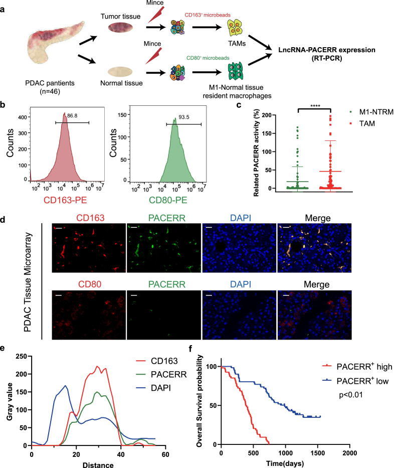
Background: LncRNA-PACERR plays critical role in the polarization of tissue-associated macrophages (TAMs). In this study, we found the function and molecular mechanism of PACERR in TAMs to regulate pancreatic ductal adenocarcinoma (PDAC) progression.

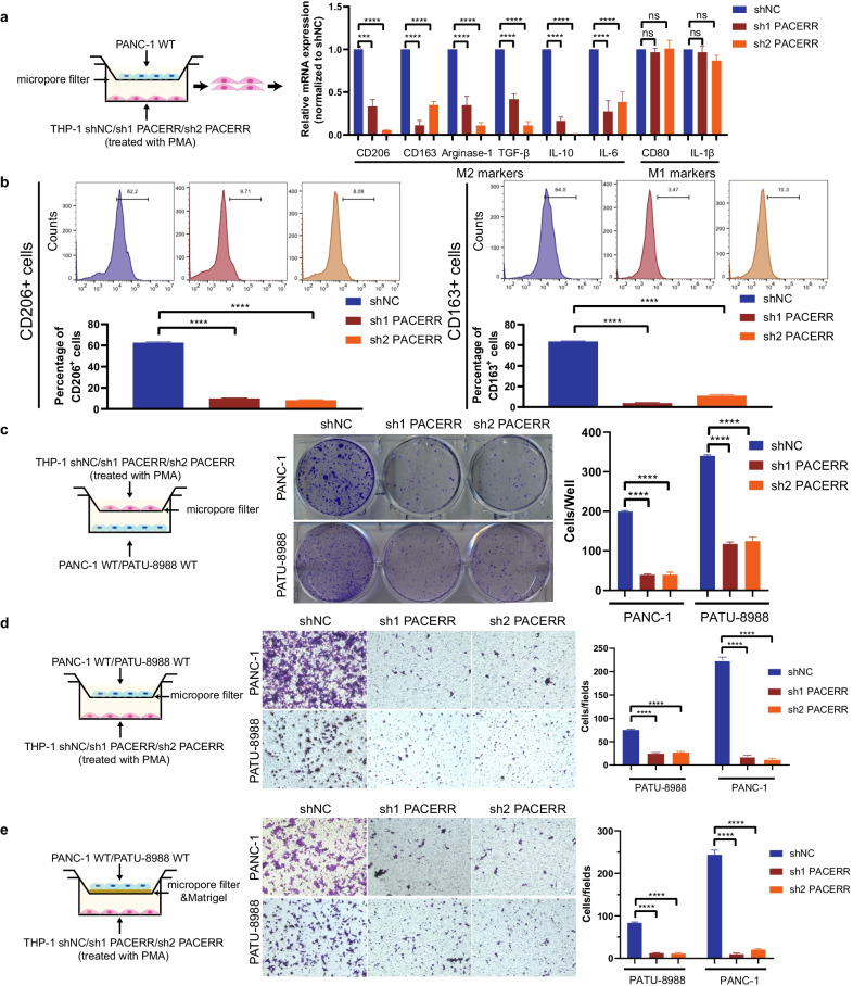
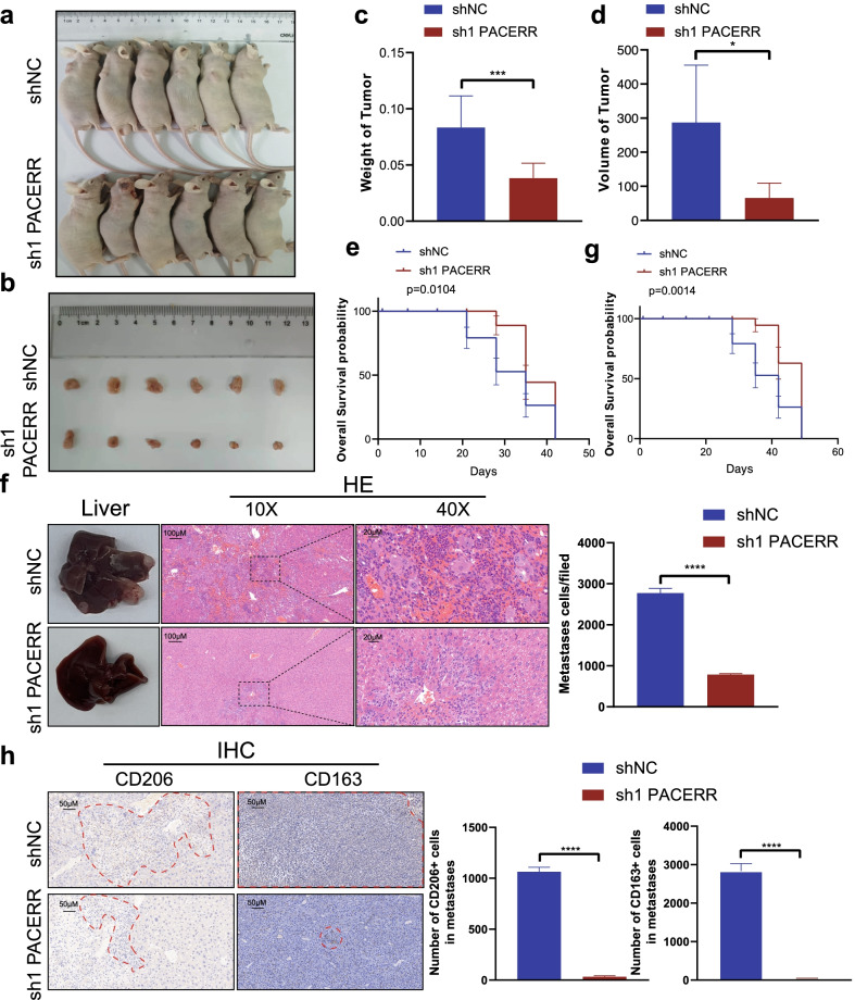
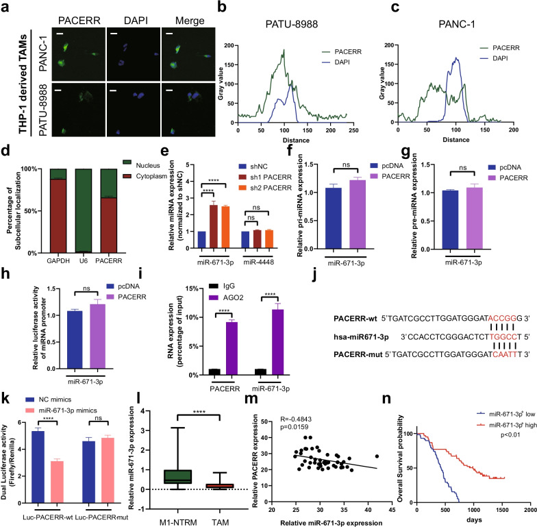
Methods: We used qPCR to analyse the expression of PACERR in TAMs and M1-tissue-resident macrophages (M1-NTRMs) which were isolated from 46 PDAC tissues. The function of PACERR on macrophages polarization and PDAC proliferation, migration and invasion were confirmed through in vivo and in vitro assays. The molecular mechanism of PACERR was discussed via fluorescence in situ hybridization (FISH), RNA pull-down, ChIP-qPCR, RIP-qPCR and luciferase assays.

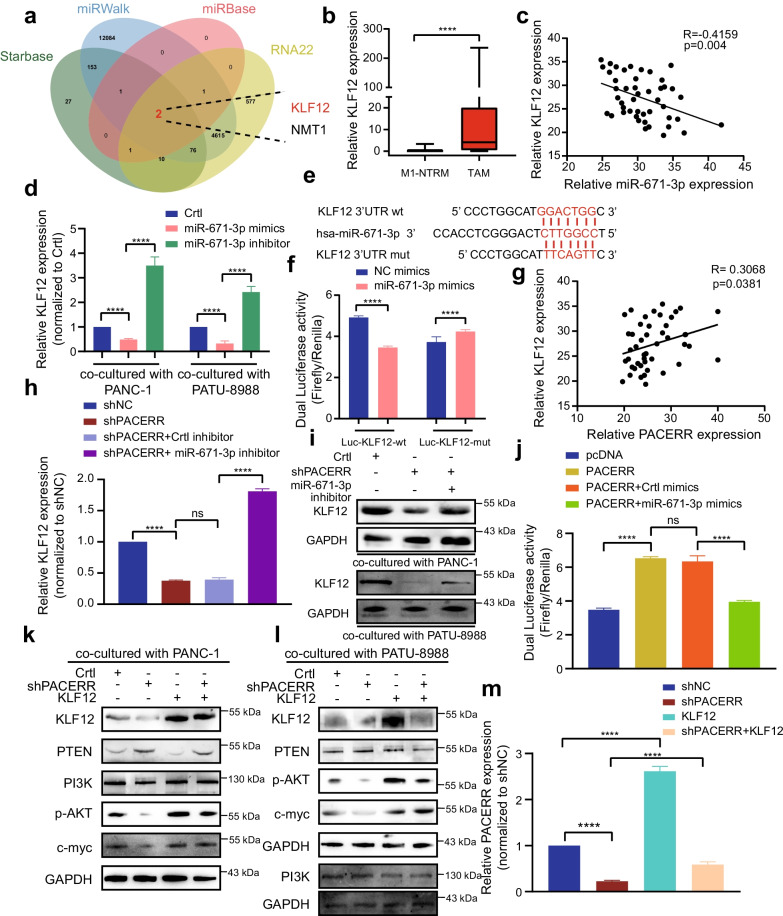
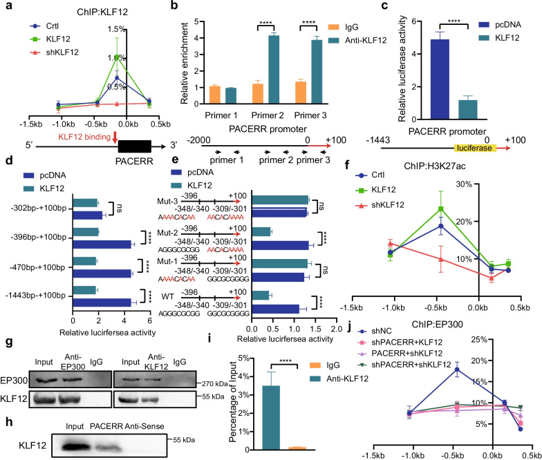
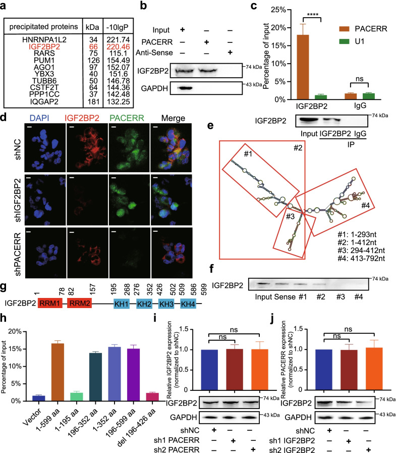
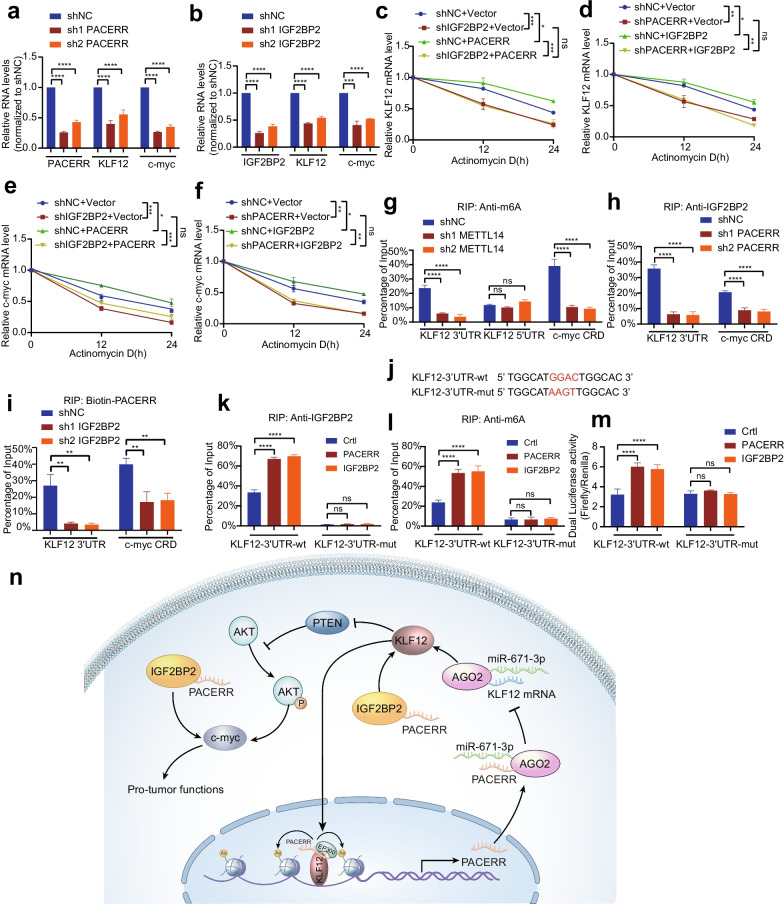
Results: LncRNA-PACERR was high expression in TAMs and associated with poor prognosis in PDAC patients. Our finding validated that LncRNA-PACERR increased the number of M2-polarized cells and facilized cell proliferation, invasion and migration in vitro and in vivo. Mechanistically, LncRNA-PACERR activate KLF12/p-AKT/c-myc pathway by binding to miR-671-3p. And LncRNA-PACERR which bound to IGF2BP2 acts as an m6A-dependent manner to enhance the stability of KLF12 and c-myc in cytoplasm. In addition, the promoter of LncRNA-PACERR was a target of KLF12 and LncRNA-PACERR recruited EP300 to increase the acetylation of histone by interacting with KLF12 in nucleus.

Conclusions: This study found that LncRNA-PACERR functions as key regulator of TAMs in PDAC microenvironment and revealed the novel mechanisms in cytoplasm and in nucleus.

Keywords: IGF2BP2; KLF12; LncRNA-PACERR; PDAC; TAMs; m6A; miR-671-3p.


Conflict of interest statement

The authors declare that they have no competing interests.

Figures

Fig. 1
Fig. 1
LncRNA-PACERR expression in TAMs is activated in PDAC tissues and is associated with poor prognosis. A Schematic illustration of qRT-PCR forty-six sample preparation (n = 46). Tumour-associated macrophages (TAMs) from forty-six PDAC tissue samples were enriched with CD163 positive selection. M1 macrophages in normal tissue-resident macrophages (M1-NTRMs) from PDAC adjacent normal tissue samples of the same patient were enriched with CD80 positive selection. B Purity of CD163+ cells and CD80+ by CD163 and CD80 microbeads sorting. C Expression of LncRNA-PACERR in 46 pairs of TAMs and M1-NTRMs from PDAC. *P < 0.05; **P < 0.01; ***P < 0.001; ****P < 0.0001. D Colocalization of LncRNA-PACERR (green) and CD163/CD80 (red) in 110 clinical samples of pancreatic ductal adenocarcinoma (PDAC) as shown by fluorescence microscopy. DAPI staining (blue) shows the nuclei (DNA). Scar bar: 50 μm. E Staining intensity of LncRNA-PACERR, CD163 and DAPI on the immunofluorescence from TMAs of 110 PDAC patients. Green represents LncRNA-PACERR. Red represents CD163. Blue represents DAPI. F Kaplan–Meier survival curve presenting the overall survival of 110 PDAC patients, grouped according to the extent of LncRNA-PACERR+ TAMs infiltration
Fig. 2
Fig. 2
Knockdown of LncRNA-PACERR hinders the M2 polarization and pro-tumour functions of THP-1-derived TAMs in vitro. A qPCR analysis of the relative expression of M2 markers (Arginase-1, CD163, TGFβ, CD206, IL-10 and IL-6) and M1 marker (CD80, IL-1β) in THP-1-derived TAMs after LncRNA-PACERR knockdown. THP-1 cells were treated with PMA and co-cultured with PANC-1 cells for two days. Data are shown as the results from three independent experiments. B Flow cytometric analysis of the expression of M2 markers (CD163 and CD206) in THP-1-derived TAMs after LncRNA-PACERR knockdown. THP-1 cells were treated with PMA and co-cultured with PANC-1 cells for two days. Data are shown as the results from two independent experiments. CE Proliferation (C), migration (D) and invasion (E) capacity of PATU-8988 or PANC-1 cells co-cultured with THP-1-derived TAMs (shNC/ shPACERR). shNC means that cells were transfected in negative control plasmids. *P < 0.05; **P < 0.01; ***P < 0.001; ****P < 0.0001
Fig. 3
Fig. 3
LncRNA-PACERR+ TAMs facilitate PDAC cell growth and liver metastasis in vivo. A Images of BALB/c nude mice which were co-injected with THP-1 cells and PATU-8988 cells subcutaneously. B Images of subcutaneous tumours. C, D Tumour weights and volumes of the subcutaneous xenografts. E, G Kaplan–Meier survival curve presenting the overall survival of BALB/c nude mice (shNC/sh1 PACERR groups) (n = 6). F Representative images of liver metastasis and the number of metastatic cells in PDAC mouse model, in which PATU-8988 cells mixed with TAMs (THP-1 shNC/sh1 LncRNA-PACERR) were injected into the spleens of BALB/c nude mice. Data iare shown as the results from three independent experiments. H Representative images of IHC (CD163 and CD206) of liver metastasis and the number of CD163+ and CD206+ cells in metastatic foci from liver tissues. *P < 0.05; **P < 0.01; ***P < 0.001; ****P < 0.0001
Fig. 4
Fig. 4
LncRNA-PACERR functions as a ceRNA to sponge miR-671-3p in TAMs. A Fluorescence in situ hybridization (FISH) of LncRNA-PACERR (green) in THP-1-derived TAMs (co-cultured with PATU-8988 or PANC-1). DAPI staining (blue) shows the nuclei. Scar bar: 10 μm. B, C Grey value of LncRNA-PACERR and DAPI on the FISH from THP-1 derived TAMs. Green represents LncRNA-PACERR. Blue represents DAPI. D Expression levels of LncRNA-PACERR in the cytoplasm and nucleus in THP-1-derived TAMs. E Expression of two potential target miRNAs (miR-671-3p and miR-4448) in THP-1 derived TAMs after LncRNA-PACERR knocked down. F, G Expression of pri-miR-671-3p (F) and pre-miR-671-3p (G) in THP-1 derived TAMs transfected with empty control or pcDNA-LncRNA-PACERR. H Promoter luciferase activity of miR-671-3p in 293-T cells overexpressing LncRNA-PACERR. I RIP assay was performed using rabbit AGO2 and IgG antibodies in THP-1 derived TAMs. Relative expression levels of LncRNA-PACERR and miR-671-3p were determined by qRT-PCR. J, K Dual luciferase activity in 293-T cells co-transfected with LncRNA-PACERR wild-type or mutant sequence and miR-671-3p mimics. L Expression of miR-671-3p in 46 pairs of TAMs and M1-NTRMs from PDAC patients. M Correlation analysis between LncRNA-PACERR and miR-671-3p using expression data from 46 pairs of TAMs. N Kaplan–Meier survival curve presenting the overall survival of 110 PDAC patients, grouped according to the extent of miR-671-3p+ TAMs infiltration
Fig. 5
Fig. 5
MiR-671-3p directly binds to the 3′UTR of KLF12 and regulates the KLF12/AKT/c-myc axis. A Predicted target genes of miR-671-3p using Starbase, miRWalk, miRbase, RNA22 and the two overlapping target genes (NMT-1 and KLF12). B Expression of KLF12 in 46 pairs of TAMs and M1-NTRMs from PDAC patients. C Correlation analysis between miR-671-3p and KLF12 using data from 46 pairs of PDAC patients. D mRNA expression of KLF12 in THP-1 derived TAMs (co-cultured with PATU-8988 or PANC-1 cells) transfected with empty control or miR-671-3p mimics or inhibitor. E, F Dual luciferase activity in HEK-293 T cells co-transfected with the KLF12 wild-type or mutant sequence (E) and miR-671-3p mimics. G Correlation analysis between LncRNA-PACERR and KLF12 in TAMs from 46 PDAC patients. H mRNA expression of KLF12 in THP-1 derived TAMs co-transfected with the LncRNA-PACERR knocked down vector and miR-671-3p inhibitor. I Protein levels of KLF12 in THP-1 derived TAMs (co-cultured with PATU-8988 or PANC-1 cells). J Dual luciferase activity in HEK-293 T cells co-transfected with LncRNA-PACERR overexpression vector and miR-671-3p mimics. K, L Potein expression of PI3K/KLF12/AKT/c-myc axis in THP-1 derived TAMs (co-cultured with PANC-1 (K) or PATU-8988 cells (L)). M LncRNA-PACERR expression was evaluated by qRT-PCR in THP-1 derived TAMs with indicated treatment. The data are presented as the mean ± SD of three independent experiments. *P < 0.05; **P < 0.01; ***P < 0.001; ****P < 0.0001
Fig. 6
Fig. 6
KLF12 binds directly to LncRNA-PACERR and recruits EP300 to the promoter region of LncRNA-PACERR in a LncRNA-PACERR-dependent manner. A Association of KLF12 with the promoter region of LncRNA-PACERR in THP-1-derived TAMs (Crtl/KLF12 OE / shKLF12) analysed by ChIP-qPCR. B ChIP assays showed endogenous KLF12 binding to the LncRNA-PACERR gene promoter. C, D HEK293T cells were co-transfected with LncRNA-PACERR promoter–luciferase truncations and KLF12 plasmids, and the luciferase activity was determined using a dual luciferase reporter assay after 48 h. E Dual luciferase assay of HEK293T cells co-transfected with firefly luciferase constructs containing the wild-type or mutant KLF12 potential binding sites of LncRNA-PACERR promoter and KLF12 plasmids were performed. F Association of H3K27ac with the promoter region of LncRNA-PACERR in THP-1-derived TAMs (Crtl/ KLF12 OE/ shKLF12) analysed by ChIP-qPCR. G Results of coimmunoprecipitation (Co-IP) in THP-1-derived TAMs. Normal rabbit IgG was used as a negative control. H WB validation of KLF12 proteins pulled down with biotin-labelled LncRNA-PACERR is shown. I RNA immunoprecipitation (RIP) was performed using a KLF12-specific antibody. Eluted KLF12-binding RNAs were reverse transcribed, and qPCR was performed with primers specific for LncRNA-PACERR. Normal rabbit IgG (IgG) was used as a negative control. Data are shown as the results from three independent experiments. J Association of EP300 with the promoter region of LncRNA-PACERR analysed by ChIP-qPCR in THP-1-derived TAMs (shNC/ shLncRNA-PACERR + KLF12 OE/shKLF12 + LncRNA-PACERR OE/ shKLF12 + shLncRNA-PACERR)
Fig. 7
Fig. 7
LncRNA-PACERR directly binds with IGF2BP2 in TAMs. A Visualization of protein bands by biotin-labelled LncRNA-PACERR RNA probes incubated with total protein extracts from THP-1 derived TAMs. B Immunoblotting to determine the specific association of IGF2BP2 with biotinylated LncRNA-PACERR. C qRT-PCR analysis of LncRNA-PACERR enriched by IGF2BP2 in THP-1 derived TAMs (top). Immunoblot of IGF2BP2 is shown (bottom). IP, immunoprecipitation. D Images showing the colocalization of LncRNA-PACERR and IGF2BP2 in the IGF2BP2 KD and LncRNA-PACERR KD THP-1 derived TAMs. Scale bars: 20 μm. E Secondary structure of LncRNA-PACERR analysed by RNAfold web server and deletion mapping of biotinylated LncRNA-PACERR motifs, as indicated. The red boxes represent the remaining fragments of LncRNA-PACERR, with the corresponding number label in the corner. F Immunoblot showing the association of IGF2BP2 with biotinylated LncRNA-PACERR RNA strands and the above-mentioned biotinylated LncRNA-PACERR motifs. G Schematic structures showing six domains in IGF2BP2. H RIP analysis for LncRNA-PACERR enrichment in HEK293T cells transfected with the FLAG-tagged full-length or truncated IGF2BP2 constructs (n = 3). aa: amino acid. I qRT-PCR analysis of IGF2BP2 mRNA levels (top) and immunoblot of IGF2BP2 (bottom) in the LncRNA-PACERR KD cells (n = 3). J qRT-PCR analysis of LncRNA-PACERR levels (top) and immunoblot of IGF2BP2 (bottom) in the IGF2BP2 KD cells (n = 3). The results are presented as the mean ± SEM. *P < 0.05; **P < 0.01; ***P < 0.001; ****P < 0.0001, ns not significant
Fig. 8
Fig. 8
LncRNA-PACERR cooperates with IGF2BP2 to regulate KLF12 and c-myc in an m6A-dependent manner. A, B qRT-PCR analysis of the LncRNA-PACERR, KLF12 and c-myc transcript levels in the LncRNA-PACERR KD (A) and IGF2BP2 KD (B) THP-1 derived TAMs (n = 3). C, D Half-life of KLF12 after treatment with 5 μmol/L actinomycin D for the indicated times in the IGF2BP2 KD THP-1 derived TAMs with ectopically expressed LncRNA-PACERR (C) and in the LncRNA-PACERR KD THP-1 derived TAMs with ectopically expressed IGF2BP2 (D). E, F Falf-life of c-myc after treatment with 5 μmol/L actinomycin D for the indicated times in the IGF2BP2 KD THP-1 derived TAMs with ectopically expressed LncRNA-PACERR (E) and in the LncRNA-PACERR KD THP-1 derived TAMs with ectopically expressed IGF2BP2 (F). G RIP qRT-PCR showing the enrichment of m6A modification in the KLF12 3′ UTR/5′ UTR and c-myc CRD regions in the METTL14 KD THP-1 derived TAMs (n = 3). H, I RIP qRT-PCR detecting the enrichment of IGF2BP2 (H) and biotin-labelled LncRNA-PACERR (I) in the KLF12 3′ UTR and c-myc CRD in LncRNA-PACERR KD (H) and IGF2BP2 KD (I) THP-1 derived TAMs (n = 3). J Schematic representation of wild-type (WT) and mutated (MUT; GGAC to AAGT) KLF12 3′ UTR of the pmirGLO vector. K, L RIP qRT-PCR detection of the enrichment of IGF2BP2 (K) and m6A (L) in the KLF12 3′ UTR WT and MUT luciferase reporters in the LncRNA-PACERR and IGF2BP2 OE cells (n = 3). M Relative luciferase activity levels of KLF12 3′ UTR WT and MUT reporters in the LncRNA-PACERR and IGF2BP2 OE cells (n = 3). N Proposed model demonstrating a positive feedback loop between LncRNA-PACERR and KLF12 in TAMs to promote proliferation and migration in PDAC. The expression of LncRNA-PACERR is activated by EP300-mediated H3K27 acylation. LncRNA-PACERR exerts its pro-tuomour function by regulating miR-671-3p/KLF12/AKT/c-myc axis and sequestering IGF2BP2

References

    1. Siegel RL, Miller KD, Fuchs HE, Jemal A. Cancer Statistics, 2021. CA Cancer J Clin 2021, 71(1). - PubMed
    1. Connor AA, Gallinger S. Pancreatic cancer evolution and heterogeneity: integrating omics and clinical data. Nat Rev Cancer 2021. - PubMed
    1. Mizrahi JD, Surana R, Valle JW, Shroff RT. Pancreatic cancer. Lancet. 2020;395(10242):2008–2020. doi: 10.1016/S0140-6736(20)30974-0. - DOI - PubMed
    1. Bear AS, Vonderheide RH, O'Hara MH. Challenges and opportunities for pancreatic cancer immunotherapy. Cancer Cell. 2020;38(6):788–802. doi: 10.1016/j.ccell.2020.08.004. - DOI - PMC - PubMed
    1. Schizas D, Charalampakis N, Kole C, Economopoulou P, Koustas E, Gkotsis E, Ziogas D, Psyrri A, Karamouzis MV. Immunotherapy for pancreatic cancer: a 2020 update. Cancer Treat Rev. 2020;86:102016. doi: 10.1016/j.ctrv.2020.102016. - DOI - PubMed

MeSH terms