. 2022 May 7;13(1):191.
doi: 10.1186/s13287-022-02861-9.

Hypoxic mesenchymal stem cell-derived extracellular vesicles ameliorate renal fibrosis after ischemia-reperfusion injure by restoring CPT1A mediated fatty acid oxidation

Affiliations

Hypoxic mesenchymal stem cell-derived extracellular vesicles ameliorate renal fibrosis after ischemia-reperfusion injure by restoring CPT1A mediated fatty acid oxidation

Zhumei Gao et al. Stem Cell Res Ther. .

Abstract

Background: Renal fibrosis is a common pathological process of chronic kidney diseases induced by multiple factors. Hypoxic pretreatment of mesenchymal stem cells can enhance the efficacy of secreted extracellular vesicles (MSC-EVs) on various diseases, but it is not clear whether they can better improve renal fibrosis. The latest research showed that recovery of fatty acid oxidation (FAO) can reduce renal fibrosis. In this study, we aimed to examine whether hypoxic pretreatment with MSC extracellular vesicles (Hypo-EVs) can improve FAO to restore renal fibrosis and to investigate the underlying mechanism.

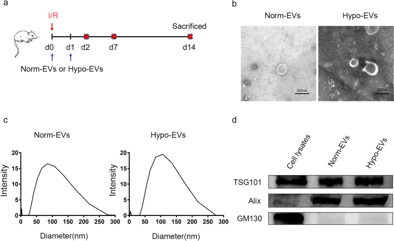
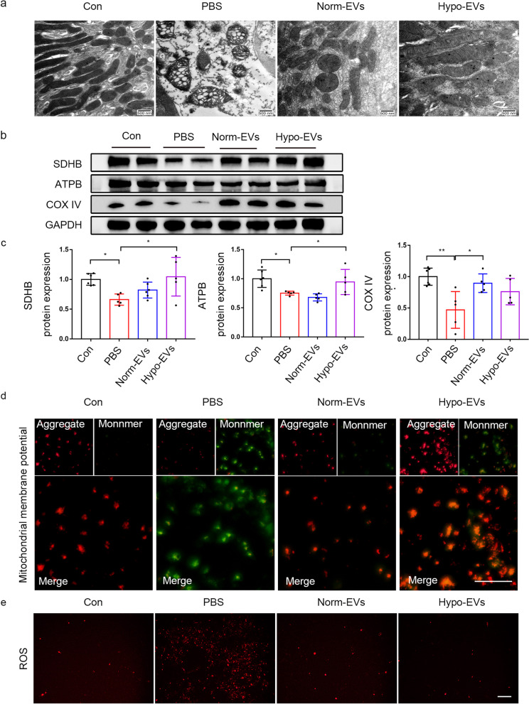
Methods: Hypo-EVs were isolated from hypoxia-pretreated human placenta-derived MSC (hP-MSC), and Norm-EVs were isolated from hP-MSC cultured under normal conditions. We used ischemia-reperfusion (I/R)-induced renal fibrosis model in vivo. The mice were injected with PBS, Hypo-EVs, or Norm-EVs immediately after the surgery and day 1 postsurgery. Renal function, kidney pathology, and renal fibrosis were assessed for kidney damage evaluation. For mechanistic exploration, fatty acid oxidation (FAO), mitochondrial morphological alterations, ATP production and mitochondrial mass proteins were detected in vivo. Mitochondrial membrane potential and reactive oxygen species (ROS) production were investigated in vitro.

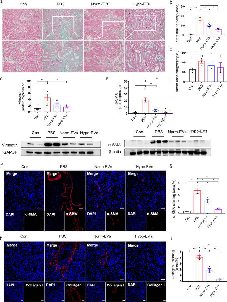
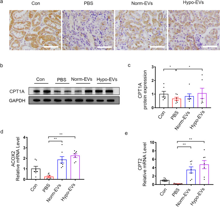
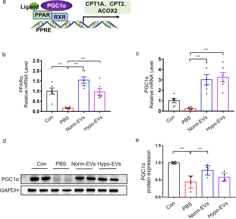
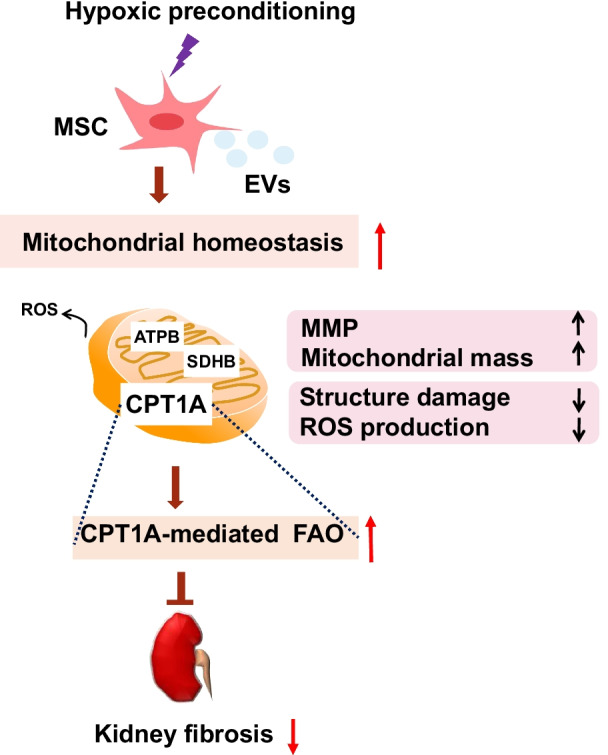
Results: We found that Hypo-EVs confer a superior therapeutic effect on recovery of renal structure damage, restoration of renal function and reduction in renal fibrosis. Meanwhile, Hypo-EVs enhanced mitochondrial FAO in kidney by restoring the expression of a FAO key rate-limiting enzyme carnitine palmitoyl-transferase 1A (CPT1A). Mechanistically, the improvement of mitochondrial homeostasis, characterized by repaired mitochondrial structure, restoration of mitochondrial mass and ATP production, inhibition of oxidative stress, and increased mitochondrial membrane potential, partially explains the effect of Hypo-EVs on improving mitochondrial FAO and thus attenuating I/R damage.

Conclusions: Hypo-EVs suppress the renal fibrosis by restoring CPT1A-mediated mitochondrial FAO, which effects may be achieved through regulation of mitochondrial homeostasis. Our findings provide further mechanism support for development cell-free therapy of renal fibrosis.

Keywords: Extracellular vesicles; Fatty acid oxidation; Hypoxic; Mesenchymal stem cell; Mitochondrial; Renal fibrosis.


Conflict of interest statement

The authors declare that the research was conducted in the absence of any commercial or financial relationships that could be construed as a potential conflict of interest.

Figures

Fig. 1
Fig. 1
Characterization of EVs. a Schematic diagram of the animal experiment. Briefly, mice were treated with Norm-EVs (100 μg), Hypo-EVs (100 μg) or PBS immediately after surgery and on D 1 postsurgery and were sacrificed at D 2, D 7, and D 14 after surgery. bTransmission electron microscopy image of both analysed EVs. Scale bar represents 200 nm. c Dynamic light scattering measuring the size distribution of both analysed EVs. d Western blot analysed the EVs-specific positive biomarkers (Alix and TSG101) and the negative marker GM130 in the EVs and their parent cell (MSC)
Fig. 2
Fig. 2
Norm-EVs and Hypo-EVs were internalized by injured kidneys. a Representative fluorescence images of I/R mice after treatment with Dil/Norm-EVs or Dil/Hypo-EVs at selected time points. b Dynamic changes in the fluorescence intensity of I/R kidneys with intravenous injection of Dil/Norm-EVs or Dil/Hypo-EVs at selected times (n = 3)
Fig. 3
Fig. 3
Hypo-EVs have a better anti-fibrosis effect than Norm-EVs in I/R-induced renal fibrosis mice. a Representative images for Masson’s staining of kidney sections on D 14 after I/R injury. b Quantification of interstitial fibrosis area as percentages of the total area (n = 6–9). c Renal function analysis. Levels of BUN in mice on D 3 after I/R injury (n = 5–6). d Western blot analysis was used to detect the protein expression of vimentin. The relative expression of vimentin was evaluated (n = 5–6). e Western blot analysis was used to detect the protein expression of α-SMA. The relative expression of α-SMA was evaluated (n = 5–6). f Immunofluorescence staining of α-SMA (red) and nuclei (blue). g Quantification of α-SMA-positive areas as percentages of the total area (n = 3). h Immunofluorescence staining of collagen I (red) and nuclei (blue). i Quantification of collagen I-positive areas as percentages of the total area (n = 3). Scale bar represents 50 μm. Data are expressed as mean ± SEM. *P < 0.05 and **P < 0.01
Fig. 4
Fig. 4
Hypo-EVs reverse CPT1A-mediated FAO defects in I/R-induced fibrotic kidneys. a The locations and expressions of CPT1A were determined by immunohistochemical staining in the kidney sections of mice from the different groups. b, c Western blot analysis was used to detect the protein expression of CPT1A. The relative expression of CPT1A was evaluated (n = 6). d The mRNA level of ACOX2 in different groups (n = 6–7). e The mRNA level of CPT2 in different groups (n = 7). Scale bar represents 50 μm. Data are expressed as mean ± SEM. *P < 0.05 and ** P < 0.01
Fig. 5
Fig. 5
Restoration of PPARα and PGC1α expression not involved in Hypo-EVs induced better recovery of FAO. a Schematic illustration for FAO regulation. PPARα and PGC1α are the major regulators of FAO. Promoting the activity of PPARα can boost FAO, and PGC1α is a coactivator of PPARα. b The mRNA level of PPARα in different groups (n = 6). c The mRNA level of PGC1α in different groups (n = 6). d, e Western blot analysis was used to detect the protein expression of PGC1α. The relative expression of PGC1α was evaluated. Data are expressed as mean ± SEM. *P < 0.05 and ** P < 0.01
Fig. 6
Fig. 6
Hypo-EVs rescued mitochondrial homeostasis in vivo. a Representative TEM images of kidneys on D 14 after I/R injury. Scale bar represents 500 nm. b, c Western blot was used to detect the protein expression of mitochondrial OXPHOS proteins (ATPB、SDHB and COX IV). The relative expression of ATPB, SDHB and COX IV were evaluated (n = 5–6). d Mitochondrial membrane potential was determined through a JC-1 probe in HK-2 cells after incubated with TGF-β1. Scale bar represents 100 μm. e Mito-SOX Red was used to observe the levels of intracellular ROS in the HK-2 cells after incubated with TGF-β1. Scale bar represents 100 μm. Data are expressed as mean ± SEM. *P < 0.05,** < 0.01
Fig. 7
Fig. 7
Possible mechanisms for preventing renal fibrosis using hypoxic mesenchymal stem cell-derived extracellular vesicles (Hypo-EVs)

Similar articles

Cited by

References

    1. Kalantar-Zadeh K, Jafar TH, Nitsch D, Neuen BL, Perkovic V. Chronic kidney disease. Lancet. 2021;398(10302):786–802. doi: 10.1016/S0140-6736(21)00519-5. - DOI - PubMed
    1. Xie Y, Bowe B, Mokdad AH, Xian H, Yan Y, Li T, Maddukuri G, Tsai CY, Floyd T, Al-Aly Z. Analysis of the Global Burden of Disease study highlights the global, regional, and national trends of chronic kidney disease epidemiology from 1990 to 2016. Kidney Int. 2018;94(3):567–581. doi: 10.1016/j.kint.2018.04.011. - DOI - PubMed
    1. Ronco C, Bellomo R, Kellum JA. Acute kidney injury. Lancet. 2019;394(10212):1949–1964. doi: 10.1016/S0140-6736(19)32563-2. - DOI - PubMed
    1. Zheng Z, Li C, Shao G, Li J, Xu K, Zhao Z, Zhang Z, Liu J, Wu H. Hippo-YAP/MCP-1 mediated tubular maladaptive repair promote inflammation in renal failed recovery after ischemic AKI. Cell Death Dis. 2021;12(8):754. doi: 10.1038/s41419-021-04041-8. - DOI - PMC - PubMed
    1. Yan H, Xu J, Xu Z, Yang B, Luo P, He Q. Defining therapeutic targets for renal fibrosis: exploiting the biology of pathogenesis. Biomed Pharmacother. 2021;143:112115. doi: 10.1016/j.biopha.2021.112115. - DOI - PubMed

Publication types