. 2022 Aug 15:257:119273.
doi: 10.1016/j.neuroimage.2022.119273. Epub 2022 May 5.

On the relationship between GABA+ and glutamate across the brain

Affiliations

On the relationship between GABA+ and glutamate across the brain

Reuben Rideaux et al. Neuroimage. .

Abstract

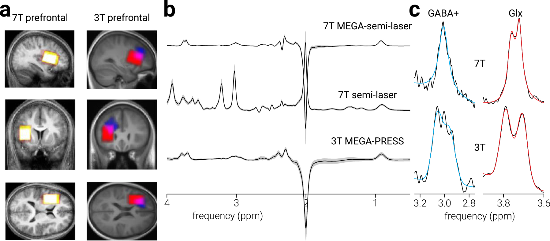
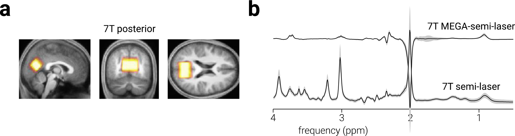
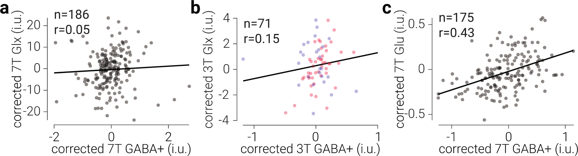
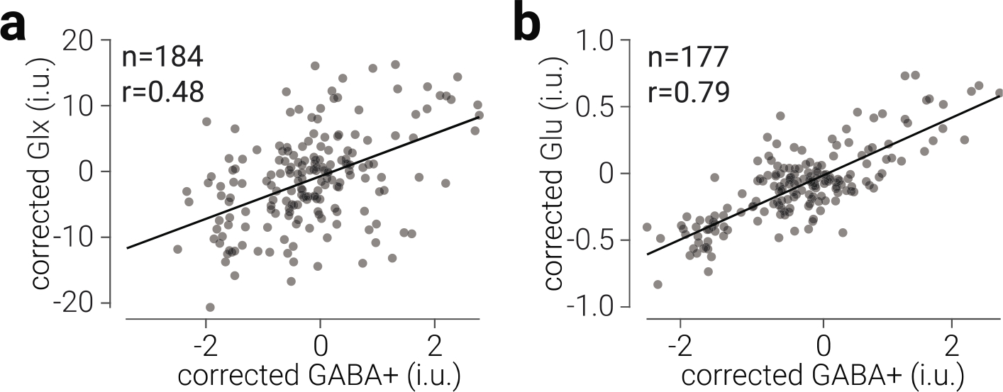
Equilibrium between excitation and inhibition (E/I balance) is key to healthy brain function. Conversely, disruption of normal E/I balance has been implicated in a range of central neurological pathologies. Magnetic resonance spectroscopy (MRS) provides a non-invasive means of quantifying in vivo concentrations of excitatory and inhibitory neurotransmitters, which could be used as diagnostic biomarkers. Using the ratio of excitatory and inhibitory neurotransmitters as an index of E/I balance is common practice in MRS work, but recent studies have shown inconsistent evidence for the validity of this proxy. This is underscored by the fact that different measures are often used in calculating E/I balance such as glutamate and Glx (glutamate and glutamine). Here we used a large MRS dataset obtained at ultra-high field (7 T) measured from 193 healthy young adults and focused on two brain regions - prefrontal and occipital cortex - to resolve this inconsistency. We find evidence that there is an inter-individual common ratio between GABA+ (γ-aminobutyric acid and macromolecules) and Glx in the occipital, but not prefrontal cortex. We further replicate the prefrontal result in a legacy dataset (n = 78) measured at high-field (3 T) strength. By contrast, with ultra-high field MRS data, we find extreme evidence that there is a common ratio between GABA+ and glutamate in both prefrontal and occipital cortices, which cannot be explained by participant demographics, signal quality, fractional tissue volume, or other metabolite concentrations. These results are consistent with previous electrophysiological and theoretical work supporting E/I balance. Our findings indicate that MRS-detected GABA+ and glutamate (but not Glx), are a reliable measure of E/I balance .

Keywords: 7T; E/I balance; GABA; Glutamate; MRS; Ultra-high field.


Conflict of interest statement

Declaration of Competing Interest The authors declare no competing interests.

Figures

Figure 1.
Figure 1.. Prefrontal cortex voxel location and spectra.
a) Sagittal, coronal, and axial views of prefrontal cortex MRS voxel placement in the current (7 T) experiment and the legacy experiments, on the average T1-weighted structural image in Montreal Neurological Institute (MNI) space. Colour intensity indicates voxel placement overlap across participants; for the 3 T data, red indicates voxels from Filmer et al. (2019) and blue indicates voxels from Rideaux et al. (2021). b) Average sLASER, MEGA-sLASER and MEGA-PRESS spectra for all participants. Shaded regions indicate ±1 standard deviation. c) Examples of GABA+ and Glx model fit to the difference-spectra for 7 T and 3 T data.
Figure 2.
Figure 2.. Occipital cortex voxel location and spectra.
a) Sagittal, coronal, and axial views of occipital cortex MRS voxel placement on the average T1-weighted structural image in MNI space. Colour intensity indicates voxel placement overlap across participants. b) Average sLASER and MEGA-sLASER spectra for all participants. Shaded regions indicate ±1 standard deviation.
Figure 3.
Figure 3.. Common ratio between GABA+ and Glu, but not Glx, in prefrontal cortex.
a) Glx concentration as a function of GABA+ concentration in prefrontal cortex, measured at 7 T. b) The same as (a), but measured at 3 T. d) Glu concentration as a function of GABA+ concentration in prefrontal cortex, measured at 7 T. Glx, Glu, and GABA+ values are tissue-corrected, referenced to water, expressed in institutional units (i.u.), and corrected for changes associated with different scanner software (7T) or studies (3T). Black lines indicate best linear fit; datapoints are semi-transparent, thus darker regions indicate overlap. Note, differences between quantification methods produced differential scaling of GABA+ and Glx relative to Glu concentration.
Figure 4.
Figure 4.. Common ratio between GABA+ and Glu and Glx in occipital cortex.
a) Glx concentration as a function of GABA+ concentration in occipital cortex. b) The same as (a), for Glu and GABA+. Glx, Glu, and GABA+ values are tissue-corrected, referenced to water, expressed in institutional units (i.u.), and corrected for changes associated with different scanner software. Black lines indicate best linear fit; datapoints are semi-transparent, thus darker regions indicate overlap.

References

    1. Anderson JS, Carandini M, & Ferster D (2000). Orientation tuning of input conductance, excitation, and inhibition in cat primary visual cortex. Journal of Neurophysiology, 84(2), 909–926. 10.1152/jn.2000.84.2.909 - DOI - PubMed
    1. Andreychenko A, Boer VO, Castro C. S. A. de, Luijten PR, & Klomp DWJ (2012). Efficient spectral editing at 7 T: GABA detection with MEGA-sLASER. Magnetic Resonance in Medicine, 68(4), 1018–1025. 10.1002/mrm.24131 - DOI - PubMed
    1. Ashburner J, & Friston KJ (2005). Unified segmentation. NeuroImage, 26(3), 839–851. 10.1016/j.neuroimage.2005.02.018 - DOI - PubMed
    1. Bang JW, Shibata K, Frank SM, Walsh EG, Greenlee MW, Watanabe T, & Sasaki Y (2018). Consolidation and reconsolidation share behavioural and neurochemical mechanisms. Nature Human Behaviour, 2(7), 507–513. 10.1038/s41562-018-0366-8 - DOI - PMC - PubMed
    1. Baumann O, & Mattingley JB (2010). Medial parietal cortex encodes perceived heading direction in humans. Journal of Neuroscience, 30(39), 12897–12901. 10.1523/JNEUROSCI.3077-10.2010 - DOI - PMC - PubMed

Publication types