Case Reports
. 2022 Dec 15;61(24):3675-3682.
doi: 10.2169/internalmedicine.9565-22. Epub 2022 May 7.

Neutrophil Infiltration and Acinar-ductal Metaplasia Are the Main Pathological Findings in Pembrolizumab-induced Pancreatitis

Affiliations
Case Reports

Neutrophil Infiltration and Acinar-ductal Metaplasia Are the Main Pathological Findings in Pembrolizumab-induced Pancreatitis

Morihisa Hirota et al. Intern Med. .

Abstract

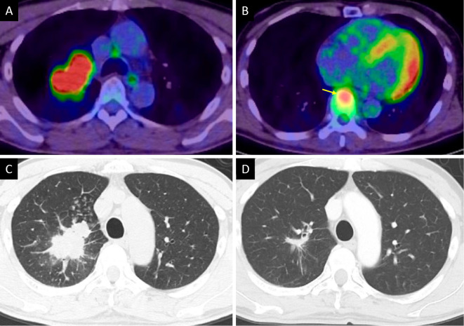
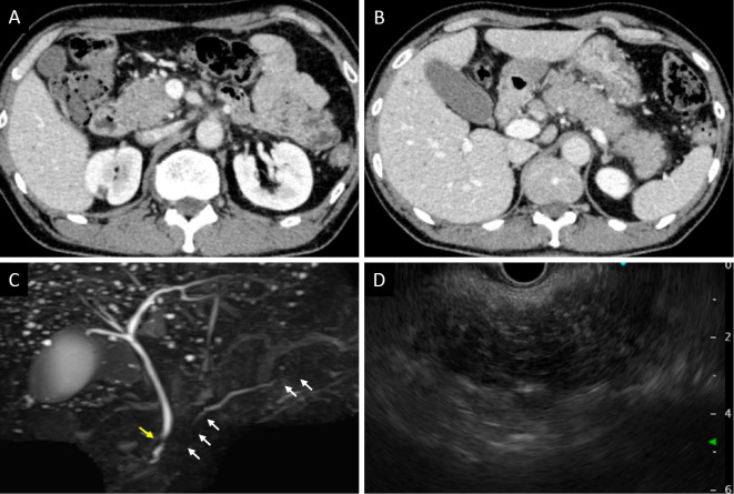
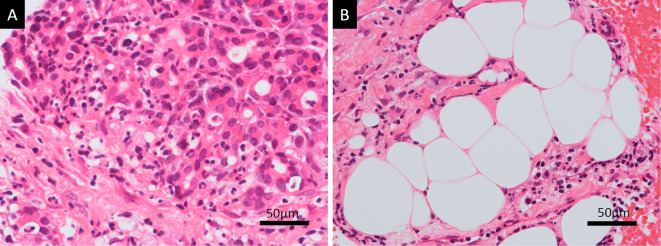
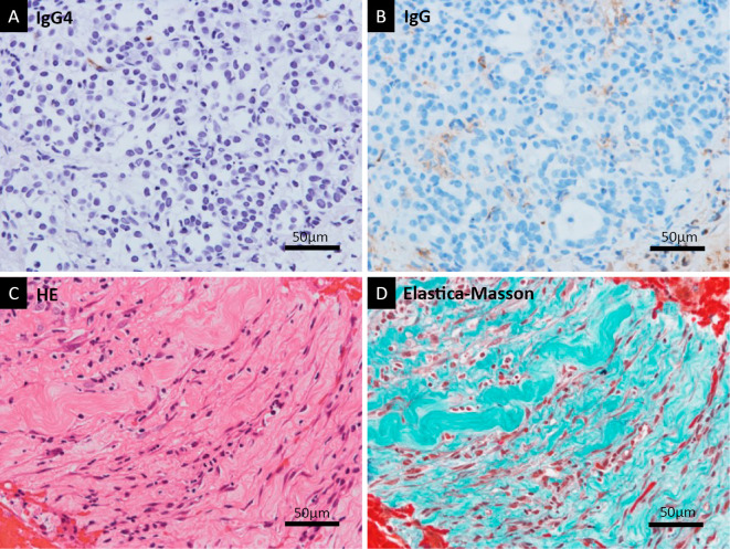
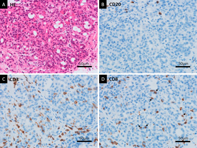
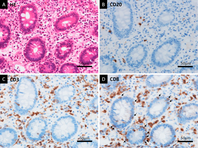
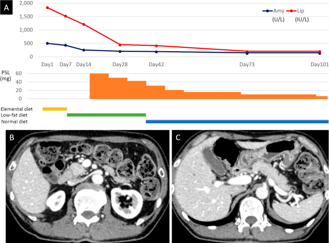
The histopathological findings of immune checkpoint inhibitor (ICI)-induced pancreatitis have rarely been reported. A 56-year-old man with squamous cell carcinoma of the lung with bone metastasis was being treated with pembrolizumab, an anti-programmed cell death protein-1 antibody. After 13 doses, he was referred to our department due to pancreatitis. Despite characteristic symptoms of acute pancreatitis, imaging findings were similar to those of autoimmune pancreatitis. However, a histological examination showed neutrophil-based inflammatory cell infiltration and acinar-ductal metaplasia. Immunostaining showed CD8-positive T lymphocyte infiltration. This case revealed the characteristic histopathology of pembrolizumab-induced pancreatitis, which was previously poorly understood.

Keywords: adverse drug event; endoscopic ultrasonography; immune checkpoint inhibitor; immunohistochemistry; pancreatitis; pembrolizumab.


Conflict of interest statement

The authors state that they have no Conflict of Interest (COI).

Figures

Figure 1.
Figure 1.
Imaging findings of lung cancer. FDG-PET CT showed a tumor with high uptake in the right upper lung (maximum SUV, 13.2) (A) and the T8 vertebra, which indicated bone metastasis (yellow arrow) (B). Axial CT showed an irregular tumor in the right upper lung (C). Eleven months after starting pembrolizumab, CT showed that the primary lung cancer lesion had decreased in size (D). FDG: 18F-fluorodeoxyglucose, PET: positron emission tomography, CT: computed tomography, SUV: standardized uptake value
Figure 2.
Figure 2.
Imaging findings of the pancreas. CECT showed diffuse enlargement of the pancreas, peripancreatic fat stranding, no acute peripancreatic fluid collection, and no capsule-like rim in the head of the pancreas (A) or from the body of the pancreas to the tail (B). MRCP showed irregular narrowing of the main pancreatic duct in the head and tail (white arrows) and localized stricture of the distal common bile duct (yellow arrow) (C). On EUS, a diffusely hypoechoic and enlarged pancreas was observed (D). CECT: contrast-enhanced computed tomography, MRCP: magnetic resonance cholangiopancreatography, EUS: endoscopic ultrasonography
Figure 3.
Figure 3.
Lack of typical histological findings for AP. Hematoxylin and Eosin staining of the EUS-FNB sample in high magnification showed that neutrophil infiltration and acinar-ductal metaplasia were the primary histological findings (A). Fat deposition was found in the pancreas, but there was no fat necrosis detected, which is a typical finding of AP (B). AP: acute pancreatitis, EUS: endoscopic ultrasonography, FNB: fine-needle biopsy
Figure 4.
Figure 4.
Lack of typical histological findings for AIP. Immunohistochemical staining of the EUS-FNB sample in high magnification showed no IgG4-positive plasma cells (A). IgG staining was a control to show background staining (B). Storiform fibrosis was not detected with HE staining (C). EM staining showed non-specific fibrosis, but no obliterative phlebitis was detected (D). AIP: autoimmune pancreatitis, EUS: endoscopic ultrasonography, FNB: fine-needle biopsy, IgG4: immunoglobulin G4, HE: Hematoxylin and Eosin staining, EM: elastica-Masson
Figure 5.
Figure 5.
T lymphocyte infiltration of the pancreas. HE staining showed neutrophil infiltration and acinar-ductal metaplasia adjacent to the immunostaining sections (A). With anti-CD20 Immunostaining, there were only a few CD20-positive B lymphocytes in a high-magnification field (B). There were abundant CD3-positive T lymphocytes detected with anti-CD3 immunostaining (C). Anti-CD8 immunostaining showed that the T lymphocytes included CD8-positive cytotoxic T lymphocytes that had infiltrated the pancreas (D). Some T lymphocytes had directly infiltrated the acinar-ductal metaplasia (arrows) (D). HE: Hematoxylin and Eosin, CD: cluster of differentiation
Figure 6.
Figure 6.
Histological findings of biopsy samples from the colonic mucosa. HE staining showed marked lymphocyte infiltration and apoptosis of crypt cells (A). With anti-CD20 immunostaining, there were a small number of CD20-positive B lymphocytes in a high-magnification field (B). In contrast, there were many CD3-positive T lymphocytes detected with anti-CD3 immunostaining (C). Anti-CD8 immunostaining showed that the T lymphocytes predominantly included CD8-positive T lymphocytes (D). Some T lymphocytes had directly infiltrated the crypts (arrows) (D). HE: Hematoxylin and Eosin, CD: cluster of differentiation
Figure 7.
Figure 7.
Improvement with steroid treatment. A diagram showing the course of steroid administration, nutrition, and pancreatic enzyme, serum amylase, and serum lipase levels (A). CECT on day 42 showed less-diffuse pancreatic enlargement and peripancreatic fat stranding in the head of the pancreas (B) and from the body to the tail of the pancreas (C). Amy: amylase, Lip: lipase, PSL: prednisolone, CECT: contrast-enhanced computed tomography

References

    1. La-Beck NM, Jean GW, Huynh C, Alzghari SK, Lowe DB. Immune checkpoint inhibitors: new insight and current place in cancer therapy. Pharmacotherapy 35: 963-976, 2015. - PubMed
    1. Weber JS, Yang JC, Atkins MB, Disis ML. Toxicities of immunotherapy for the practitioner. J Clin Oncol 33: 2092-2099, 2015. - PMC - PubMed
    1. Hofmann L, Forschner A, Loquai C, et al. . Cutaneous, gastrointestinal, hepatic, endocrine, and renal side-effect of anti-PD-1 therapy. Eur J Cancer 60: 190-209, 2016. - PubMed
    1. Abu-Sbeih H, Tang T, Lu Y, et al. . Clinical characteristics and outcomes of immune checkpoint inhibitor-induced pancreatic injury. J Immunother Cancer 7: 31, 2019. - PMC - PubMed
    1. Hsu C, Marshall JL, He AR. Workup and management of immune-mediated hepatobiliary pancreatic toxicities that develop during immune checkpoint inhibitor treatment. Oncologist 25: 105-111, 2020. - PMC - PubMed

Publication types