

[A 3D hydrogel loaded with exosomes derived from bone marrow stem cells promotes cartilage repair in rats by modulating immunological microenvironment]
- PMID: 35527488
- PMCID: PMC9085588
- DOI: 10.12122/j.issn.1673-4254.2022.04.08
[A 3D hydrogel loaded with exosomes derived from bone marrow stem cells promotes cartilage repair in rats by modulating immunological microenvironment]
Abstract
Objective: To assess the efficacy of GelMA hydrogel loaded with bone marrow stem cell-derived exosomes for repairing injured rat knee articular cartilage.
Methods: The supernatant of cultured bone marrow stem cells was subjected to ultracentrifugation separate and extract the exosomes, which were characterized by transmission electron microscopy, particle size analysis and Western blotting of the surface markers. The changes in rheology and electron microscopic features of GelMA hydrogel were examined after loading the exosomes. We assessed exosome release from the hydrogel was detected by BCA protein detection method, and labeled the exosomes with PKH26 red fluorescent dye to observe their phagocytosis by RAW264.7 cells. The effects of the exosomes alone, unloaded hydrogel, and exosome-loaded hydrogel on the polarization of RAW264.7 cells were detected by q-PCR and immunofluorescence assay. We further tested the effect of the exosome-loaded hydrogel on cartilage repair in a Transwell co-culture cell model of RAW264.7 cells and chondrocytes in a rat model of knee cartilage injury using q-PCR and immunofluorescence assay and HE and Masson staining.
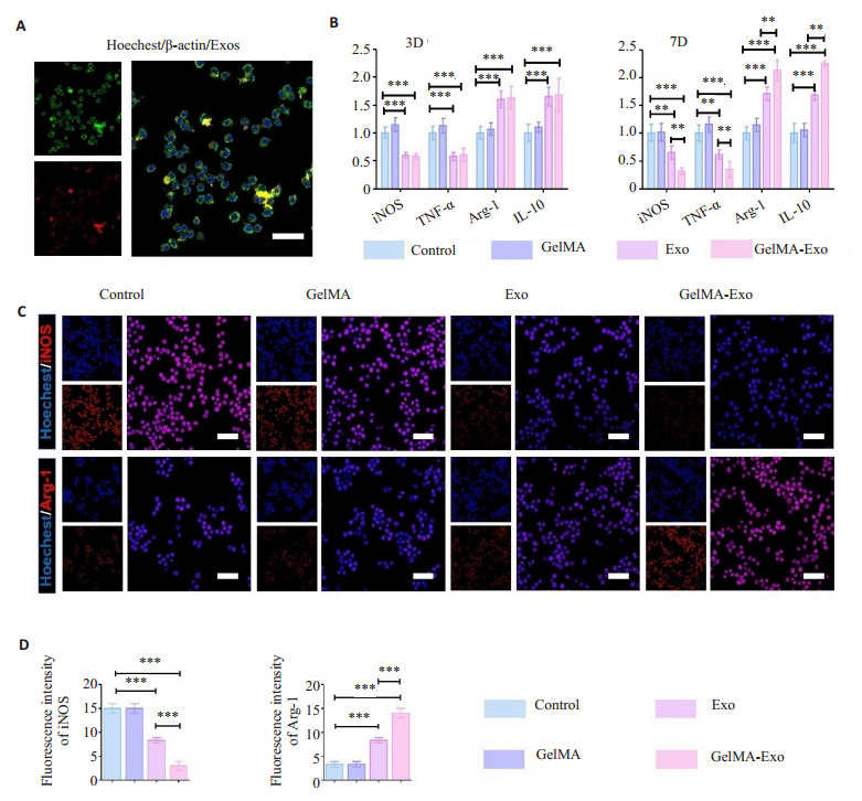
Results: GelMA hydrogel loaded with exosomes significantly promoted M2-type polarization of RAW264.7 cells (P < 0.05). In the Transwell co-culture model, the exosome-loaded GelMA hydrogel significantly promoted the repair of injured chondrocytes by regulating RAW264.7 cell transformation from M1 to M2 (P < 0.05). HE and Masson staining showed that the exosome-loaded hydrogel obviously promoted cartilage repair in the rat models damage.
Conclusion: GelMA hydrogel loaded with bone marrow stem cell-derived exosomes can significantly promote the repair of cartilage damage in rats by improving the immune microenvironment.
目的: 分探究负载骨髓干细胞来源外泌体的GelMA水凝胶对软骨损伤的修复情况。
方法: 首先利用超速离心法分离提取出骨髓干细胞上清液中的外泌体,并通过外泌体透射电镜、粒径分析以及Western blot检测外泌体表面marker。检测负载外泌体的GelMA水凝胶相关性能(包括流变以及电镜)。通过BCA测蛋白法检测外泌体的释放情况,以及通过PKH26红色荧光染料标记外泌体,观察外泌体被RAW264.7细胞吞噬情况。将RAW264.7细胞种在孔板表面,分为空白对照组(Control组)、单纯外泌体组(Exo组)、单纯水凝胶组(GelMA组)、负载外泌体的水凝胶组(GelMA-Exo组)。通过q-PCR以及免疫荧光检测负载外泌体的GelMA水凝胶对于RAW264.7细胞极化的影响。建立材料/RAW264.7细胞/软骨细胞Transwell共培养模型,上室为软骨细胞,下室为材料/ RAW264.7细胞,分组与上面一致,通过q-PCR以及免疫荧光检测损伤软骨细胞的修复情况。构建大鼠膝关节软骨损伤模型,分为假手术组(Sham组)、单纯损伤组(SI组)、单纯水凝胶组(GelMA组)以及负载外泌体的水凝胶组(GelMA-Exo组),通过HE和Masson染色检测软骨修复情况。
结果: 通过对提取的细胞进行多系分化能力的检测,成功获取了骨髓干细胞。通过透射电镜、粒径分析以及Western blot检测,成功分离出骨髓干细胞外泌体。负载外泌体的GelMA水凝胶明显促进RAW264.7细胞向M2型极化(P<0.05)。体外共培养模型表明负载外泌体的GelMA水凝胶能显著促进损伤软骨细胞的修复通过调节RAW264.7细胞由M1型向M2型转化(P<0.05)。HE和Masson染色表明负载骨髓干细胞来源外泌体的GelMA水凝胶明显促进软骨损伤的修复。
结论: 负载骨髓干细胞来源外泌体的GelMA水凝胶能够通过改善免疫微环境明显促进大鼠软骨损伤的修复。
Keywords: GleMA hydrogel; cartilage damage; exosomes; immunoregulation.
Figures


References
-
- Li MZ, Yin H, Yan ZN, et al. The immune microenvironment in cartilage injury and repair. Acta Biomater. 2022;140(7):23–42. - PubMed
-
-
Kulkarni P, Martson A, Vidya R, et al. Pathophysiological landscape of osteoarthritis[M] //Advances in Clinical Chemistry. Amsterdam: Elsevier, 2021: 37-90.
-
MeSH terms
Substances
LinkOut - more resources
Full Text Sources