. 2022 Jun;23(6):184.
doi: 10.3892/ol.2022.13304. Epub 2022 Apr 21.

Tanshinone IIA inhibits cell growth by suppressing SIX1-induced aerobic glycolysis in non-small cell lung cancer cells

Affiliations

Tanshinone IIA inhibits cell growth by suppressing SIX1-induced aerobic glycolysis in non-small cell lung cancer cells

Hailiang Qi et al. Oncol Lett. 2022 Jun.

Abstract

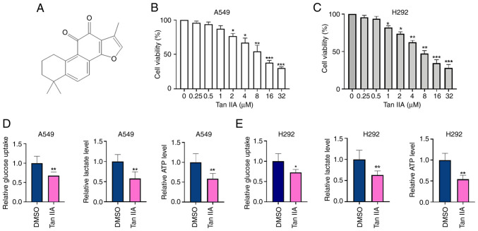
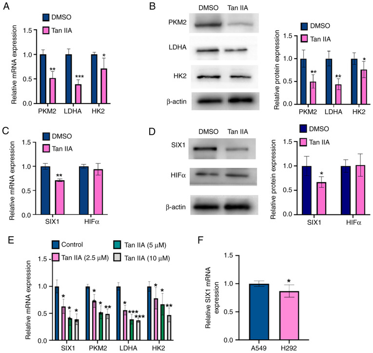
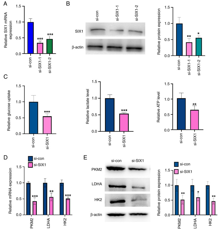
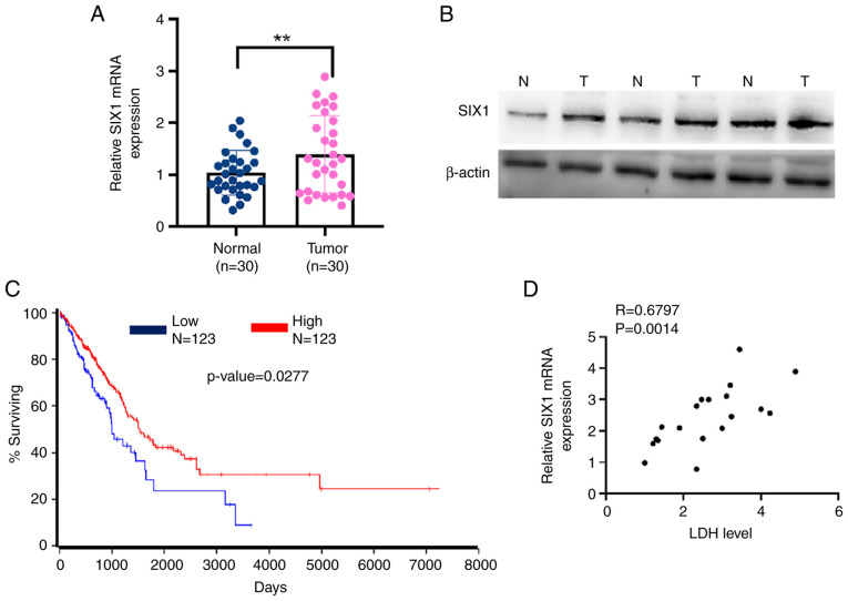
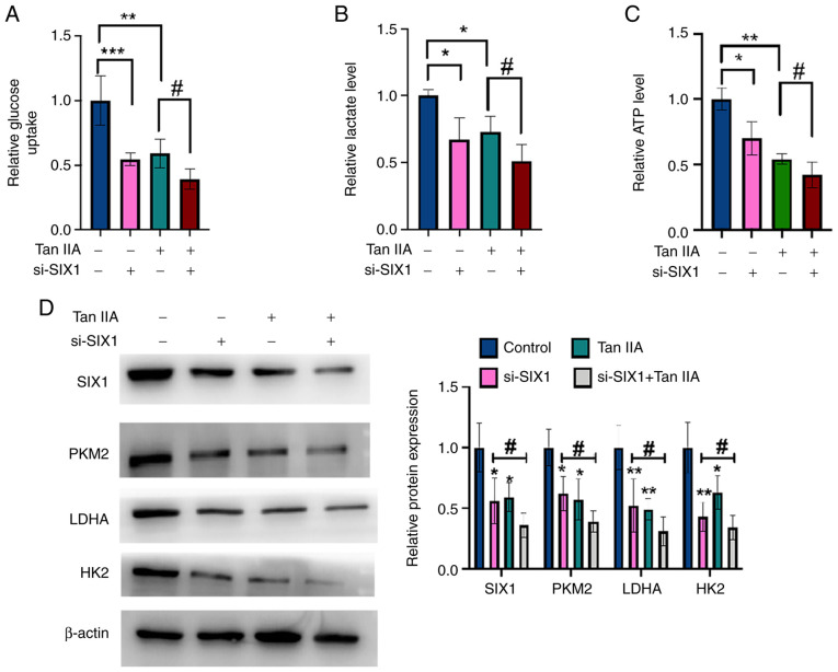
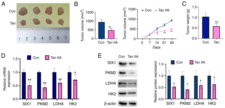
Aerobic glycolysis plays a key role in cancer cell metabolism and contributes to tumorigenesis, including that of non-small cell lung cancer (NSCLC). Tanshinone IIA (Tan IIA), an active compound of Salvia miltiorrhiza, exhibits antitumor properties. Multiple mechanisms are involved in the antitumor action of Tan IIA in lung cancer, such as inhibiting cell growth, promoting cell apoptosis and influencing cellular metabolism. However, the effects of Tan IIA on NSCLC cells and its mechanisms of action remain unclear. The present study shows Tan IIA dose-dependently attenuated the growth of NSCLC cells and in vitro in a dose-dependent manner. Moreover, Tan IIA markedly decreased the ATP level, glucose uptake and lactate production in the NSCLC cells in vitro. Tan IIA also inhibited tumor growth in a xenograft model in vivo. Mechanically, Tan IIA treatment decreased sine oculis homeobox homolog 1 (SIX1) mRNA and protein levels, thus leading to the downregulation of pyruvate kinase isozyme M2, hexokinase 2 and lactate dehydrogenase A (LDHA) expression in A549 cells. SIX1 knockdown with small interfering-RNA inhibited glycolysis in NSCLC cells, suggesting that SIX1 plays a role in the antitumor effect of Tan IIA on NSCLC cells. More importantly, it was demonstrated that SIX1 expression was stimulated in patients with NSCLC and was positively correlated with the LDH serum level. Finally, SIX1 low expression levels predicted the poor prognosis of patients with NSCLC. In conclusion, the present study showed that Tan IIA functioned as an anti-glycolysis agent in NSCLC cells by downregulating SIX1 expression and inhibiting cell proliferation.

Keywords: SIX1; Tan IIA; aerobic glycolysis; non-small cell lung cancer; proliferation.


Conflict of interest statement

The authors declare that they have no competing interests.

Figures

Figure 1.
Figure 1.
Tan IIA inhibits cell proliferation and glycolysis in non-small cell lung cancer cells. (A) The structure of Tan IIA. (B and C) A549 and H292 cells were treated with different concentrations of Tan IIA for 48 h. CCK-8 assay was used to detect cell viability. *P<0.05, **P<0.01 and ***P<0.001 vs. DMSO. (D and E) A549 cells and H292 cells were treated with 5 µM Tan IIA for 48 h. Glucoses uptake, lactate production and ATP level were measured by corresponding assays. *P<0.05 and **P<0.01 vs. DMSO. Tan IIA, Tanshinone IIA.
Figure 2.
Figure 2.
Tan IIA downregulates SIX1 expression in NSCLC cells. (A) A549 cells were treated with 5 µM Tan IIA for 48 h. The mRNA levels of PKM2, LDHA and HK2 were detected by RT-qPCR. *P<0.05, **P<0.01 and ***P<0.001 vs. DMSO. (B) A549 cells were treated as in (A), and the protein levels of PKM2, LDHA and HK2 were detected by western blot analysis. The right panel shows the densitometric analysis of three independent experiments. *P<0.05 and **P<0.01 vs. DMSO. (C) A549 cells were treated as in (A), and the mRNA levels of SIX1 and HIF1α were detected by RT-qPCR. **P<0.01 vs. DMSO. (D) A549 cells were treated as in (A), and the protein levels of SIX1 and HIF1α were detected by western blot analysis. The right panel shows the densitometric analysis of three independent experiments. *P<0.05 vs. DMSO. (E) A549 cells were treated with Tan IIA (2.5, 5 and 10 µM) and mRNA levels of PKM2, LDHA, HK2 and SIX1 were detected by RT-qPCR. *P<0.05, **P<0.01 and ***P<0.001 vs. DMSO. (F) RT-qPCR was used to detect the mRNA level of SIX1 in A549 and H292 cell lines. *P<0.05 vs. A549 cell line. Tan IIA, Tanshinone IIA; SIX1, sine oculis homeobox homolog 1; PKM2, pyruvate kinase subtype M2; HK2, hexokinases 2; LDHA, lactate dehydrogenase A; HIF1α, hypoxia inducible factor 1α; RT-qPCR, reverse transcription-quantitative PCR.
Figure 3.
Figure 3.
Knockdown of SIX1 represses glycolysis in non-small cell lung cancer cells. (A) A549 cells were transfected with si-SIX1 (si-SIX1-1 and si-SIX1-2) or si-con, respectively. The mRNA level of SIX1 was detected by RT-qPCR. ***P<0.001 vs. si-con. (B) A549 cells were treated as in (A), and the protein level of SIX1 was measured by western blot analysis. The right panel shows the densitometric analysis of three independent experiments. *P<0.05 and **P<0.01 vs. si-con. (C) A549 cells were transfected with si-SIX1 or si-con respectively. Glucose uptake, lactate production and ATP level were measured by corresponding assays. **P<0.01 and ***P<0.001 vs. si-con. (D) A549 cells were treated as in (C), and the mRNA levels of PKM2, LDHA and HK2 were detected by RT-qPCR. **P<0.01 and ***P<0.001 vs. si-con. (E) A549 cells were treated as in (C), and the protein levels of PKM2, LDHA and HK2 were detected by western blot analysis. The right panel shows the densitometric analysis of three independent experiments. *P<0.05 and **P<0.01 vs. si-con. Tan IIA, Tanshinone IIA; SIX1, sine oculis homeobox homolog 1; PKM2, pyruvate kinase subtype M2; HK2, hexokinases 2; LDHA, lactate dehydrogenase A; RT-qPCR, reverse transcription-quantitative PCR; si-, small interfering; con, control.
Figure 4.
Figure 4.
SIX1 is upregulated in NSCLC tissues and correlates with LDH level in patients with NSCLC. (A) mRNA levels of SIX1 in NSCLC tissues (n=30) and normal lung tissues (n=30) were detected by reverse transcription-quantitative PCR. **P<0.01 vs. normal lung tissue. (B) The protein levels of SIX1 in NSCLC tissues and normal lung tissues were detected by western blot analysis. (C) The overall survival of NSCLC patients with low or high levels of SIX1 was analyzed by Kaplan-Meier analysis of data from The Cancer Genome Atlas database (P=0.00319). (D) The correlation between mRNA expression level of SIX1 and serum LDH level in patients with NSCLC was analyzed by Pearson's correlation analysis (R=0.6796, P=0.0014). NSCLC, non-small cell lung cancer; SIX1, sine oculis homeobox homolog 1; T, tumor; N, normal; LDH, lactate dehydrogenase.
Figure 5.
Figure 5.
Tan IIA decreases glycolysis in non-small cell lung cancer cells through SIX1. (A) A549 cells were transfected with si-SIX1 or si-con and then treated with Tan IIA or DMSO. Glucose uptake was detected by glucose assay kit. (B) A549 cells were treated as in (A), and lactate level was detected by lactate assay kit. (C) A549 cells were treated as in (A), and ATP level was detected by ATP assay kit. (D) A549 cells were treated as in (A), and the protein levels of SIX1, PKM2, LDHA and HK2 were detected by western blot analysis. The right panel shows the densitometric analysis of three independent experiments. *P<0.05, ***P<0.001, **P<0.01 and ##P<0.05 vs. corresponding control. SIX1, sine oculis homeobox homolog 1; Tan IIA, Tanshinone IIA; si-, small interfering; con, control; PKM2, pyruvate kinase subtype M2; HK2, hexokinases 2; LDHA, lactate dehydrogenase A.
Figure 6.
Figure 6.
Tan IIA suppresses non-small cell lung cancer cell growth in vivo. A549 cells were injected into the right posterior ankle of the nude mice to establish xenograft tumors. From the eighth day, mice were intraperitoneally injected with Tan IIA (20 mg/kg) for 2 weeks. (A) Representative tumor sizes in each group of mice. (B) Tumor volumes were monitored by direct measurement. *P<0.05 and **P<0.01 vs. control group. (C) Xenograft tumor wet weight in each group of mice. **P<0.01 vs. control group. (D) The mRNA levels of SIX1, PKM2, LDHA and HK2 in xenograft tumor tissues were detected by reverse transcription-quantitative PCR. *P<0.05 and **P<0.01 vs. control group. (E) The protein levels of SIX1, PKM2, LDHA and HK2 in xenograft tumor tissues were detected by western blot analysis. The right panel shows the densitometric analysis of three independent experiments. *P<0.05 vs. control group. SIX1, sine oculis homeobox homolog 1; Tan IIA, Tanshinone IIAcon, control; PKM2, pyruvate kinase subtype M2; HK2, hexokinases 2; LDHA, lactate dehydrogenase A.

References

    1. Siegel RL, Miller KD, Jemal A. Cancer statistics, 2020. CA Cancer J Clin. 2020;70:7–30. doi: 10.3322/caac.21601. - DOI - PubMed
    1. Ettinger DS, Wood DE, Aggarwal C, Aisner DL, Akerley W, Bauman JR, Bharat A, Bruno DS, Chang JY, Chirieac LR, et al. NCCN guidelines insights: Non-small cell lung cancer, version 1.2020. J Natl Compr Canc Netw. 2019;17:1464–1472. doi: 10.6004/jnccn.2019.0059. - DOI - PubMed
    1. Chen Z, Fillmore CM, Hammerman PS, Kim CF, Wong KK. Non-small-cell lung cancers: A heterogeneous set of diseases. Nat Rev Cancer. 2014;14:535–546. doi: 10.1038/nrc3775. - DOI - PMC - PubMed
    1. Herbst RS, Morgensztern D, Boshoff C. The biology and management of non-small cell lung cancer. Nature. 2018;553:446–454. doi: 10.1038/nature25183. - DOI - PubMed
    1. Rosner S, Reuss JE, Forde PM. PD-1 blockade in early-stage lung cancer. Annu Rev Med. 2019;70:425–435. doi: 10.1146/annurev-med-050217-025205. - DOI - PubMed