. 2019 Oct 2;9(54):31284-31295.
doi: 10.1039/c9ra04964a. eCollection 2019 Oct 1.

Computational design of enhanced detoxification activity of a zearalenone lactonase from Clonostachys rosea in acidic medium

Affiliations

Computational design of enhanced detoxification activity of a zearalenone lactonase from Clonostachys rosea in acidic medium

Min Lin et al. RSC Adv. .

Abstract

Computational design of pH-activity profiles for enzymes is of great importance in industrial applications. In this research, a computational strategy was developed to engineer the pH-activity profile of a zearalenone lactonase (ZHD101) from Clonostachys rosea to promote its activity in acidic medium. The active site pK a values of ZHD101 were computationally designed by introducing positively charged lysine mutations on the enzyme surface, and the experimental results showed that two variants, M2(D157K) and M9(E171K), increased the catalytic efficiencies of ZHD101 modestly under acidic conditions. Moreover, two variants, M8(D133K) and M9(E171K), were shown to increase the turnover numbers by 2.73 and 2.06-fold with respect to wild type, respectively, though their apparent Michaelis constants were concomitantly increased. These results imply that the active site pK a value change might affect the pH-activity profile of the enzyme. Our computational strategy for pH-activity profile engineering considers protein stability; therefore, limited experimental validation is needed to discover beneficial mutations under shifted pH conditions.


Conflict of interest statement

There are no conflicts of interest to declare.

Figures

Fig. 1
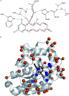
Fig. 1. Mechanism of zearalenone hydrolysis catalyzed by ZHD101. (A) Reaction scheme. (B) Tetrahedral intermediate at the acylation step. The dashed lines indicate hydrogen bonds, and the hashed line represents the covalent bond generated in the nucleophilic attack by S102.
Fig. 2
Fig. 2. ZEN rotamer generation and design sites on ZHD101. (A) Targeted ZEN placement. The dashed lines indicate hydrogen bonds, the hashed line represents the acylation covalent bond, and the bold line between atoms C7′ and C8′ of ZEN denotes the closure position of the lactone loop. The red arrows show the torsion angles that are variated during placing. (B) Locations of sequence selection positions and ZEN on ZHD101. The secondary structure of ZHD101 is shown in cartoon and colored in gray. ZEN and His242 are shown as stick model. The O, N, C atoms are colored in red, slate and gray, respectively. Design positions on enzyme surface are presented as orange spheres, while design positions in active pocket are shown as blue spheres.
Fig. 3
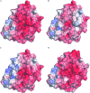
Fig. 3. Electrostatic potential surface of (A) wild type ZHD101; (B) M2(D157K); (C) M8(D133K); (D) M9(E171K). Positive, negative, and neutral potentials are colored in blue, red, and white, respectively. His242 and residues at mutation sites 133, 157, and 171 are shown as stick models.
Fig. 4
Fig. 4. Stereochemical relationship between charges on mutated lysines, ZEN and His242 for (A) M2(D157K); (B) M8(D133K); (C) M9(E171K). ZEN is shown as ball-stick model while the mutated lysines and H242 are shown as stick models. The distances between designated atoms are shown as yellow dashed lines and values are given in angstrom.
Fig. 5
Fig. 5. (A) Circular dichroism spectroscopy measurement of wild type ZHD101 at 25 °C. (B) Thermo unfolding curves of wild type ZHD101 and variants M2, M8, and M9, which are determined at 220 nm from 20 to 95 °C. (C) Catalytic efficiencies of wild type ZHD101 and variants M2, M8, M9 at different pH values (pH 5.5, 7.0, and 8.5).
Fig. 6
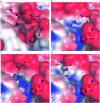
Fig. 6. Electrostatic potential surface at active site of (A) wild type ZHD101; (B) M10(V158H); (C) M11(M154H); (D) M12(V153H). Positive, negative, and neutral potentials are colored in blue, red, and white, respectively. His242 and residues at mutation sites 153, 154, and 158 are shown as stick models.

Similar articles

Cited by

References

    1. Ito S. Kobayashi T. Ara K. Ozaki K. Kawai S. Hatada Y. Extremophiles. 1998;2:185–190. doi: 10.1007/s007920050059. - DOI - PubMed
    1. Shaw A. Bott R. Day A. G. Curr. Opin. Biotechnol. 1999;10:349–352. doi: 10.1016/S0958-1669(99)80063-9. - DOI - PubMed
    1. Cherry J. R. Lamsa M. H. Schneider P. Vind J. Svendsen A. Jones A. Pedersen A. H. Nat. Biotechnol. 1999;17:379–384. doi: 10.1038/7939. - DOI - PubMed
    1. Tynan-Connolly B. M. Nielsen J. E. Protein Sci. 2007;16:239–249. doi: 10.1110/ps.062538707. - DOI - PMC - PubMed
    1. Russell A. J. Fersht A. R. Nature. 1987;328:496–500. doi: 10.1038/328496a0. - DOI - PubMed