

Metabolic Regulation of Fibroblast Activation and Proliferation during Organ Fibrosis
- PMID: 35527985
- PMCID: PMC9021660
- DOI: 10.1159/000522417
Metabolic Regulation of Fibroblast Activation and Proliferation during Organ Fibrosis
Abstract
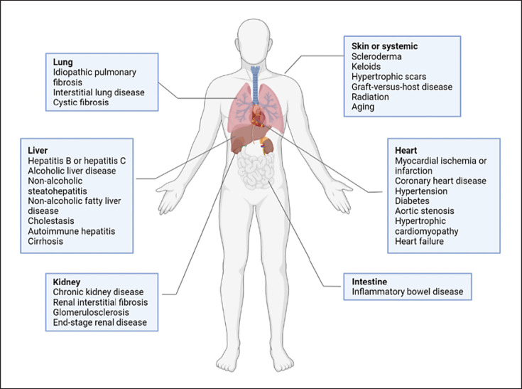
Background: Activated fibroblasts are present in the injury response, tumorigenesis, fibrosis, and inflammation in a variety of tissues and myriad disease types.
Summary: During normal tissue repair, quiescent fibroblasts transform into a proliferative and contractile phenotype termed myofibroblasts and are then lost as repair resolves to form a scar. When excessive levels are reached, activated fibroblasts proliferate and produce large amounts of extracellular matrix, which accumulates in the interstitial space of different organs. This accumulation leads to fibrotic dysfunction and multiple-organ dysfunction syndrome. To date, there are limited effective treatments for these conditions. Cellular metabolism is the cornerstone of all biological activities. Emerging evidence shows that metabolic alterations in fibroblasts are important for the activation process and illness progression. These discoveries, along with current clinical advances showing decreased lung fibrosis after targeting specific metabolic pathways, thus offer new possibilities for therapeutic interventions. The purpose of this review was to summarize the most recent knowledge of the major metabolic changes that occur during fibroblast transition from quiescent to activated states and the evidence linking alterations in fibroblast metabolism to the pathobiology of several common fibrotic diseases and tumor-related diseases.
Key messages: Metabolic disorders are associated with the progression of chronic kidney diseases. Interfering with fibroblast metabolism may be a promising therapeutic strategy for renal fibrosis and other fibrosis-related diseases.
Keywords: Fibroblast activation; Fibroblast proliferation; Metabolic pathways; Renal fibrosis.
Copyright © 2022 by S. Karger AG, Basel.
Conflict of interest statement
Chunsun Dai serves as the associate editor of the Journal of “Kidney Diseases.” The authors have no conflicts of interest to declare.
Figures

References
-
- Ginès P, Castera L, Lammert F, Graupera I, Serra-Burriel M, Allen A, et al. Baltimore, MD: Hepatology; 2021. Population screening for liver fibrosis: towards early diagnosis and intervention for chronic liver diseases. - PubMed
-
- Bos S, Laukens D. Metabolic modulation during intestinal fibrosis. J Dig Dis. 2020;21((6)):319–325. - PubMed
-
- Djudjaj S, Boor P. Cellular and molecular mechanisms of kidney fibrosis. Mol Aspects Med. 2019;65:16–36. - PubMed
Publication types
LinkOut - more resources
Full Text Sources
