

Effects and formulation of silver nanoscaffolds on cytotoxicity dependent ion release kinetics towards enhanced excision wound healing patterns in Wistar albino rats
- PMID: 35528070
- PMCID: PMC9074428
- DOI: 10.1039/c9ra06913e
Effects and formulation of silver nanoscaffolds on cytotoxicity dependent ion release kinetics towards enhanced excision wound healing patterns in Wistar albino rats
Abstract
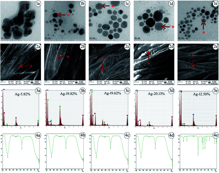
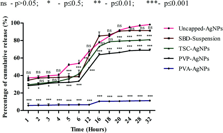
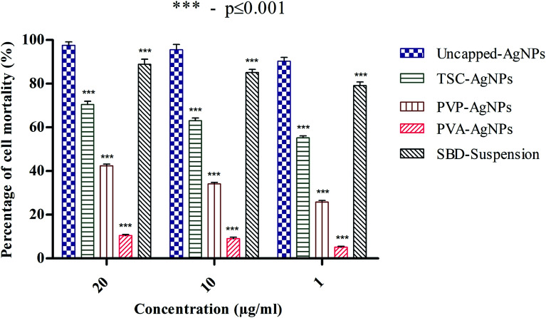
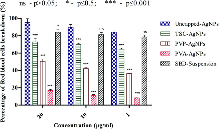
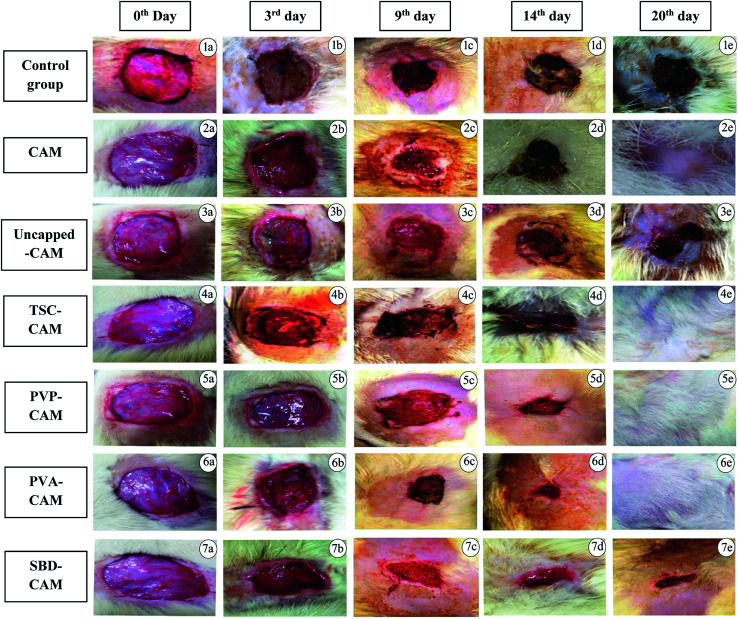
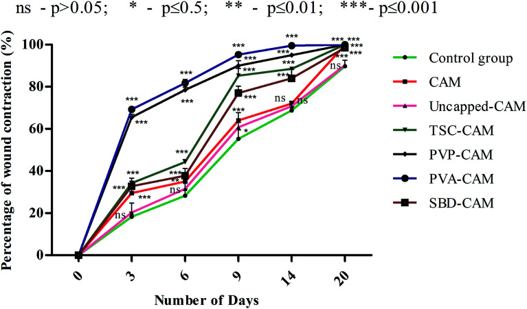
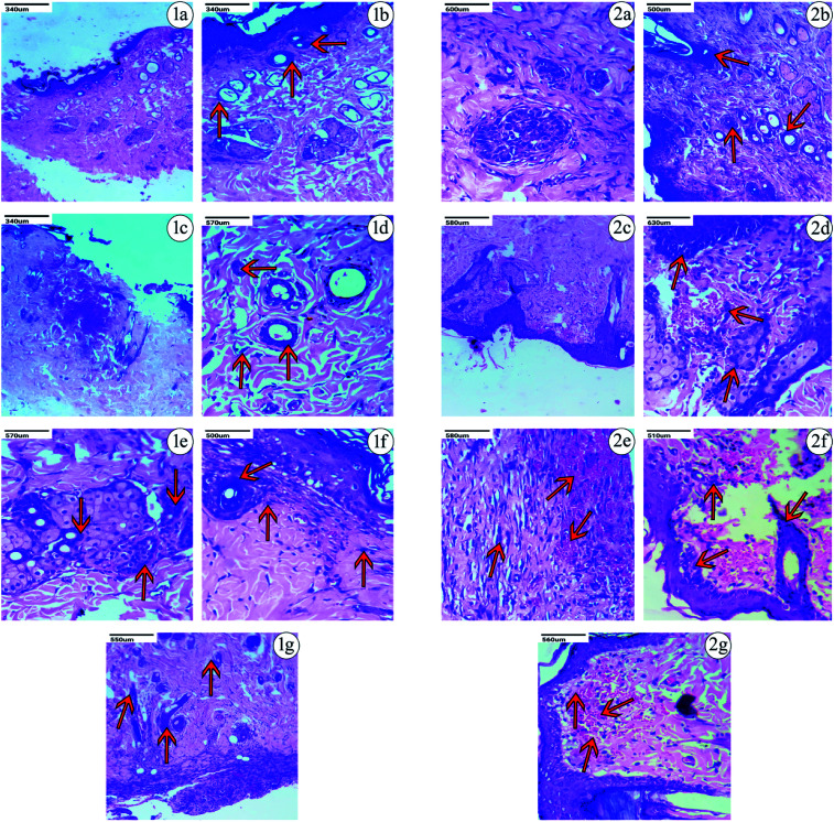
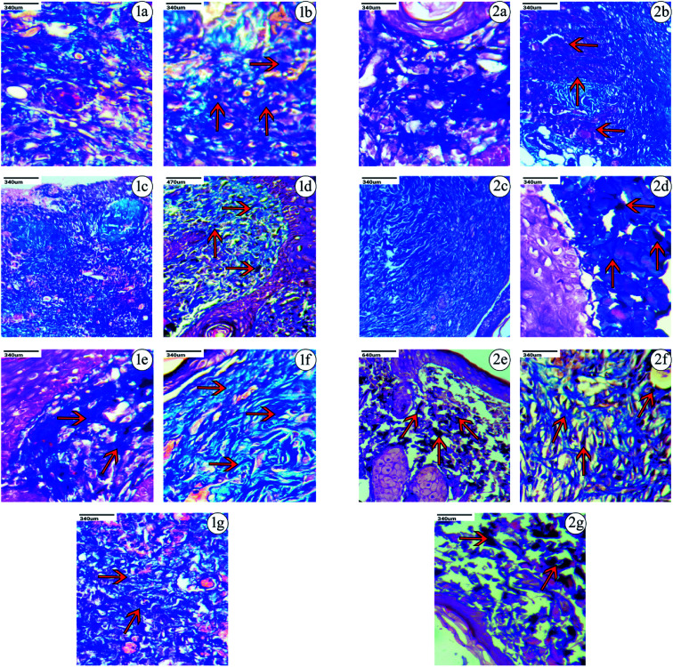
Wound tissue regeneration and angiogenesis are dynamic processes that send physiological signals to the body. Thus, designing novel nanoscaffolds by understanding their surface modifications and toxicological response in a biological system with a potent anti-inflammatory response is a viable solution. In this respect, inspired by the surface chemistry, in the present work we focus on the chemical optimization of silver nanoscaffolds using surface cappings in order to understand their kinetic release behaviour in simulated wound fluids (SWF), to analyze their blood compatibility in human lymphocytes and erythrocytes and then embed them in a chitosan-agarose matrix (CAM) as a productive drug delivery system to evaluate in vivo excision wound tissue regeneration efficiency in Wistar rats. In this regard, polyvinyl alcohol capped silver nanocomposites (PVA-AgNPs) exhibit a dominant antibacterial efficacy with the sustained and controlled release of silver ions and percentage cell mortality and percentage hemolysis of only 10% and 16% compared with uncapped-AgNPs or silver bandaids (SBDs). Also, PVA-AgNP impregnated CAM (PVA-CAM) shows positive effects through their anti-inflammatory and angiogenic properties, with a nearly 95% healing effect within 9 days. The complete development of collagen and fibroblast constituents was also monitored in PVA-CAM by hematoxylin & eosin (H & E) and Masson trichrome (MT) staining. These results provide a clear insight into the development of a potent therapeutic formulation using CAM as a scaffold incorporated with surface functionalized PVA-AgNPs as a bioeffective and biocompatible polymer for the fabrication of efficacious silver wound dressing scaffolds in clinical practice.
This journal is © The Royal Society of Chemistry.
Conflict of interest statement
All authors declare no personal or professional conflicts of interest.
Figures
Similar articles
-
Silver nanoparticle/chitosan oligosaccharide/poly(vinyl alcohol) nanofibers as wound dressings: a preclinical study.Int J Nanomedicine. 2013;8:4131-45. doi: 10.2147/IJN.S51679. Epub 2013 Nov 1. Int J Nanomedicine. 2013. PMID: 24204142 Free PMC article.
-
Biomedical potential of chitosan-silver nanoparticles with special reference to antioxidant, antibacterial, hemolytic and in vivo cutaneous wound healing effects.Biochim Biophys Acta Gen Subj. 2019 Jan;1863(1):241-254. doi: 10.1016/j.bbagen.2018.10.010. Epub 2018 Oct 16. Biochim Biophys Acta Gen Subj. 2019. PMID: 30339915
-
Kaolin-loaded chitosan/polyvinyl alcohol electrospun scaffold as a wound dressing material: in vitro and in vivo studies.J Wound Care. 2020 May 2;29(5):270-280. doi: 10.12968/jowc.2020.29.5.270. J Wound Care. 2020. PMID: 32421483
-
Silver nanoparticles/chitosan oligosaccharide/poly(vinyl alcohol) nanofiber promotes wound healing by activating TGFβ1/Smad signaling pathway.Int J Nanomedicine. 2016 Jan 21;11:373-86. doi: 10.2147/IJN.S91975. eCollection 2016. Int J Nanomedicine. 2016. PMID: 26855575 Free PMC article.
-
Production and Application of Biomaterials Based on Polyvinyl alcohol (PVA) as Wound Dressing.Chem Asian J. 2022 Nov 2;17(21):e202200595. doi: 10.1002/asia.202200595. Epub 2022 Sep 19. Chem Asian J. 2022. PMID: 36066570 Review.
Cited by
-
Significance of Capping Agents of Colloidal Nanoparticles from the Perspective of Drug and Gene Delivery, Bioimaging, and Biosensing: An Insight.Int J Mol Sci. 2022 Sep 10;23(18):10521. doi: 10.3390/ijms231810521. Int J Mol Sci. 2022. PMID: 36142435 Free PMC article. Review.
-
Fabrication of polymeric sorafenib coated chitosan and fucoidan nanoparticles: Investigation of anticancer activity and apoptosis in colorectal cancer cells.Heliyon. 2024 Jul 11;10(14):e34316. doi: 10.1016/j.heliyon.2024.e34316. eCollection 2024 Jul 30. Heliyon. 2024. PMID: 39130440 Free PMC article.
-
Role of medicinal herbs and phytochemicals in post burn management.Inflammopharmacology. 2023 Aug;31(4):1695-1714. doi: 10.1007/s10787-023-01246-5. Epub 2023 May 19. Inflammopharmacology. 2023. PMID: 37204694 Review.
-
Electrical aspects of skin as a pathway to engineering skin devices.APL Bioeng. 2021 Nov 18;5(4):041509. doi: 10.1063/5.0064529. eCollection 2021 Dec. APL Bioeng. 2021. PMID: 34849444 Free PMC article. Review.
-
Fabrication and evaluation of vitamin doped Zno/AgNPs nanocomposite based wheat gluten films: a promising findings for burn wound treatment.Sci Rep. 2023 Sep 26;13(1):16072. doi: 10.1038/s41598-023-43413-2. Sci Rep. 2023. PMID: 37752271 Free PMC article.
References
-
- Paul W., Advances in Wound Healing Materials, Smithers Rapra, Jun 11, 2015
-
- Sezer A. D. and Cevher E., Biopolymers as wound healing materials: challenges and new strategies, Biomaterials Applications for Nanomedicine, Nov 16, 2011, pp. 383–414
-
- Hutchinson J., The wound programme, Centre for Medical Education, Dundee, 1992, vol. 1(2)
-
- Mukherjee S., Nethi S. K. and Patra C., Particulate Technology for Delivery of Therapeutics, 2017, pp. 359–393
LinkOut - more resources
Full Text Sources
Miscellaneous