

Concomitant presentation of spontaneous coronary artery dissection with Takotsubo syndrome: a case report
- PMID: 35528117
- PMCID: PMC9071323
- DOI: 10.1093/ehjcr/ytac172
Concomitant presentation of spontaneous coronary artery dissection with Takotsubo syndrome: a case report
Abstract
Background: Spontaneous coronary artery dissection (SCAD) is still an underdiagnosed condition that requires a detailed assessment of angiographic signs. It also shares similar clinical presentations with Takotsubo syndrome (TTS). The concomitant presentation of SCAD with TTS is a possible occurrence, making it difficult for clinicians to treat and manage.
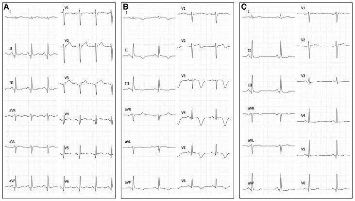
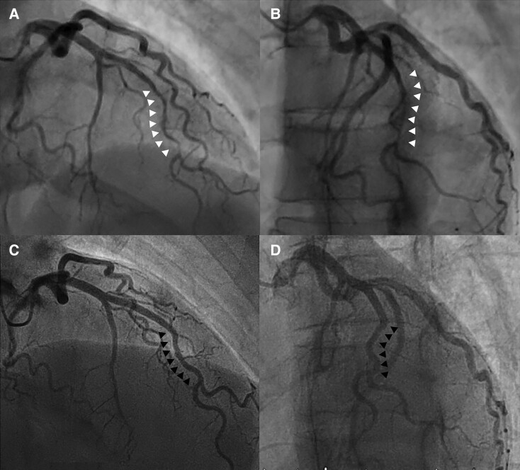
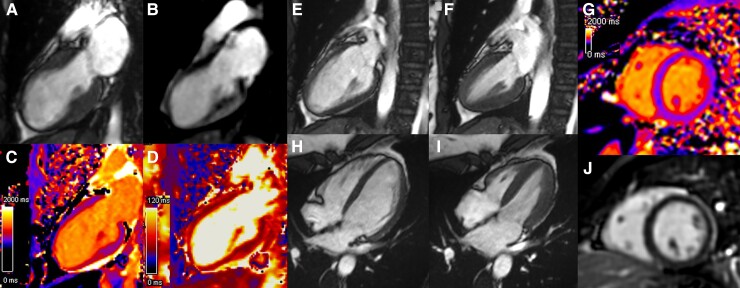
Case summary: This study included a 49-year-old woman with retrosternal chest pain who was admitted to the emergency department. Coronary angiography indicated Type 2A SCAD involving the middle part of the left anterior descending artery, while the left ventriculography indicated a typical left ventricular apical ballooning compatible with TTS. A conservative approach to the management of SCAD was observed. After a 3-month follow-up, the control coronary angiography showed a complete angiographic resolution. The results of the transthoracic echocardiogram (TTE) and cardiac magnetic resonance revealed a complete normalization of the pathological features. The patient remained asymptomatic and showed no recurrence of chest pain.
Discussion: Although TTS and SCAD are commonly observed in patients who share certain characteristics (women, without atheromatous terrain, stress-related factors), it is difficult to establish a pathophysiological link between them. This observation confirms the non-random association of two rare entities of myocardial infarction with no obstructive coronary arteries. Although TTS can be easily diagnosed via non-invasive imaging, the diagnosis of SCAD is more difficult. The findings of this study suggest a concomitant presentation between SCAD and TTS. Although the treatment approach to SCAD is usually conservative, severe forms of this disease require early diagnosis and appropriate treatment.
Keywords: Acute coronary syndrome; Case report; Left ventricular apical ballooning; Spontaneous coronary artery dissection; Takotsubo syndrome.
© The Author(s) 2022. Published by Oxford University Press on behalf of the European Society of Cardiology.
Figures
References
-
- Gori T, Anadol R, Jung F. Tako-Tsubo syndrome, spontaneous coronary dissection and microvascular disease: sex-differences. Clin Hemorheol Microcirc 2019;70:375–379. - PubMed
-
- Combaret N, Gerbaud E, Dérimay F, Souteyrand G, Cassagnes L, Bouajila S, Berrandou T, Rangé G, Meneveau N, Harbaoui B, Lattuca B, Bouatia-Naji N, Motreff P. National French registry of spontaneous coronary artery dissections: prevalence of fibromuscular dysplasia and genetic analyses. EuroIntervention 2021;17:508–515. - PMC - PubMed
-
- Ghadri J-R, Wittstein IS, Prasad A, Sharkey S, Dote K, Akashi YJ, Cammann VL, Crea F, Galiuto L, Desmet W, Yoshida T, Manfredini R, Eitel I, Kosuge M, Nef HM, Deshmukh A, Lerman A, Bossone E, Citro R, Ueyama T, Corrado D, Kurisu S, Ruschitzka F, Winchester D, Lyon AR, Omerovic E, Bax JJ, Meimoun P, Tarantini G, Rihal C, Y.-Hassan S, Migliore F, Horowitz JD, Shimokawa H, Lüscher TF, Templin C. International expert consensus document on Takotsubo syndrome (Part I): clinical characteristics, diagnostic criteria, and pathophysiology. Eur Heart J 2018;39:2032–2046. - PMC - PubMed
-
- Tweet MS, Gulati R, Hayes SN. Spontaneous coronary artery dissection. Curr Cardiol Rep 2016;18:60. - PubMed
-
- Motreff P, Malcles G, Combaret N, Barber-Chamoux N, Bouajila S, Pereira B, Amonchot A, Citron B, Lusson J-R, Eschalier R, Souteyrand G. How and when to suspect spontaneous coronary artery dissection: novel insights from a single-centre series on prevalence and angiographic appearance. EuroIntervention 2017;12:e2236–e2243. - PubMed
Publication types
LinkOut - more resources
Full Text Sources