Osteoblast Derived Exosomes Alleviate Radiation- Induced Hematopoietic Injury
- PMID: 35528209
- PMCID: PMC9070646
- DOI: 10.3389/fbioe.2022.850303
Osteoblast Derived Exosomes Alleviate Radiation- Induced Hematopoietic Injury
Abstract
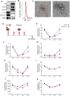
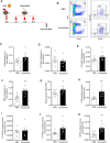
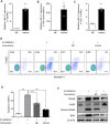
As hematopoietic stem cells can differentiate into all hematopoietic lineages, mitigating the damage to hematopoietic stem cells is important for recovery from overdose radiation injury. Cells in bone marrow microenvironment are essential for hematopoietic stem cells maintenance and protection, and many of the paracrine mediators have been discovered in shaping hematopoietic function. Several recent reports support exosomes as effective regulators of hematopoietic stem cells, but the role of osteoblast derived exosomes in hematopoietic stem cells protection is less understood. Here, we investigated that osteoblast derived exosomes could alleviate radiation damage to hematopoietic stem cells. We show that intravenous injection of osteoblast derived exosomes promoted WBC, lymphocyte, monocyte and hematopoietic stem cells recovery after irradiation significantly. By sequencing osteoblast derived exosomes derived miRNAs and verified in vitro, we identified miR-21 is involved in hematopoietic stem cells protection via targeting PDCD4. Collectively, our data demonstrate that osteoblast derived exosomes derived miR-21 is a resultful regulator to radio-protection of hematopoietic stem cells and provide a new strategy for reducing radiation induced hematopoietic injury.
Keywords: apoptosis resistance; hematopoietic injury; irradiation; miR-21; osteoblast derived exosomes.
Copyright © 2022 Xue, Du, Ling, Song, Yuan, Liu, Sun, Li, Zhong, Wang, Yuan, Jin, Liu, Zhao, Li, Xing, Fan, Liu, Pan, Zhen, Zhao, Yang, Li, Chang and Li.
Conflict of interest statement
The authors declare that the research was conducted in the absence of any commercial or financial relationships that could be construed as a potential conflict of interest.
Figures


Similar articles
-
microRNA-935-modified bone marrow mesenchymal stem cells-derived exosomes enhance osteoblast proliferation and differentiation in osteoporotic rats.Life Sci. 2021 May 1;272:119204. doi: 10.1016/j.lfs.2021.119204. Epub 2021 Feb 10. Life Sci. 2021. PMID: 33581127
-
Bone Marrow Mesenchymal Stem Cells-Derived Exosomal MicroRNA-150-3p Promotes Osteoblast Proliferation and Differentiation in Osteoporosis.Hum Gene Ther. 2021 Jul;32(13-14):717-729. doi: 10.1089/hum.2020.005. Epub 2021 Jan 22. Hum Gene Ther. 2021. PMID: 33107350
-
Cardiac progenitor cell-derived exosomes prevent cardiomyocytes apoptosis through exosomal miR-21 by targeting PDCD4.Cell Death Dis. 2016 Jun 23;7(6):e2277. doi: 10.1038/cddis.2016.181. Cell Death Dis. 2016. PMID: 27336721 Free PMC article.
-
Therapeutic Potential of Hematopoietic Stem Cell-Derived Exosomes in Cardiovascular Disease.Adv Exp Med Biol. 2017;998:221-235. doi: 10.1007/978-981-10-4397-0_15. Adv Exp Med Biol. 2017. PMID: 28936743 Review.
-
Therapeutic applications of stem cell-derived exosomes in radiation-induced lung injury.Cancer Cell Int. 2024 Dec 18;24(1):403. doi: 10.1186/s12935-024-03595-9. Cancer Cell Int. 2024. PMID: 39695650 Free PMC article. Review.
Cited by
-
Remodeling of the bone marrow microenvironment during acute myeloid leukemia progression.Ann Transl Med. 2024 Aug 1;12(4):63. doi: 10.21037/atm-23-1824. Epub 2024 Jan 15. Ann Transl Med. 2024. PMID: 39118939 Free PMC article. Review.
-
Protection of the hematopoietic system against radiation-induced damage: drugs, mechanisms, and developments.Arch Pharm Res. 2022 Aug;45(8):558-571. doi: 10.1007/s12272-022-01400-7. Epub 2022 Aug 11. Arch Pharm Res. 2022. PMID: 35951164 Review.
-
New Properties of a Well-Known Antioxidant: Pleiotropic Effects of Human Lactoferrin in Mice Exposed to Gamma Irradiation in a Sublethal Dose.Antioxidants (Basel). 2022 Sep 18;11(9):1833. doi: 10.3390/antiox11091833. Antioxidants (Basel). 2022. PMID: 36139907 Free PMC article.
References
-
- Alessio N., Del Gaudio S., Capasso S., Di Bernardo G., Cappabianca S., Cipollaro M., et al. (2015). Low Dose Radiation Induced Senescence of Human Mesenchymal Stromal Cells and Impaired the Autophagy Process. Oncotarget 6 (10), 8155–8166. 10.18632/oncotarget.2692 PubMed Abstract | 10.18632/oncotarget.2692 | Google Scholar - DOI - DOI - PMC - PubMed
-
- Alessio N., Esposito G., Galano G., De Rosa R., Anello P., Peluso G., et al. (2017). Irradiation of Mesenchymal Stromal Cells with Low and High Doses of Alpha Particles Induces Senescence And/or Apoptosis. J. Cel. Biochem. 118 (9), 2993–3002. 10.1002/jcb.25961 PubMed Abstract | 10.1002/jcb.25961 | Google Scholar - DOI - DOI - PubMed
-
- Arai F., Suda T. (2007). Maintenance of Quiescent Hematopoietic Stem Cells in the Osteoblastic Niche. Ann. New York Acad. Sci. 1106, 41–53. 10.1196/annals.1392.005 PubMed Abstract | 10.1196/annals.1392.005 | Google Scholar - DOI - DOI - PubMed
-
- Arora R., Chawla R., Marwah R., Kumar V., Goel R., Arora P., et al. (2010). Medical Radiation Countermeasures for Nuclear and Radiological Emergencies: Current Status and Future Perspectives. J. Pharm. Bioall Sci. 2 (3), 202–212. 10.4103/0975-7406.68502 PubMed Abstract | 10.4103/0975-7406.68502 | Google Scholar - DOI - DOI - PMC - PubMed
-
- Bai J., Wang Y., Wang J., Zhai J., He F., Zhu G. (2020). Irradiation-induced Senescence of Bone Marrow Mesenchymal Stem Cells Aggravates Osteogenic Differentiation Dysfunction via Paracrine Signaling. Am. J. Physiology-Cell Physiol. 318 (5), C1005–c1017. 10.1152/ajpcell.00520.2019 10.1152/ajpcell.00520.2019 | Google Scholar - DOI - DOI - PubMed
LinkOut - more resources
Full Text Sources
Research Materials

