. 2022 Mar;2(3):231-242.
doi: 10.1038/s43587-022-00187-y. Epub 2022 Mar 18.

Age-associated impairment of T cell immunity is linked to sex-dimorphic elevation of N-glycan branching

Affiliations

Age-associated impairment of T cell immunity is linked to sex-dimorphic elevation of N-glycan branching

Haik Mkhikian et al. Nat Aging. 2022 Mar.

Abstract

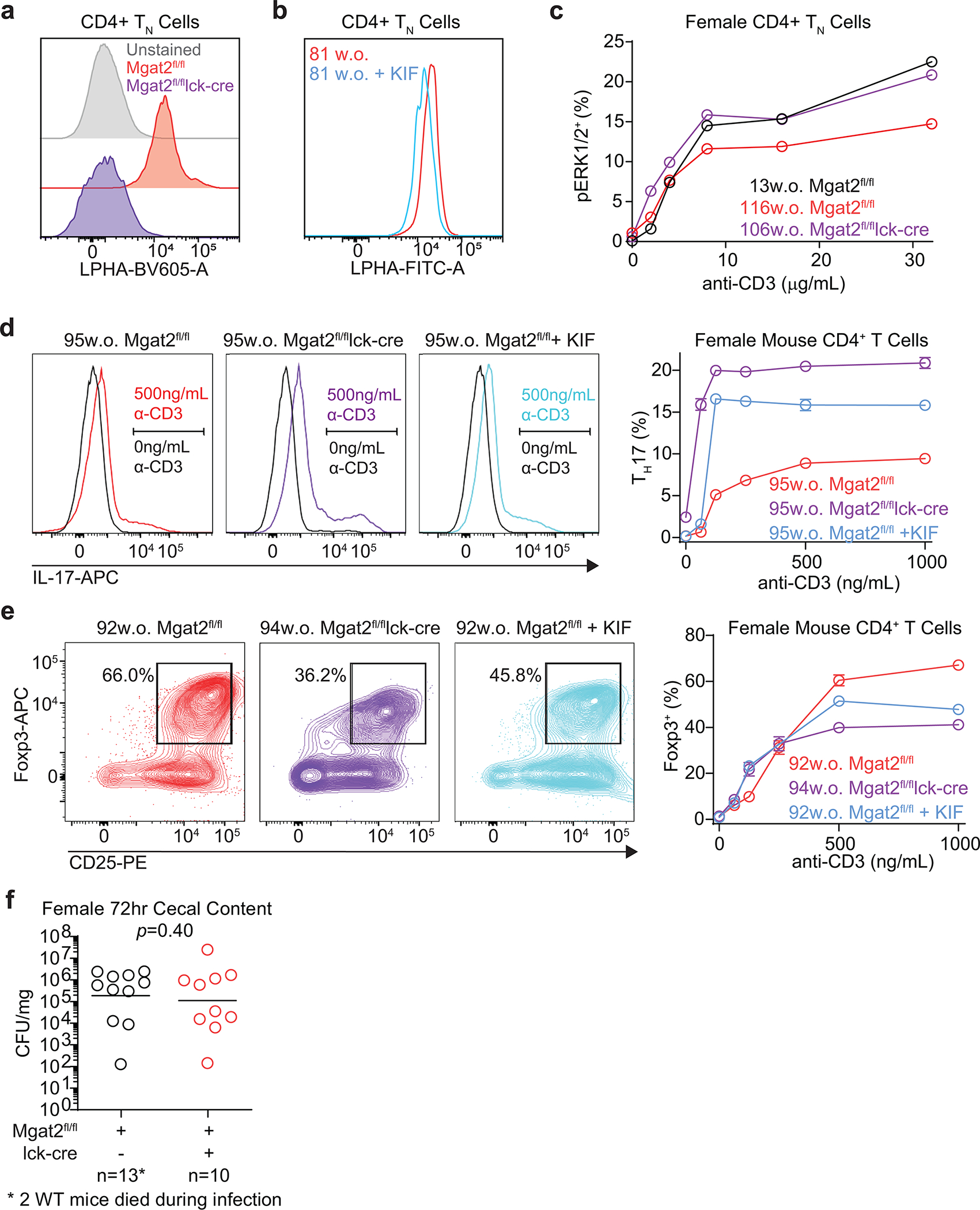
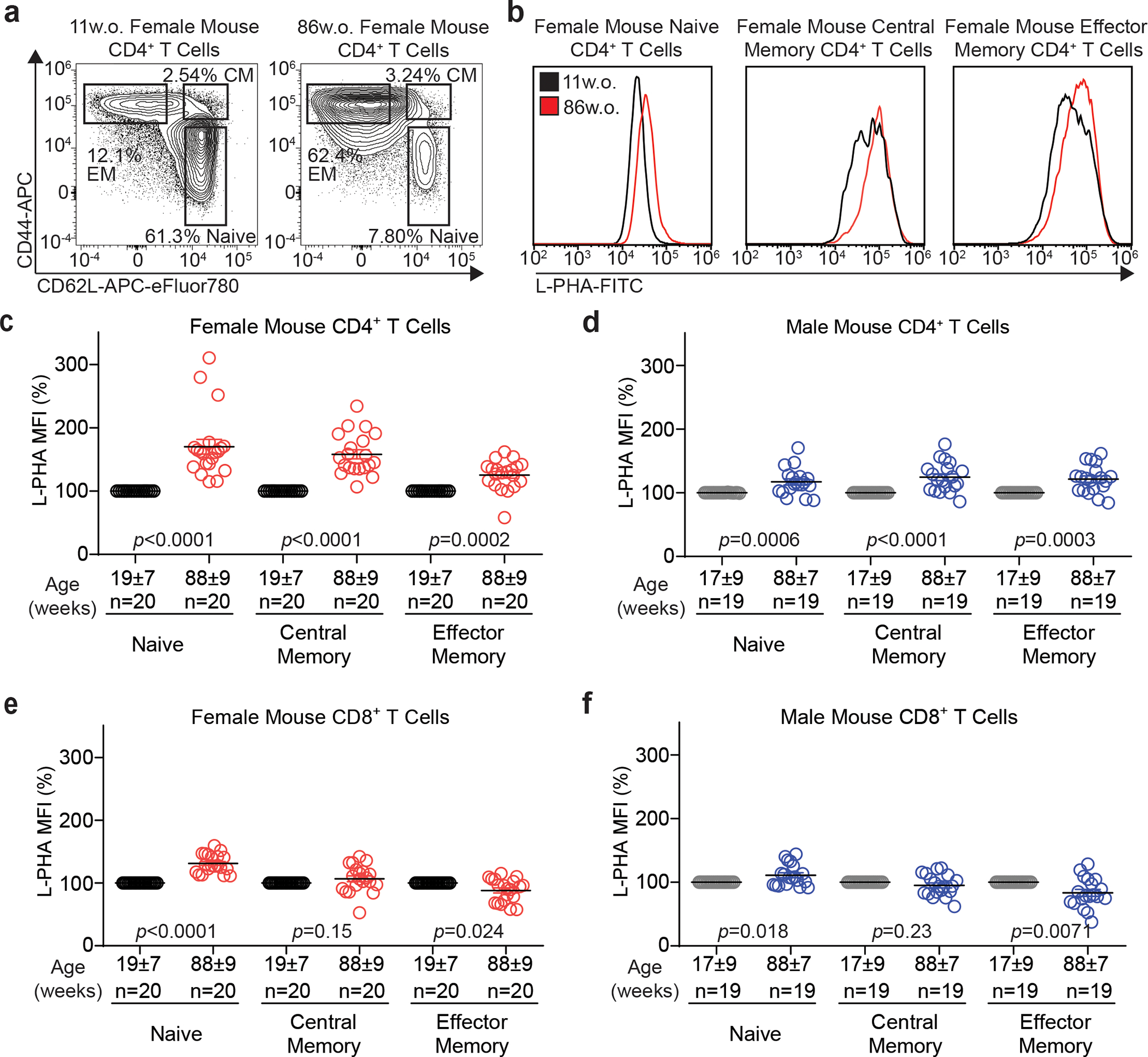
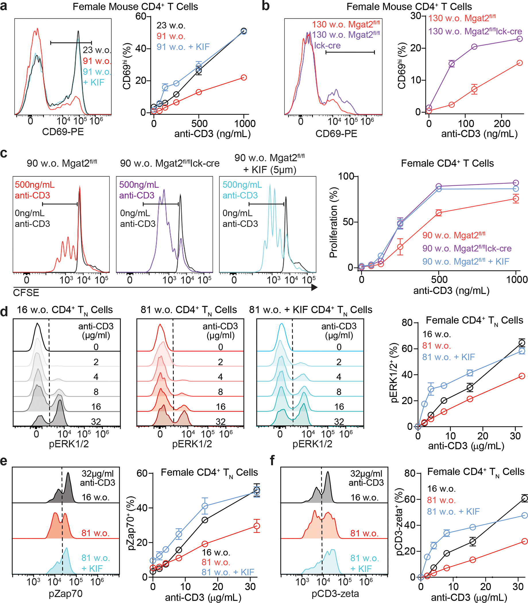
Impaired T cell immunity with aging increases mortality from infectious disease. The branching of Asparagine-linked glycans is a critical negative regulator of T cell immunity. Here we show that branching increases with age in females more than males, in naïve more than memory T cells, and in CD4+ more than CD8+ T cells. Female sex hormones and thymic output of naïve T cells (TN) decrease with age, however neither thymectomy nor ovariectomy altered branching. Interleukin-7 (IL-7) signaling was increased in old female more than male mouse TN cells, and triggered increased branching. N-acetylglucosamine, a rate-limiting metabolite for branching, increased with age in humans and synergized with IL-7 to raise branching. Reversing elevated branching rejuvenated T cell function and reduced severity of Salmonella infection in old female mice. These data suggest sex-dimorphic antagonistic pleiotropy, where IL-7 initially benefits immunity through TN maintenance but inhibits TN function by raising branching synergistically with age-dependent increases in N-acetylglucosamine.

Keywords: Immunosenescence; N-acetyglucosamine; N-glycan branching; N-glycosylation; T cell, infection; aging; immunity; interleukin-7.


Conflict of interest statement

Competing Interests Statement JD and MD are named as inventors on a patent application that describes GlcNAc as a biomarker for multiple sclerosis. JD and MD are named as inventors on a patent for use of GlcNAc in MS. The remaining authors declare no competing interests.

Figures

Extended Data Figure 1.
Extended Data Figure 1.. Mouse T cells display a sex-dimorphic increase in N-glycan branching with age.
a) Fructose 6-phosphate may be metabolized by glycolysis or enter the hexosamine pathway to supply UDP-GlcNAc to the Golgi branching enzymes Mgat1, 2, 4 and 5, which generate mono-, bi-, tri-, and tetra-antennary GlcNAc branched glycans, respectively. The branching enzymes utilize UDP-GlcNAc with declining efficiency such that both Mgat4 and Mgat5 are limited for branching by the metabolic production of UDP-GlcNAc. Small molecule inhibitor kifunensine (KIF) can be used to eliminate N-glycan branching. Plant lectin L-PHA (Phaseolus vulgaris, leukoagglutinin) binding sites are also shown. Abbreviations: OT, oligosaccharyltransferases; GI, glucosidase I; GII, glucosidase II; MI, mannosidase I; MII, mannosidase II; Mgat, N-acetylglucosaminyltransferase; GalT3, galactosyltransferase 3; iGnT, i-branching enzyme β1,3-N-acetylglucosaminyltransferase; KIF, kifunensine; GlcNAc, N-acetylglucosamine; UDP, uridine diphosphate; Km, Michaelis constant of the enzyme. b) The gating strategy is demonstrated for CD4+ TN cells. Lymphocytes were first gated on singlets, followed by gating on CD3+CD4+CD8CD25CD62L+CD44 cells by sequential steps. c, d) Splenocytes from five young and old mice were analyzed for L-PHA (c) or ConA (d) binding by flow cytometry, gating on the indicated CD4+ T cell subsets. Absolute geometric mean fluorescence intensity (MFI) is shown to allow direct comparison between naïve and memory subsets. e-i) CD4+ TN cells (e-g) CD19+ B cells (h) or thymocytes (i) were obtained from the lymph node (e), spleen (f-h) or thymus (i) of female (e, g-i) or male (f-h) mice of the indicated ages, and analyzed for L-PHA binding by flow cytometry. Absolute or normalized geometric mean fluorescence intensity (MFI) are shown. Each symbol represents a single mouse. P-values by two-tailed Mann-Whitney (c, d) or two-tailed Wilcoxon (e, h, i). Error bars indicate mean ± s.e.m.
Extended Data Figure 2.
Extended Data Figure 2.. Elevated IL-7 signaling increases N-glycan branching in old female naïve T cells.
a) Representative flow cytometry plots of donor and recipient cells post-adoptive transfer. b) Negatively selected CD4+ T cells were FACS sorted for TN (CD62L+CD44) and TEM (CD62LCD44+) populations. Representative flow cytometry demonstrating purity of sorted cells used for RNA-seq. c) Principal component analysis (PCA) of RNA-seq data comparing gene expression in CD4+ TN and CD4+ TEM cells from young male (7–8 weeks old), young female (10–11 weeks old), old male (83–86 weeks old) and old female (85 weeks old) mice. Three biological replicates were performed for each group. d) Out of 24062 genes analyzed by RNAseq, 158 DEGs were identified when comparing young and old CD4+ TN cells in females, 192 DEGs were identified in males, and 44 DEGs were shared. e) Young and old naïve CD4+ T cell mRNA expression of N-glycan pathway genes by real-time qPCR. f) Flow cytometric analysis of IL7Rα in ex vivo CD4+ TN cells from young and old male mice. g) L-PHA versus IL7Rα expression in ex vivo CD4+ TN cells from old male and female mice. h, i) Flow cytometric analysis of IL7Rα in ex vivo young and old CD4+ TEM cells from female (h) and male (i) mice. j-l) C57BL/6 mice of the indicated ages were injected intraperitoneal with either isotype control (1.5mg) or anti-IL-7 antibody (M25, 1.5mg) three times per week for two (j) or four (k, l) weeks, and then analyzed for L-PHA or IL7Rα expression in blood (k) or spleen (j, l). Each symbol represents a single mouse unless specified otherwise. P-values determined by one-tailed Wilcoxon (f), linear regression (g), two-tailed Wilcoxon (h, i), Kruskal-Wallis with Dunn’s multiple comparisons test (j), or one-tailed Mann-Whitney (k, l). Error bars indicate mean ± s.e.m.
Extended Data Figure 3.
Extended Data Figure 3.. Thymectomy and ovariectomy are insufficient to drive increases in N-glycan branching.
a-i) Female mice underwent thymectomy (a-c), ovariectomy (d-g), both surgeries (h, i), or corresponding sham procedures at the age of 9 weeks. Flow cytometry on blood at the indicated time points post-procedure was performed to detect percentage of CD4+ TN cells (a), IL7Rα expression (b, d, g, h), or L-PHA binding (c, e, f, i), gating on CD4+ TN cells. Absolute or normalized geometric mean fluorescence intensity (MFI) are shown. Each symbol at a particular time point represents a single mouse. P-values determined by one-tailed Mann-Whitney (b, d, g, h). Error bars indicate mean ± s.e.m
Extended Data Figure 4.
Extended Data Figure 4.. Age-dependent increases in N-glycan branching suppress pro-inflammatory T cell function.
a) L-PHA binding of splenocytes from Mgat2fl/fl and Mgat2fl/fllck-cre female mice, gated on CD4+ TN cells. b) Splenocytes from an old mouse were treated with or without kifunensine for 24 hours, then analyzed for L-PHA binding by flow cytometry, gating on CD4+ TN cells. c) Splenocytes from female mice of the indicated ages and genotypes were activated with plate bound anti-CD3 for 15 minutes. Following fixation and permeabilization, phospho-ERK1/2 induction was analyzed in CD4+ TN cells by flow cytometry, gating additionally on L-PHA negative cells in Mgat2fl/fl/lck-cre mice. d, e) Flow cytometry analysis of purified mouse splenic CD4+ T cells activated with anti-CD3 and anti-CD28 for 4 days with TH17 (TGFβ+IL-6+IL-23) or iTreg (TGFβ) inducing conditions, gating additionally on L-PHA negative cells in Mgat2fl/fl/lck-cre mice. f) Female Mgat2fl/fl and Mgat2fl/fllck-cre mice were inoculated with streptomycin (0.1ml of a 200mg/ml solution in sterile water) intragastrically one day prior to inoculation with S. Typhimurium (5×10⁸ colony forming units, CFU per mouse) by oral gavage. CFU in the cecal content was determined 72 hours after infection. Data shown are representative of two (c), or at least three (a, b, d, e) independent experiments. P-values by one-tailed Mann-Whitney (f). Error bars indicate mean ± s.e.m (c, d, e) or geometric mean (f).
Extended Data Figure 5.
Extended Data Figure 5.. Age-dependent increases in N-glycan branching suppress T cell activity in human females.
a-d) Human PBMCs from healthy females (a, c) or males (b, d) as indicated were analyzed for L-PHA binding by flow cytometry gating on CD8+ T cells (a, b) or CD19+ B cells (c, d). e, f) CD4+ TN cells (CD45RA+CD45RO) from healthy females (e) and males (f) under the age of 65 were analyzed for L-PHA binding by flow cytometry. g) Human PBMCs from young (22–38 years old) and old (90–94 years old) female subjects were analyzed for L-PHA binding on CD4+ TN cells (CD45RA+CD45RO) before or after 96 hours of culture in complete media. Shown is the ratio or each old subject over the average of the young at the two timepoints. Each symbol represents a single individual. R2 and p-values by linear regression (a-f) or by paired one-tailed t test, following passage of Shapiro-Wilk normality test (g). Error bars indicate mean ± s.e.m.
Extended Data Figure 6.
Extended Data Figure 6.. N-acetylglucosamine and IL-7 synergize to raise N-glycan branching in human T cells.
a, b) PBMCs from nine healthy female donors (28–45 years old) were cultured with or without rhIL-7 (50ng/ml) and/or GlcNAc (10mM or 40mM) for 9 days, then analyzed for L-PHA binding by flow cytometry, gating on CD4+ TEM (CD45RACD45RO+CCR7) cells (a) or CD8+ TEM (CD45RACD45RO+CCR7) cells (b). c) Mouse plasma from female mice of the indicated ages was analyzed for HexNAc levels by LC-MS/MS. d, e) Flow cytometric analysis of human PBMCs stimulated by anti-CD3 in the presence or absence of kifunensine as indicated for 24 hours to analyze for activation marker CD69 (d) or 72 hours to assess proliferation by CFSE dilution (e), gating on CD4+ T cells. f) Female human PBMCs were treated in vitro with kifunensine for 24 hours, followed by analysis of L-PHA binding on CD4+ TN cells by flow cytometry. Data shown is representative of three independent experiments with different donors. g) Female human PBMCs were treated in vitro with kifunensine for up to four days, followed by analysis of L-PHA binding on CD4+ TN cells by flow cytometry. P-values by Kruskal-Wallis with Dunn’s multiple comparisons test (a, b), two-tailed Mann-Whitney (c), and one-tailed Mann-Whitney (d, e). Error bars indicate mean ± s.e.m.
Figure 1.
Figure 1.. Mouse T cells display a sex-dimorphic increase in N-glycan branching with age.
a) Splenic T cells from representative young and old female mice were stained for naïve and memory markers, demonstrating naïve (CD62L+CD44), central memory (CD62L+CD44+), and effector memory (CD62LCD44) subsets. b) L-PHA staining histograms of young and old female CD4+ naïve, central memory, and effector memory T cell subsets. c-f) Splenic T cells from 20 young (range 7–32 weeks) and 20 old (range 74–113 weeks) female, as well as 19 young (range 7–31 weeks) and 19 old (range 80–100 weeks) male mice were analyzed in pairs by flow cytometry for L-PHA binding on naïve, central memory, or effector memory subsets. Analyses of female CD4+ (c), male CD4+ (d), female CD8+ (e), and male CD8+ (f) T cells are shown. Each symbol represents a single mouse. Each old mouse was normalized to its young control. Age of mice in weeks with standard deviation is shown. P-values by two-tailed Wilcoxen test. Error bars indicate mean ± s.e.m.
Figure 2.
Figure 2.. Elevated IL-7 signaling increases N-glycan branching in old female CD4+ TN cells.
a) L-PHA flow cytometry of splenocytes gated on CD4+ TN cells from young (12±4 weeks old) and old (86±3 weeks old) female mice immediately ex vivo and after 72 hours of rest in culture. Shown is L-PHA MFI ratio in old over young. b) Old and young female CD45.2+ CD4+ TN cells were adoptively transferred into young female CD45.1+ recipient mice and analyzed by flow cytometry for L-PHA binding pre- and 2 weeks post-transfer, gating on CD4+ TN cells. c, d) Flow cytometric analysis of IL7Rα (c) and pSTAT5 (d) in ex vivo naïve (c, d) and total memory (CD44+ ) (d) CD4+ T cells from young and old female mice. e, f) Flow cytometric analysis of L-PHA binding on CD4+ TN cells from young (e) and old (f) female (red) and male (blue) mice treated with or without rhIL-7 (50ng/ml) in vitro for 72 hours. g) L-PHA flow cytometry gating on splenic CD4+ TN cells from young female mice following intraperitoneal injections of isotype control (1.5μg) or rhIL-7/M25 complex on days 1, 3, and 5. h-k) Flow cytometry gating on CD4+ TN cells from the blood (h) or spleen (i-k) of female mice following intraperitoneal injections of either isotype control (1.5mg) or anti-IL-7 antibody (M25, 1.5mg) 3 times a week for two (h, i) or four (j, k) weeks. Normalized geometric mean fluorescence intensity (MFI) is shown. Each symbol represents a single mouse. P-values determined by two-tailed Wilcoxon (a, e, f) Kruskal-Wallis with Dunn’s multiple comparisons test (b, g, i), one-tailed Wilcoxon (c, d) or one-tailed Mann-Whitney (h, j, k). In panel (h) p-values indicate comparison of 12 w.o. isotype control group to 82 w.o. anti-IL7 treatment group. Error bars indicate mean ± s.e.m.
Figure 3.
Figure 3.. Age dependent increases in N-glycan branching suppress T cell function in female mice.
a-c) Splenocytes from female mice of the indicated ages and genotypes were activated with plate bound anti-CD3e for 24 (a, b) or 72 (c) hours in the presence or absence of 5μM kifunensine. Total CD4+ T cells were analyzed for CD69 expression (a, b) or 5, 6-carboxyfluorescein diacetate succinimidyl ester (CFSE) dilution (c) by flow cytometry, gating additionally on L-PHA negative cells in Mgat2fl/fl/lck-cre mice. d-f) Splenocytes from female mice of the indicated ages were pretreated with or without kifunensine for 24 hours, followed by activation with plate bound anti-CD3e for 15 minutes. Following fixation and permeabilization, phospho-ERK (d), phospho-Zap70 (e) or phospho-CD3zeta (f) induction was analyzed in CD4+ TN cells by flow cytometry. Data are representative of at least three independent experiments. Error bars indicate mean ± s.e.m.
Figure 4.
Figure 4.. Elevated N-glycan branching in T cells suppresses immune response to Salmonella in old female mice.
a-e) Mgat2fl/fl and Mgat2fl/fllck-cre female mice were pre-treated with streptomycin intragastrically one day prior to inoculation with S. Typhimurium (5×10⁸ colony forming units (CFU) per mouse). CFU in Peyer’s patches (a), mesenteric lymph nodes (MLN; b), and spleen (c) were enumerated 72 hours after infection. Total IL-17+ (d) and Foxp3+ (e) cells within the gut were detected by flow cytometry and normalized to the average number of cells from Mgat2fl/fl mice. Each symbol represents a single mouse. P-values by one-tailed Mann-Whitney. Bars indicate geometric mean (a-c) or mean ± s.e.m (d, e).
Figure 5.
Figure 5.. N-glycan branching increases with age in human T cells.
a-e) L-PHA flow cytometry of human PBMCs from healthy females (a-c) and males (d, e), gating on naïve CD4+ (CD45RA+ CD45RO; b, d) or memory CD4+ (CD45RACD45RO+; c, e) T cells. Data were normalized to a reference control across experiments. Each symbol represents a single individual. R2 and p-values by linear regression. Error bars indicate mean ± s.e.m.
Figure 6.
Figure 6.. Serum N-acetylglucosamine increases with age and synergizes with IL-7 to raise N-glycan branching in human T cells.
a, b) PBMCs from healthy female donors (28–45 years old) were cultured with or without rhIL-7 (50ng/ml) and/or GlcNAc (10mM or 40mM) for 9 days, then analyzed for L-PHA binding by flow cytometry, gating on CD4+ TN cells (a) or CD8+ TN cells (b). c, d) Human serum from healthy female (c; 19–98 years old; 45±20 years old) or male (d; 20–92 years old; 38±19 years old) donors of the indicated ages was analyzed for HexNAc levels by LC-MS/MS. e, f) Correlation of serum HexNAc with L-PHA binding on CD4+ TN cells in healthy female (e; 19–98 years old; 44±20 years old) or male (f; 20–92 years old; 40±20 years old) donors. g) Flow cytometric analysis of intracellular pERK1/2 in 90–94 year old female human CD4+ TN cells stimulated with plate bound anti-CD3 for 15 minutes. Cells were pre-treated with or without 10μM kifunensine for 24 hours prior to stimulation. Each symbol represents a single individual. Normalized geometric mean fluorescence intensity (MFI) is shown (a, b, e, f). P-values by Kruskal-Wallis with Dunn’s multiple comparisons test (a,b), linear regression (c-f), and one-tailed Mann-Whitney (g). Error bars indicate mean ± s.e.m.

Comment in

  • Time isn't kind to female T-cells.
    McGill CJ, Benayoun BA. McGill CJ, et al. Nat Aging. 2022 Mar;2(3):189-191. doi: 10.1038/s43587-022-00185-0. Epub 2022 Mar 18. Nat Aging. 2022. PMID: 36157102 Free PMC article.

Similar articles

Cited by

References

    1. Goronzy JJ & Weyand CM Mechanisms underlying T cell ageing. Nat Rev Immunol 19, 573–583, doi:10.1038/s41577-019-0180-1 (2019). - DOI - PMC - PubMed
    1. Nikolich-Zugich J The twilight of immunity: emerging concepts in aging of the immune system. Nature immunology 19, 10–19, doi:10.1038/s41590-017-0006-x (2018). - DOI - PubMed
    1. Centers for Disease, C. & Prevention. Estimates of deaths associated with seasonal influenza --- United States, 1976–2007. MMWR Morb Mortal Wkly Rep 59, 1057–1062 (2010). - PubMed
    1. Wiersinga WJ, Rhodes A, Cheng AC, Peacock SJ & Prescott HC Pathophysiology, Transmission, Diagnosis, and Treatment of Coronavirus Disease 2019 (COVID-19): A Review. JAMA 324, 782–793, doi:10.1001/jama.2020.12839 (2020). - DOI - PubMed
    1. Chen PL et al. Non-typhoidal Salmonella bacteraemia in elderly patients: an increased risk for endovascular infections, osteomyelitis and mortality. Epidemiol Infect 140, 2037–2044, doi:10.1017/S0950268811002901 (2012). - DOI - PubMed

Publication types