. 2019 Aug 13;9(43):25107-25118.
doi: 10.1039/c9ra05037j. eCollection 2019 Aug 8.

Ginsenoside Rk1 inhibits cell proliferation and promotes apoptosis in lung squamous cell carcinoma by calcium signaling pathway

Affiliations

Ginsenoside Rk1 inhibits cell proliferation and promotes apoptosis in lung squamous cell carcinoma by calcium signaling pathway

Xining An et al. RSC Adv. .

Abstract

Ginsenoside Rk1 (Rk1) is a rare saponin extracted from Sun Ginseng (SG) and has been shown to have an anti-tumor effect; however, the potential role of its in lung squamous cell carcinoma remains elusive. In this study, we investigated the anti-proliferative activity and involved mechanism of Rk1 against lung squamous cell carcinoma in vitro and in vivo. First, MTT assay, cell colony formation assay and cell cycle assay showed that Rk1 effectively inhibited cell proliferation and colony formation, and induced cell arrest at G1 phase. Following AV/PI staining, JC-10 staining, Western blot and immunohistochemistry indicated that Rk1 induced caspase-dependent apoptosis. In addition, Rk1 induced ER stress, causing the release of Ca2+, resulting in intracellular calcium and mitochondrial calcium overload. Intracellular calcium overload activated the calpain-caspase-12 and calpain-caspase-7-PARP pathways, while mitochondrial calcium overload caused mitochondrial membrane potential reduced, and the release of cytochrome c. BAPTA-AM (Ca2+ scavengers) and calpeptin (calpain inhibitors) significantly attenuated Rk1-induced apoptosis. Moreover, Rk1 significantly inhibited the growth of SK-MES-1 xenograft tumors with low toxic side effects. In summary, this study for the first time demonstrated that Rk1 had significant antitumor effects against lung squamous cell carcinoma and great potential to serve as a novel anticancer agent.


Conflict of interest statement

The authors declare that they have no competing interests.

Figures

Fig. 1
Fig. 1. Rk1 significantly suppressed lung squamous cell carcinoma growth in vitro. (A) The chemical structure of the ginsenoside Rk1. (B) The cell viability of SK-MES-1 and H226 cells was measured via the MTT assay after Rk1 treatment. (C) Giemsa-stained colonies were observed under an inverted microscope. Data are presented as the means ± SD of triplicate experiments, *p < 0.05, **p < 0.01, ***p < 0.001 compared with the control.
Fig. 2
Fig. 2. Rk1 induced G1 cell cycle arrest in lung squamous cell carcinoma cell lines. (A) The cell cycle distribution was analyzed via flow cytometry. (B) Cells were treated with the indicated concentrations of Rk1 for 24 h, and the expression levels of G1 transition-related proteins were evaluated via immunoblot analysis. Values are presented as the means ± SD, n = 3; *p < 0.05, **p < 0.01 compared with the control.
Fig. 3
Fig. 3. Rk1 induced apoptosis in SK-MES-1 and H226 cells. (A) Hoechst 33342 staining was used to evaluate cell morphology. Luminous cells are indicative of apoptotic cells. Scale bars = 200 μm. (B) Apoptosis was analyzed via flow cytometry after Annexin V/PI staining. (C) Cells were treated with the indicated concentrations of Rk1 for 24 h, and the levels of apoptosis-related proteins were determined by Western blot. (D) Immunohistochemical staining was performed to analyze the expression level of cleaved caspase 3 in tumor tissues. Scale bars = 200 μm (E) SK-MES-1 cells were pretreated with 50 μM Z-VAD-fmk for 2 h and then treated with 100 μM Rk1 for 48 h. Cell inhibition was determined via MTT assay. Values are presented as the means ± SD, n = 3, *p < 0.05, **p < 0.01, ***p < 0.001 compared with the control.
Fig. 4
Fig. 4. Rk1 induced endoplasmic reticulum stress to release calcium ions in SK-MES-1 and H226 cells. (A) Cells were treated with the indicated concentrations of Rk1 for 24 h. The levels of GRP78, ATF-6, IRE1α and CHOP were determined via Western blot. (B) Intracellular calcium levels were analyzed via flow cytometry after Fluo-3/AM staining. (C) SK-MES-1 cells were pretreated with 100 μM 2-APB (IP3Rs channel inhibitor), 40 μM BAPTA-AM (intracellular Ca2+ chelating agent) or 50 μM calpeptin (calpain inhibitor) for 2 h and then treated with 100 μM Rk1 for 24 h. Cell inhibition was determined via MTT. Values are presented as the means ± SD, n = 3; *p < 0.05, **p < 0.01 compared with the control, #p < 0.05, ##p < 0.01, ###p < 0.001 in contrast to the Rk1-treated group.
Fig. 5
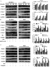
Fig. 5. Rk1 increased intracellular calcium levels to activate calpain-induced apoptosis in SK-MES-1 and H226 cells. (A) Cells were treated with the indicated concentrations of Rk1 for 24 h. The levels of calpain, cleaved caspase 12, 7 and cleaved PARP were determined via Western blotting. (B) BAPTA-AM pretreated cells using the above methods, and calpain, cleaved caspase 12, 7, 3, cleaved PARP and Bax were analyzed by Western blotting. (C) Calpeptin pretreated cells using the above methods, and calpain, cleaved caspase -12, -7, -3 and cleaved PARP were analyzed by Western blotting. Values are presented as the means ± SD, n = 3, *p < 0.05, **p < 0.01, ***p < 0.001 compared with the control, #p < 0.05, ##p < 0.01, ###p < 0.001 in contrast to the Rk1-treated group.
Fig. 6
Fig. 6. Rk1 induced mitochondria-mediated apoptosis via mitochondrial Ca2+ overload in SK-MES-1 and H226 cells. (A) Mitochondrial calcium levels were analyzed via flow cytometry after Rhod-2/AM staining. (B) MMP was assessed with the fluorescent mitochondrial probe JC-10, and the red/green fluorescence intensity was analyzed via flow cytometry. (C) SK-MES-1 cells were pretreated with 40 μM BAPTA-AM (intracellular Ca2+ chelating agent) for 2 h and then treated with 100 μM Rk1 for 24 h. MMP was assessed with the fluorescent mitochondrial probe JC-10, and the red/green fluorescence intensity was analyzed via fluorescence microscopy. Scale bars = 200 μm. Values are presented as the means ± SD, n = 3.
Fig. 7
Fig. 7. Rk1 significantly suppressed lung squamous cell carcinoma growth in vivo. SK-MES-1 lung squamous cell carcinoma xenografts were treated with solvent or Rk1 (10 and 20 mg kg−1 d−1) or with Gefitinib (20 mg kg−1 d−1). (A) Representative image of SK-MES-1 xenograft tumors from the control group, Rk1-treated groups and Gefitinib-treated group. (B) Tumor volume and (C) body weight were measured every three days. (D) H&E staining of tumor specimens and major organs. Scale bars = 200 μm.
Fig. 8
Fig. 8. Schematic representation of the hypothesized molecular mechanism underlying the anti-cancer activity of Rk1. The calcium signaling pathway was involved in Rk1-induced apoptosis.

Similar articles

Cited by

References

    1. Aggarwal A. Lewison G. Idir S. et al., The State of Lung Cancer Research: A Global Analysis. J. Thorac. Oncol. 2016;11:1040–1050. doi: 10.1016/j.jtho.2016.03.010. - DOI - PubMed
    1. Yan L. Yao Y. Wang L. H. Wang M. L. Fu X. H. Detection of CK19, LUNX, and KS1/4 mRNA expression in the peripheral blood for diagnosis of micrometastases in patients with non-small cell lung cancer and their clinical implications. Genet. Mol. Res. 2015;14:15090–15095. doi: 10.4238/2015.November.24.17. - DOI - PubMed
    1. Network T. C. G. A. R. Comprehensive genomic characterization of squamous cell lung cancers. Nature. 2012;517:576. doi: 10.1038/nature14129. - DOI - PMC - PubMed
    1. Scagliotti G. V., Novello S., Rapetti S. and Papotti M., Current State-of-the-Art Therapy for Advanced Squamous Cell Lung Cancer, American Society of Clinical Oncology Educational Book, 2013, vol. 354 - PubMed
    1. Sanders H. R. Albitar M. Somatic mutations of signaling genes in non-small-cell lung cancer. Cancer Genet. Cytogenet. 2010;203:7–15. doi: 10.1016/j.cancergencyto.2010.07.134. - DOI - PubMed