Lower Extremity Varicose Veins: An Unusual Presentation of Small Bowel Leiomyosarcoma
- PMID: 35528748
- PMCID: PMC9021616
- DOI: 10.1159/000520802
Lower Extremity Varicose Veins: An Unusual Presentation of Small Bowel Leiomyosarcoma
Abstract
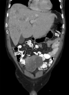
Leiomyosarcomas (LMSs) are extremely rare and comprise only 1.2% of small bowel malignancies. Advancements in immunohistochemical techniques have allowed for the differentiation between LMSs and gastrointestinal stromal tumors. LMSs remain difficult to detect via endoscopy and require a more intricate diagnostic approach. The staging and sizing of these tumors are important prognostic indicators. We report a case of a 67-year-old male who presented with bulging lower extremity veins, abdominal bloating, and weight loss. A CT of the abdomen and pelvis revealed a pelvic mass arising from the small bowel and a metastatic hepatic lesion, which was found to be compressing the inferior vena cava. A biopsy of the hepatic lesion confirmed the diagnosis of metastatic LMS.
Keywords: Gastrointestinal stromal tumor; Leiomyosarcoma; Small intestine; Stromal tumors.
Copyright © 2021 by S. Karger AG, Basel.
Conflict of interest statement
The authors have no conflicts of interest to declare.
Figures


References
-
- Jemal A, Siegel R, Ward E, Hao Y, Xu J, Murray T, et al. Cancer statistics, 2008. CA Cancer J Clin. 2008;58((2)):71–96. - PubMed
-
- Bilimoria KY, Bentrem DJ, Wayne JD, Ko CY, Bennett CL, Talamonti MS, et al. Small bowel cancer in the United States: changes in epidemiology, treatment, and survival over the last 20 years. Ann Surg. 2009;249:63–71. - PubMed
-
- Aggarwal G, Sharma S, Zheng M, Reid MD, Crosby JH, Chamberlain SM, et al. Primary leiomyosarcomas of the gastrointestinal tract in the post-gastrointestinal stromal tumor era. Ann Diagn Pathol. 2012;16((6)):532–540. - PubMed
-
- Akwari OE, Dozois RR, Weiland LH, Beahrs OH. Leiomyosarcoma of the small and large bowel. Cancer. 1978;42:1375–1384. - PubMed
-
- Evans HL. Smooth muscle tumors of the gastrointestinal tract: a study of 56 cases followed for a minimum of 10 years. Cancer. 1985;56:2242–2250. - PubMed
Publication types
LinkOut - more resources
Full Text Sources

