

Small-Intestinal Metastasis from Lung Carcinoma
- PMID: 35528768
- PMCID: PMC9035918
- DOI: 10.1159/000523663
Small-Intestinal Metastasis from Lung Carcinoma
Abstract
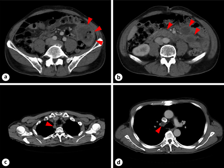
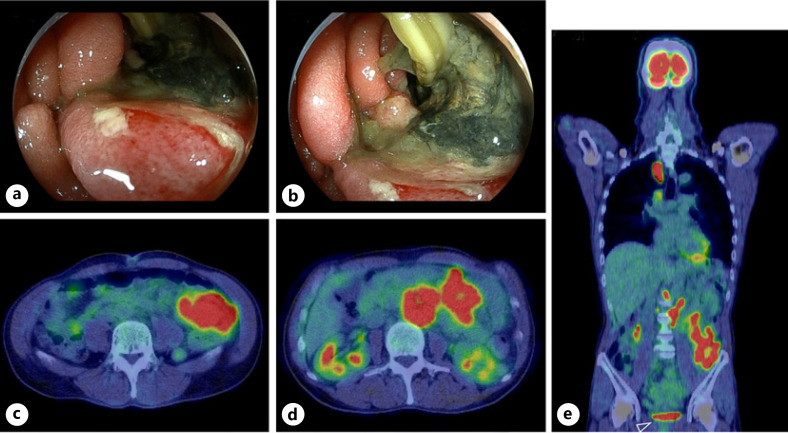
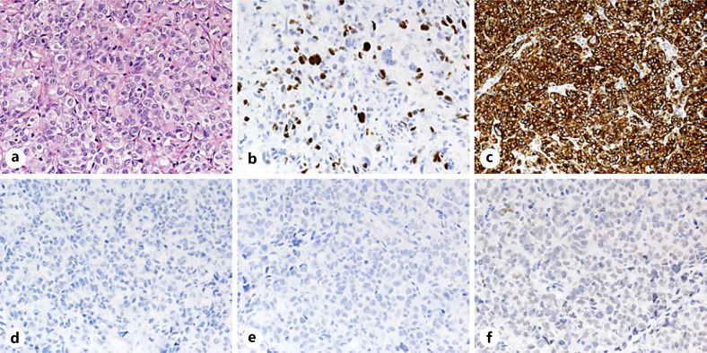
A 62-year-old man was referred to our hospital because of abdominal pain. Computed tomography revealed an approximately 7-cm-diameter tumor in the left abdomen with metastatic lymph nodes, an approximately 1-cm-diameter round tumor in contact with the subclavian artery in the apical lobe of the right lung, and mediastinal lymph node enlargement in contact with the superior vena cava. Esophagogastroduodenoscopy and colonoscopy revealed no abnormalities. Double-balloon endoscopy revealed a whole circumferential ulcer in the jejunum approximately 20 cm from the ligament of Treitz. Biopsy analysis of an ulcer specimen revealed a poorly differentiated carcinoma. Immunohistochemical staining of the specimen showed that it was positive for thyroid transcription factor 1 and cytokeratin 7 and negative for cytokeratin 20, GATA-binding protein 3, caudal-type homeobox protein 2, and paired box 8. Positron emission tomography revealed positive findings in the small-intestinal tumor, nearby mesenteric lymph nodes, lymph nodes around the abdominal aorta, lung tumor, and mediastinal lymph node in the apical lobe of the right lung. Accordingly, the patient was diagnosed as having a lung carcinoma with small-intestinal metastasis (T1b, N3, M1c; cStage IVB). Pathological examination helped distinguish the primary small-intestinal tumor from the metastatic small-intestinal tumor and detect the tumor origin.
Keywords: Immunohistochemistry; Lung cancer; Metastasis; Small-intestinal metastasis.
Copyright © 2022 by S. Karger AG, Basel.
Conflict of interest statement
The authors declare no conflicts of interest pertaining to this manuscript.
Figures
Similar articles
-
Pedunculated-type T1 colorectal carcinoma with lung carcinoma metastasis at the deepest invasive portion.Clin J Gastroenterol. 2016 Aug;9(4):208-14. doi: 10.1007/s12328-016-0659-2. Epub 2016 Jun 3. Clin J Gastroenterol. 2016. PMID: 27259703
-
[A case of cStage III B non-squamous non-small-cell lung cancer completely resected after downstaging with chemotherapy].Gan To Kagaku Ryoho. 2014 Nov;41(11):1429-32. Gan To Kagaku Ryoho. 2014. PMID: 25434449 Japanese.
-
Small Intestinal Metastasis from Esophageal Squamous Cell Carcinoma Presenting with Perforated Peritonitis.Tokai J Exp Clin Med. 2015 Jun 20;40(2):63-8. Tokai J Exp Clin Med. 2015. PMID: 26150186
-
Thyroid metastasis from small cell lung carcinoma: a case report and review of the literature.J Med Case Rep. 2015 Oct 7;9:231. doi: 10.1186/s13256-015-0707-4. J Med Case Rep. 2015. PMID: 26445938 Free PMC article. Review.
-
Primary pulmonary adenocarcinoma with intestinal differentiation mimicking metastatic colorectal carcinoma: case report and review of literature.Am J Clin Pathol. 2009 Jan;131(1):129-33. doi: 10.1309/AJCPB04XWICTFERL. Am J Clin Pathol. 2009. PMID: 19095576 Review.
Cited by
-
Case Report: Genetic profiling of small intestine metastasis from poorly differentiated non-small cell lung cancer: report of 2 cases and literature review of the past 5 years.Front Oncol. 2023 Nov 21;13:1265749. doi: 10.3389/fonc.2023.1265749. eCollection 2023. Front Oncol. 2023. PMID: 38074661 Free PMC article.
-
Intraluminal Small Bowel Metastasis From Primary Lung Cancer.World J Oncol. 2022 Dec;13(6):409-416. doi: 10.14740/wjon1532. Epub 2022 Dec 24. World J Oncol. 2022. PMID: 36660214 Free PMC article.
-
Obscure Bleeding from a Metastatic Small Bowel Tumor Diagnosed Using Motorized Spiral Enteroscopy: A Case Study and a Literature Review.Diagnostics (Basel). 2024 Apr 26;14(9):904. doi: 10.3390/diagnostics14090904. Diagnostics (Basel). 2024. PMID: 38732318 Free PMC article.
References
-
- Hoffman PC, Mauer AM, Vokes EE. Lung cancer. Lancet. 2000;355:479–85. - PubMed
-
- National Cancer Institute . Common toxicity criteria, version 2.0. National Cancer Institute; 1999. - PubMed
-
- Yang CJ, Hwang JJ, Kang WY, Chong IW, Wang TH, Sheu CC, et al. Gastro-intestinal metastasis of primary lung carcinoma: clinical presentations and outcome. Lung Cancer. 2006;54:319–23. - PubMed
-
- Yoshimoto A, Kasahara K, Kawashima A. Gastrointestinal metastases from primary lung cancer. Eur J Cancer. 2006;42:3157–60. - PubMed
Publication types
LinkOut - more resources
Full Text Sources
Research Materials
