. 2022 Feb 1:9:rbac001.
doi: 10.1093/rb/rbac001. eCollection 2022.

Preparation of multigradient hydroxyapatite scaffolds and evaluation of their osteoinduction properties

Affiliations

Preparation of multigradient hydroxyapatite scaffolds and evaluation of their osteoinduction properties

Hao Huang et al. Regen Biomater. .

Abstract

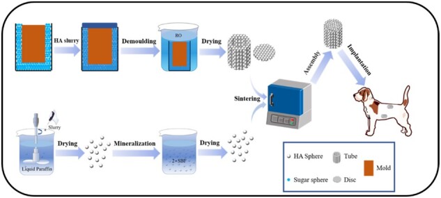
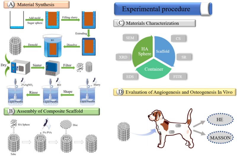
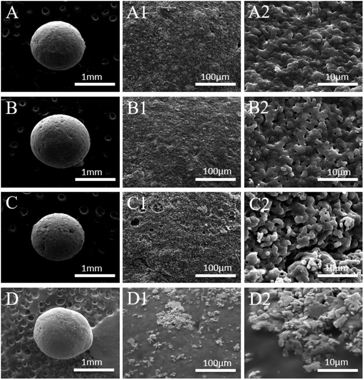
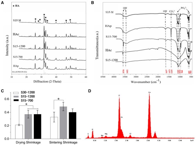
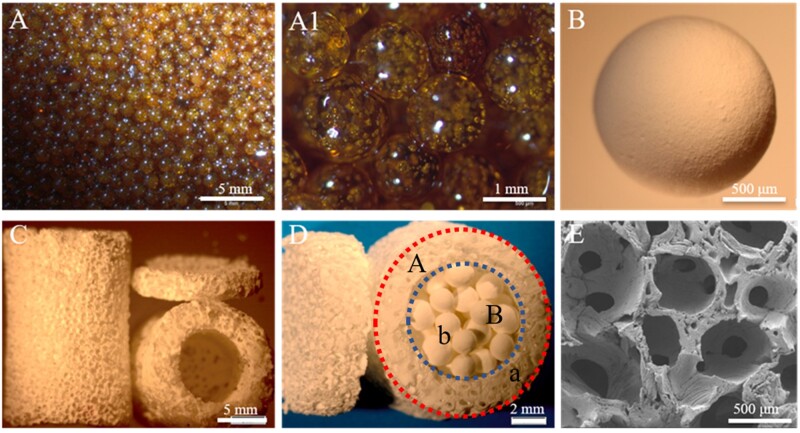
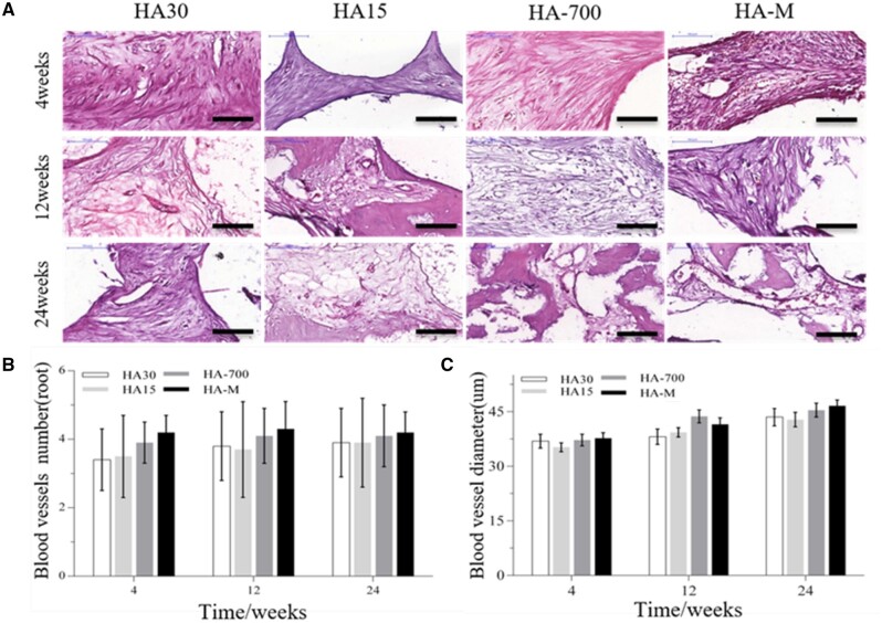
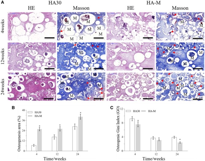
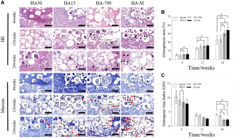
Porous hydroxyapatite (HA) scaffolds are often used as bone repair materials, owing to their good biocompatibility, osteoconductivity and low cost. Vascularization and osteoinductivity of porous HA scaffolds were limited in clinical application, and these disadvantages were need to be improved urgently. We used water-in-oil gelation and pore former methods to prepare HA spheres and a porous cylindrical HA container, respectively. The prepared HA spheres were filled in container to assemble into composite scaffold. By adjusting the solid content of the slurry (solid mixture of chitin sol and HA powder) and the sintering temperature, the porosity and crystallinity of the HA spheres could be significantly improved; and mineralization of the HA spheres significantly improved the biological activity of the composite scaffold. The multigradient (porosity, crystallinity and mineralization) scaffold (HA-700) filled with the mineralized HA spheres exhibited a lower compressive strength; however, in vivo results showed that their vascularization ability were higher than those of other groups, and their osteogenic Gini index (Go: an index of bone mass, and inversely proportional to bone mass) showed a continuous decrease with the implantation time. This study provides a new method to improve porous HA scaffolds and meet the demands of bone tissue engineering applications.

Keywords: biocompatibility; bone tissue engineering; osteoinduction; porous hydroxyapatite; scaffold.


Figures

None
Graphical abstract
Figure 1.
Figure 1.
Flow diagrams of experimental procedure
Figure 2.
Figure 2.
SEM images of HA spheres: S30-1200 (A, A1, A2), S15-1200 (B, B1, B2), S15-700 (C, C1, C2) and S15-M (D, D1, D2)
Figure 3.
Figure 3.
Cross-section SEM images of HA spheres: (A, A1) S30-1200, (B, B1) S15-1200 and (C, C1) S15-700
Figure 4.
Figure 4.
(A) XRD patterns and (B) FTIR spectra of HAp, HA spheres and HA container; (C) drying and sintering shrinkages of HA spheres; (D) surface elemental analysis of S15-M. *P <0.05 indicates statistically significant differences
Figure 5.
Figure 5.
Optical and SEM images of (A, A1) sucrose spheres, (B) HA spheres, (C, E) HA container and (D) composite scaffold
Figure 6.
Figure 6.
Masson’s trichrome and HE staining images of implant histological sections in abdominal cavity, showing scaffold changes and new bone formation at 4, 12 and 24 weeks post-operation; scale bars = 2 mm
Figure 7.
Figure 7.
Masson’s trichrome and HE staining images of implant histological sections in dorsal muscle, showing scaffold changes and new bone formation at 4, 12 and 24 weeks post-operation; scale bars = 2 mm
Figure 8.
Figure 8.
(A) HE staining images of blood vessels in scaffolds implanted in abdominal cavity at 4, 12 and 24 weeks (scale bars = 100 μm); (B) number of blood vessels; (C) diameter of blood vessels
Figure 9.
Figure 9.
(A) HE staining images of blood vessels in scaffolds implanted in back muscles at 4, 12 and 24 weeks (scale bars = 100 μm); (B) number of blood vessels; (C) diameter of blood vessels. *P <0.05 indicates statistically significant differences
Figure 10.
Figure 10.
Bone regeneration activity of scaffolds at 4, 12 and 24 weeks after implantation in abdominal cavity. (A) HE staining and Masson’s trichrome images (scale bars = 1 mm), red arrow: new bone, M: materials; (B) osteogenesis area; (C) osteogenic Gini index. *P <0.05 indicates statistically significant differences
Figure 11.
Figure 11.
Bone regeneration activity of scaffolds at 4, 12 and 24 weeks after implantation in dorsal muscles. (A) HE staining and Masson’s trichrome images (scale bars = 1 mm), black arrow: Havers systems, red arrow: new bone, M: materials; (B) osteogenesis area; (C) osteogenic Gini index. *P <0.05 indicates statistically significant differences

Similar articles

Cited by

References

    1. Lin S, Yang G, Jiang F, Zhou M, Yin S, Tang Y, Tang T, Zhang Z, Zhang W, Jiang X.. A magnesium-enriched 3D culture system that mimics the bone development microenvironment for vascularized bone regeneration. Adv Sci (Weinh) 2019;6:1900209. - PMC - PubMed
    1. O’Neill E, Awale G, Daneshmandi L, Umerah O, Lo KW-H.. The roles of ions on bone regeneration. Drug Discov Today 2018;23:879–90. - PubMed
    1. Habraken W, Habibovic P, Epple M, Bohner M.. Calcium phosphates in biomedical applications: materials for the future? Mater Today 2016;19:69–87.
    1. Nair MB, Suresh Babu S, Varma HK, John A.. A triphasic ceramic-coated porous hydroxyapatite for tissue engineering application. Acta Biomater 2008;4:173–81. - PubMed
    1. Wang Q, Yan J, Yang J, Li B.. Nanomaterials promise better bone repair. Mater Today 2016;19:451–63.