

A sustained release of BMP2 in urine-derived stem cells enhances the osteogenic differentiation and the potential of bone regeneration
- PMID: 35529046
- PMCID: PMC9070791
- DOI: 10.1093/rb/rbac015
A sustained release of BMP2 in urine-derived stem cells enhances the osteogenic differentiation and the potential of bone regeneration
Abstract
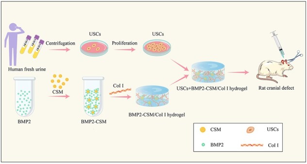
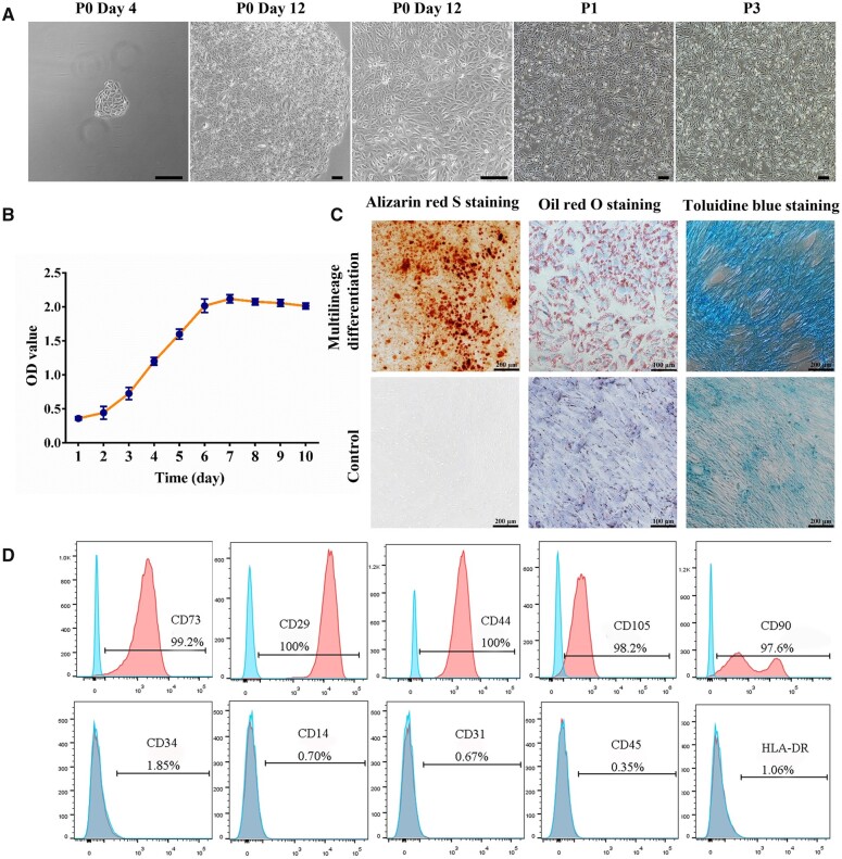
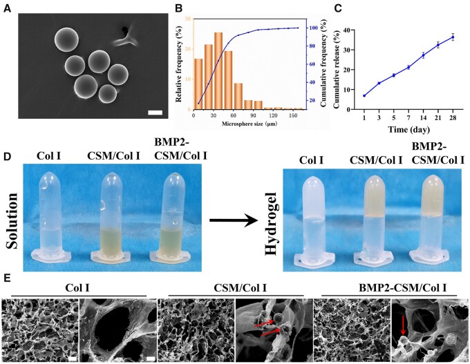
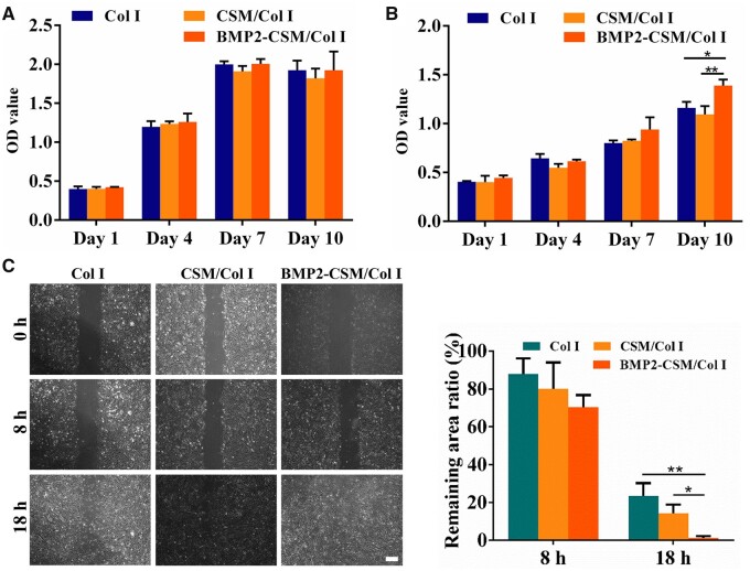
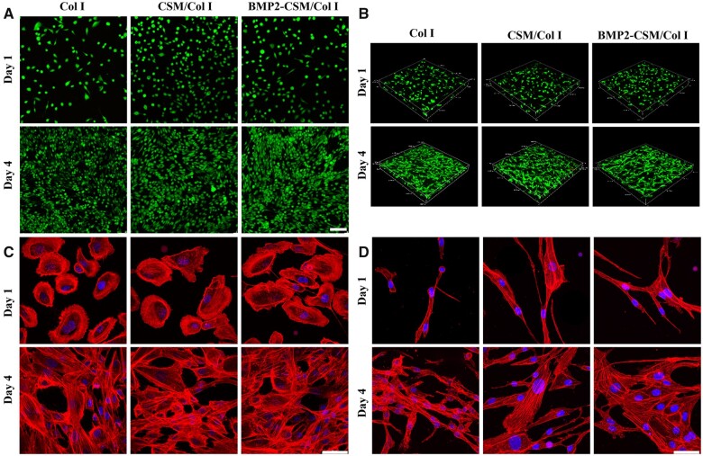
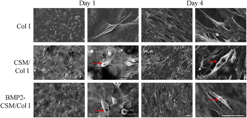
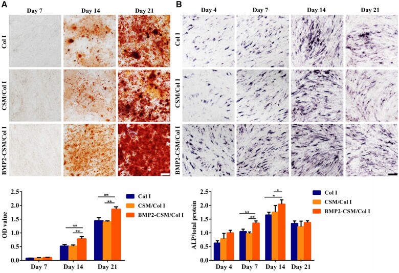
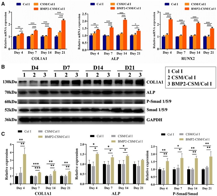
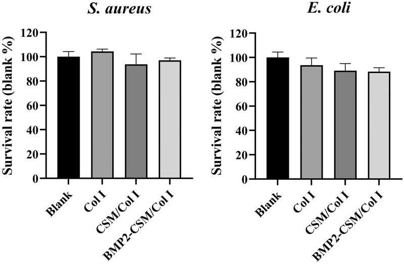
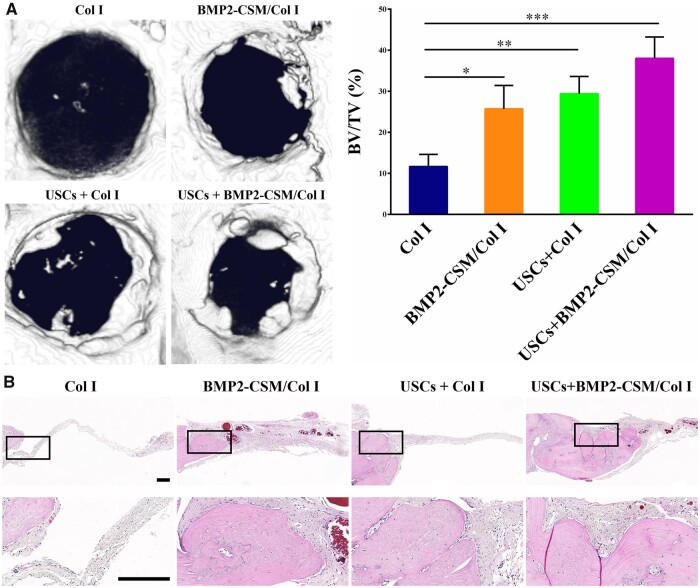
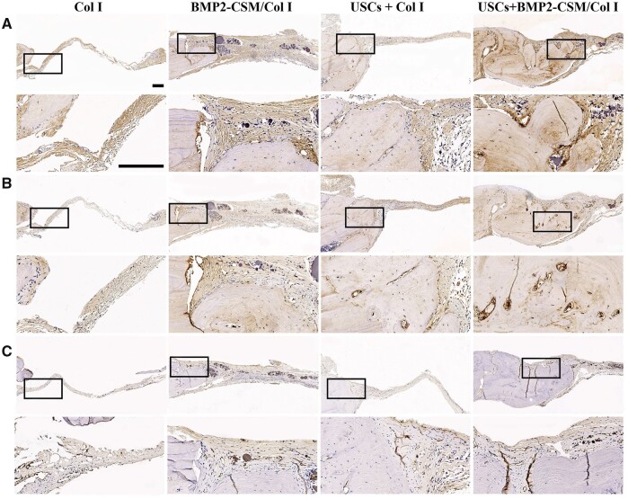
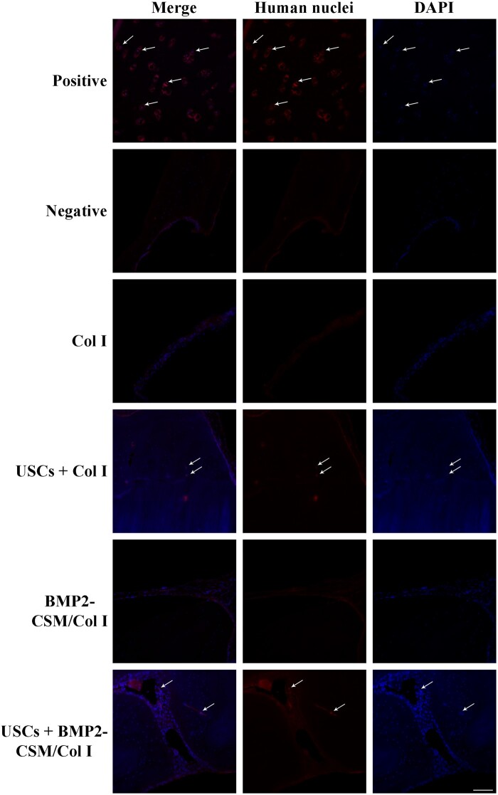
Cell-based tissue engineering is one of the optimistic approaches to replace current treatments for bone defects. Urine-derived stem cells (USCs) are obtained non-invasively and become one of the promising seed cells for bone regeneration. An injectable BMP2-releasing chitosan microspheres/type I collagen hydrogel (BMP2-CSM/Col I hydrogel) was fabricated. USCs proliferated in a time-dependent fashion, spread with good extension and interconnected with each other in different hydrogels both for 2D and 3D models. BMP2 was released in a sustained mode for more than 28 days. Sustained-released BMP2 increased the ALP activities and mineral depositions of USCs in 2D culture, and enhanced the expression of osteogenic genes and proteins in 3D culture. In vivo, the mixture of USCs and BMP2-CSM/Col I hydrogels effectively enhanced bone regeneration, and the ratio of new bone volume to total bone volume was 38% after 8 weeks of implantation. Our results suggested that BMP2-CSM/Col I hydrogels promoted osteogenic differentiation of USCs in 2D and 3D culture in vitro and USCs provided a promising cell source for bone tissue engineering in vivo. As such, USCs-seeded hydrogel scaffolds are regarded as an alternative approach in the repair of bone defects.
Keywords: BMP2; bone tissue engineering; chitosan microspheres; urine-derived stem cells.
© The Author(s) 2022. Published by Oxford University Press.
Figures
Similar articles
-
Bone morphogenetic protein 2 gene transduction enhances the osteogenic potential of human urine-derived stem cells.Stem Cell Res Ther. 2015 Jan 7;6(1):5. doi: 10.1186/scrt539. Stem Cell Res Ther. 2015. PMID: 25567327 Free PMC article.
-
Surface mineralized biphasic calcium phosphate ceramics loaded with urine-derived stem cells are effective in bone regeneration.J Orthop Surg Res. 2019 Dec 9;14(1):419. doi: 10.1186/s13018-019-1500-7. J Orthop Surg Res. 2019. PMID: 31818319 Free PMC article.
-
Human Urine Derived Stem Cells in Combination with β-TCP Can Be Applied for Bone Regeneration.PLoS One. 2015 May 13;10(5):e0125253. doi: 10.1371/journal.pone.0125253. eCollection 2015. PLoS One. 2015. PMID: 25970295 Free PMC article.
-
[The application of urine derived stem cells in regeneration of musculoskeletal system].Zhongguo Xiu Fu Chong Jian Wai Ke Za Zhi. 2018 Nov 15;32(11):1477-1482. doi: 10.7507/1002-1892.201804024. Zhongguo Xiu Fu Chong Jian Wai Ke Za Zhi. 2018. PMID: 30417628 Free PMC article. Review. Chinese.
-
Urine-derived stem cells: Promising advancements and applications in regenerative medicine and beyond.Heliyon. 2024 Mar 10;10(6):e27306. doi: 10.1016/j.heliyon.2024.e27306. eCollection 2024 Mar 30. Heliyon. 2024. PMID: 38509987 Free PMC article. Review.
Cited by
-
Hydrogel-Based Scaffolds: Advancing Bone Regeneration Through Tissue Engineering.Gels. 2025 Feb 27;11(3):175. doi: 10.3390/gels11030175. Gels. 2025. PMID: 40136878 Free PMC article. Review.
-
Recent advances in regenerative biomaterials.Regen Biomater. 2022 Dec 5;9:rbac098. doi: 10.1093/rb/rbac098. eCollection 2022. Regen Biomater. 2022. PMID: 36518879 Free PMC article. Review.
-
Nanotopographical 3D-Printed Poly(ε-caprolactone) Scaffolds Enhance Proliferation and Osteogenic Differentiation of Urine-Derived Stem Cells for Bone Regeneration.Pharmaceutics. 2022 Jul 8;14(7):1437. doi: 10.3390/pharmaceutics14071437. Pharmaceutics. 2022. PMID: 35890332 Free PMC article.
-
A construct of adipose-derived mesenchymal stem cells-laden collagen scaffold for fertility restoration by inhibiting fibrosis in a rat model of endometrial injury.Regen Biomater. 2023 Sep 7;10:rbad080. doi: 10.1093/rb/rbad080. eCollection 2023. Regen Biomater. 2023. PMID: 37808957 Free PMC article.
-
Stem Cells and Bone Tissue Engineering.Life (Basel). 2024 Feb 21;14(3):287. doi: 10.3390/life14030287. Life (Basel). 2024. PMID: 38541613 Free PMC article. Review.
References
-
- Raftery RM, Mencia Castano I, Chen G. et al. Translating the role of osteogenic-angiogenic coupling in bone formation: highly efficient chitosan-pDNA activated scaffolds can accelerate bone regeneration in critical-sized bone defects. Biomaterials 2017;149:116–27. - PubMed
-
- Diallo AM, Rota S, Boissiere M. et al. Osteoformation potential of an allogenic partially demineralized bone matrix in critical-size defects in the rat calvarium. Mater Sci Eng C Mater Biol Appl 2021;127:112207. - PubMed
LinkOut - more resources
Full Text Sources
Research Materials
