. 2022 Apr 30:2022:7169353.
doi: 10.1155/2022/7169353. eCollection 2022.

Role of Long Noncoding RNAs in Smoking-Induced Lung Cancer: An In Silico Study

Affiliations

Role of Long Noncoding RNAs in Smoking-Induced Lung Cancer: An In Silico Study

Lei Ge et al. Comput Math Methods Med. .

Abstract

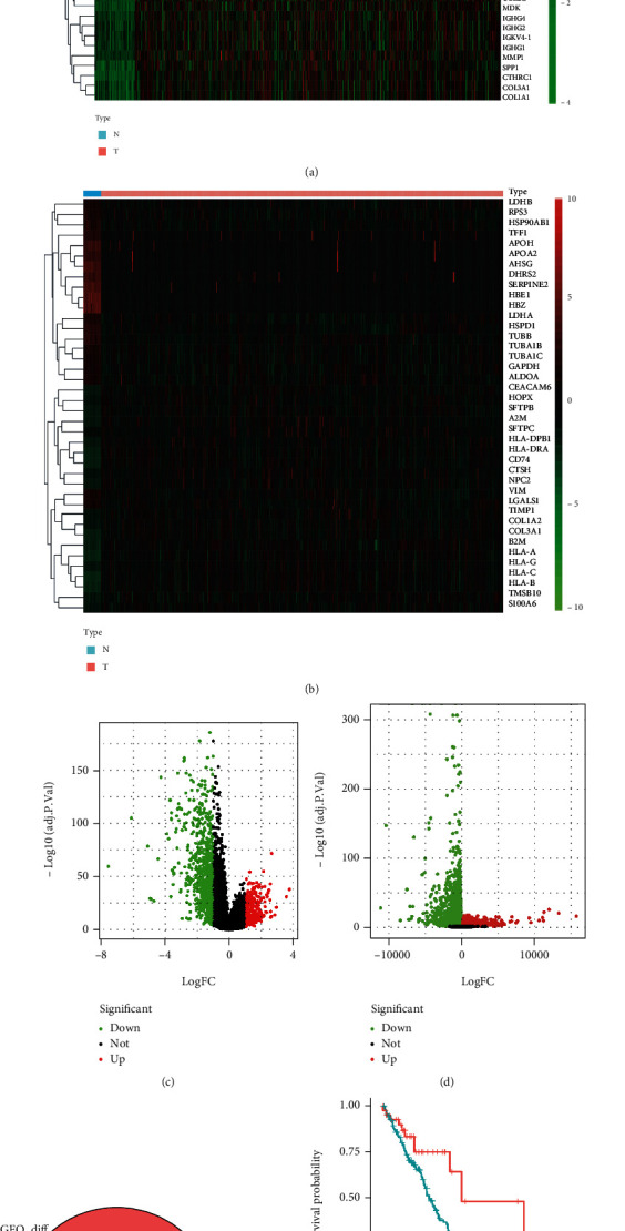
The prevalence of lung cancer induced by cigarette smoking has increased over time. Long noncoding (lnc) RNAs, regulatory factors that play a role in human diseases, are commonly dysregulated in lung cancer. Cigarette smoking is closely related to changes in lncRNA expression, which can affect lung cancer. Herein, we assess the mechanism of lung cancer initiation induced by smoking. To calculate the impact of smoking on the survival of patients with lung cancer, we extracted data from The Cancer Genome Atlas and Gene Expression Omnibus databases and identified the differentially expressed genes in the lung cancer tissue compared to the normal lung tissue. Genes positively and negatively associated with smoking were identified. Gene Ontology, Kyoto Encyclopedia of Genes and Genomes, and Cytoscape analyses were performed to determine the function of the genes and the effects of smoking on the immune microenvironment. lncRNAs corresponding to smoking-associated genes were identified, and a smoking-related lncRNA model was constructed using univariate and multivariate Cox analyses. This model was used to assess the survival of and potential risk in patients who smoked. During screening, 562 differentially expressed genes were identified, and we elucidated that smoking affected the survival of patients 4.5 years after the diagnosis of lung cancer. Furthermore, genes negatively associated with smoking were closely associated with immunity. Twelve immune cell types were also found to infiltrate differentially in smokers and nonsmokers. Thus, the smoking-associated lncRNA model is a good predictor of survival and risk in smokers and may be used as an independent prognostic factor for lung cancer.


Conflict of interest statement

The authors declare that there is no conflict of interest regarding the publication of this paper.

Figures

Figure 1
Figure 1
Differential expression of genes in lung cancer and effect of smoking on the prognosis of patients with lung cancer. (a) Genes obtained from The Cancer Genome Atlas (TCGA) dataset; blue denotes healthy subjects, pink represents patients with lung cancer, red represents positive association with high gene expression, and green represents negative association with low gene expression. The top 20 genes with the greatest positive and negative correlations were selected. (b) Genes obtained from the Gene Expression Omnibus (GEO) dataset, and others are similar to those in (a). (c) Volcano map of all differentially expressed genes identified through TCGA database; downregulated genes are indicated in green, and upregulated genes are indicated in red. (d) Differentially expressed genes identified in GEO. (e) The Venn diagram indicates the genes identified in TCGA and GEO databases. The differentially expressed genes are present in the intersection. Between the two databases, 562 differentially expressed genes were common. (f) Survival curve comparison for smoking and nonsmoking patients with lung cancer.
Figure 2
Figure 2
Effects of smoking on lung cancer. (a and b) The Cancer Genome Atlas (TCGA) and Gene Expression Omnibus (GEO) datasets; each tree fork represents a collection of genes for which the band is a series of modules. (c and d) Relationship between gene modules and traits in the GEO and TCGA datasets; pink represents positive and green denotes negative relation between traits and genes. The two databases contain 44 genes that are positively (e) and 80 genes that are negatively (f) associated with smoking.
Figure 3
Figure 3
Gene Ontology (GO) analysis for functional enrichment of genes positively (a) and negatively (c) associated with smoking. Kyoto Encyclopedia of Genes and Genomes (KEGG) pathway enrichment analysis for determining the function of genes positively (b) and negatively (d) associated with smoking. (e) Cytoscape analysis showing the major functions of genes negatively associated with smoking.
Figure 4
Figure 4
Effects of smoking on the immune microenvironment. (a) Infiltration rate of immune cells in the smoking and nonsmoking patient groups. (b) Relationship between different immune cell types. A pie diagram showing the positive and negative correlations in red and blue, respectively. (c) Relationship of different immune cell types observed in smoking and nonsmoking patients with lung cancer.
Figure 5
Figure 5
Construction of long noncoding (lnc) RNA models and analysis of their influence on smoking-induced lung cancer prognosis. (a) Cytoscape was used to associate the genes identified in the study with their subsequent expression of lncRNAs. Univariate Cox regression analysis was performed on the expression of lncRNAs for genes positively (b) and negatively (c) associated with smoking in The Cancer Genome Atlas (TCGA) and Gene Expression Omnibus (GEO) databases. Multivariate Cox regression analysis was performed on lncRNAs screened by a single factor positive (d) and negative (e) correlation in TCGA and GEO databases. Survival curves for high- and low-risk lncRNAs positively (f) and negatively (g) associated with smoking. p values < 0.001 were considered to indicate statistical significance.
Figure 6
Figure 6
Comparison and risk assessment of the smoking-associated lung cancer prognostic models. (a) Sankey diagram. Half are high risk, while half are low risk. There was higher survival than death in the low-risk sample. (b) Receiver operating characteristic curve analysis for the prognostic accuracy of the model. (c and d) Patient risk scores for positive and negative long noncoding (lnc) RNAs. (e and f) Survival rates in the high- and low-risk patient groups for positive and negative lncRNAs. (g and h) Heat maps of gene expression in the risk model for the high- and low-risk groups with positive and negative lncRNAs.
Figure 7
Figure 7
Relationship between the risk model and clinical factors. (a and b) Single factor prognostic analysis included age, sex, tumor-node-metastasis (TNM) stage, and the risk scores of patients with lung cancer with positive and negative long noncoding (lnc) RNAs. (c and d) Multifactor prognostic analysis included age, sex, TNM stage, and the risk scores of patients with lung cancer with positive and negative lncRNAs. (e and f) Pathways identified using the positive and negative lncRNA model.

Similar articles

Cited by

References

    1. Sung H., Ferlay J., Siegel R. L., et al. Global cancer statistics 2020: GLOBOCAN estimates of incidence and mortality worldwide for 36 cancers in 185 countries. CA: a Cancer Journal for Clinicians . 2021;71(3):209–249. doi: 10.3322/caac.21660. - DOI - PubMed
    1. Goldstraw P., Ball D., Jett J. R., et al. Non-small-cell lung cancer. Lancet . 2011;378(9804):1727–1740. doi: 10.1016/S0140-6736(10)62101-0. - DOI - PubMed
    1. Herbst R. S., Heymach J. V., Lippman S. M. Lung cancer. The New England Journal of Medicine . 2008;359(13):1367–1380. doi: 10.1056/NEJMra0802714. - DOI - PMC - PubMed
    1. Allemani C., Matsuda T., Di Carlo V., et al. Global surveillance of trends in cancer survival 2000-14 (CONCORD-3): analysis of individual records for 37 513 025 patients diagnosed with one of 18 cancers from 322 population-based registries in 71 countries. Lancet . 2018;391(10125):1023–1075. doi: 10.1016/S0140-6736(17)33326-3. - DOI - PMC - PubMed
    1. National Cancer Institute. SEER stat fact sheets: lung and bronchus cancer. 2018, http://seer.cancer.gov/statfacts/html/lungb.html.