Case Reports
. 2022 Mar 18;15(1):267-276.
doi: 10.1159/000522152. eCollection 2022 Jan-Apr.

Ewing Sarcoma and Primitive Neuroectodermal Tumor of the Thoracic Esophagus: Case Report and Comprehensive Literature Review

Affiliations
Case Reports

Ewing Sarcoma and Primitive Neuroectodermal Tumor of the Thoracic Esophagus: Case Report and Comprehensive Literature Review

Jie Li et al. Case Rep Oncol. .

Abstract

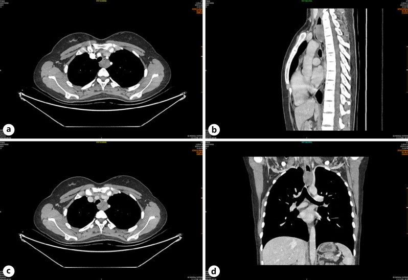
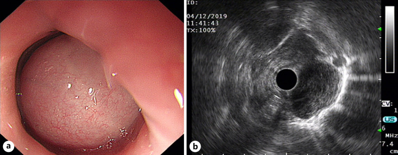
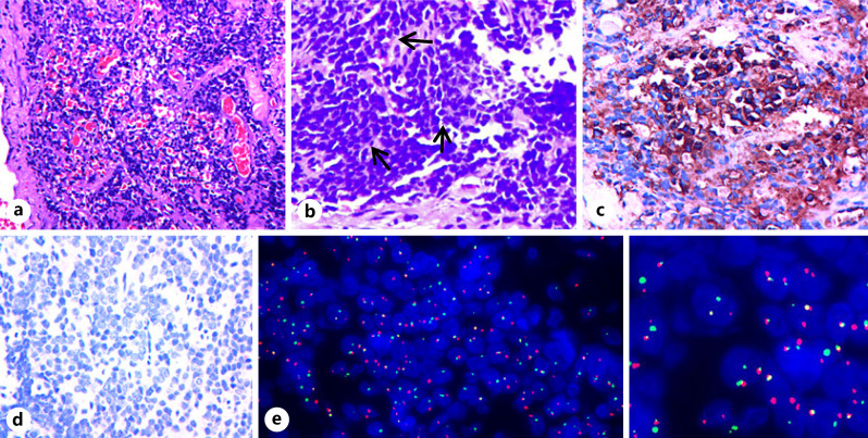
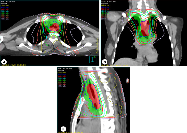
Ewing sarcoma and primitive neuroectodermal tumors (ES/PNETs) are rare tumors that belong to a family of round-cell neuroectodermally derived tumors, and their optimal treatment remains a great challenge. This study presented a case of ES/PNET, arising in the esophagus of a 21-year-old female patient presented with progressive dysphagia. Computed tomography and endoscopic ultrasonography showed a well-defined, submucosal solid mass in the superthoracic esophagus. The accurate diagnosis after surgery was obtained through immunohistochemistry and genetic studies, namely the CD99 immunopositivity as well as the EWSR1/FLI1 gene rearrangement associated with t(11;22)(q24;q12) in tumor cells. The patient underwent localized tumor resection followed by chemotherapy and chest radiotherapy. The patient is doing well with no evidence of tumor recurrence or metastasis 18 months after surgery. Although the esophagus is a rare site for ES/pPNET, we can speculate that the treatment protocol of ES/pPNET should include multi-agent chemotherapy, surgery, and local radiotherapy in order to improve the prognosis based on our report.

Keywords: Diagnosis; Esophagus; Extraosseous Ewing sarcoma; Primitive neuroectodermal tumor; Treatment.


Conflict of interest statement

The authors have no conflicts of interest.

Figures

Fig. 1
Fig. 1
Axial (a–c), coronal (c, d), and sagittal (b, d) CT showing a well-defined, spherical solid mass in the upper thoracic esophagus presenting with mild uniform enhancement, which caused proximal esophagus dilatation.
Fig. 2
Fig. 2
Endoscopic ultrasonography of the esophagus before operation. a Hemispherical protruding lesion in the upper thoracic esophagus with an intact mucosal surface, presented with the formation of mucosal bridges. b Lesion originated from the esophageal muscularis propria, and the adventitia is smooth and continuous.
Fig. 3
Fig. 3
The pathological and FISH results of pPNET of the upper thoracic esophagus. a H&E stain showed a uniform population of small round blue cells with markedly vesicular nuclei and finely dispersed chromatin, accompanied by some remarkable hemorrhage. b Homer-Wright rosettes (arrow) were seen on H&E. c, d Immunohistochemistry of CD99 (c) showed a diffuse strong membrane staining, but synaptophysin (d) was negative in the tumor cells. e FISH by the EWSR1/FLI1 Fusion Translocation t(11;22) Probe illustrating the EWSR1/FLI1 gene rearrangement associated with t(11;22)(q24;q12). Note: green signal (e) is GSP EWSR1, and red signal (e) is GSP FLI1 (original magnification ×20 (a), ×40 (b–d)). H&E, hematoxylin and eosin.
Fig. 4
Fig. 4
Computed dosimetric reconstruction images used for IMRT. a–c PGTVtb (red color wash image) and PTV (green color wash image) were administered 56 Gy (shown in blue isodose line) and 50.4 Gy (shown in red isodose line), respectively. Note: GTVtb refers to gross tumor volume of TB, and CTV refers to GTVtb with a radial margin of 0.5–1.0 cm and a longitudinal margin of 3 cm; CTV and GTVtb with a margin of 0.5 cm in three dimensions formed the PTV and PGTVtb. IMRT, intensity-modulated radiation therapy; PGTVtb, planning gross tumor volume of the tumor bed; PTV, planning target volume.

Similar articles

Cited by

References

    1. Kumar V, Singh A, Sharma V, Kumar M. Primary intracranial dural-based Ewing sarcoma/peripheral primitive neuroectodermal tumor mimicking a meningioma: a rare tumor with review of literature. Asian J Neurosurg. 2017 Jul–Sep;12((3)):351–7. - PMC - PubMed
    1. Malek A, Ziaee V, Kompani F, Moradinejad MH, Afzali N. Primitive neuroectodermal tumor, a rare cause of musculoskeletal manifestations in a child. Iran J Pediatr. 2014 Apr;24((2)):221–2. - PMC - PubMed
    1. Vogel H, Fuller GN. Primitive neuroectodermal tumors, embryonal tumors, and other small cell and poorly differentiated malignant neoplasms of the central and peripheral nervous systems. Ann Diagn Pathol. 2003 Dec;7((6)):387–98. - PubMed
    1. Batsakis JG, MacKay B, El-Naggar AK. Ewing's sarcoma and peripheral primitive neuroectodermal tumor: an interim report. Ann Otol Rhinol Laryngol. 1996 Oct;105((10)):838–43. - PubMed
    1. Yang MJ, Whelan R, Madden J, Mulcahy Levy JM, Kleinschmidt-DeMasters BK, Hankinson TC, et al. Intracranial Ewing sarcoma: four pediatric examples. Childs Nerv Syst. 2018 Mar;34((3)):441–8. - PMC - PubMed

Publication types