

Hybrid microfluidic sorting of rare cells based on high throughput inertial focusing and high accuracy acoustic manipulation
- PMID: 35529382
- PMCID: PMC9072550
- DOI: 10.1039/c9ra01792e
Hybrid microfluidic sorting of rare cells based on high throughput inertial focusing and high accuracy acoustic manipulation
Abstract
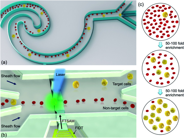
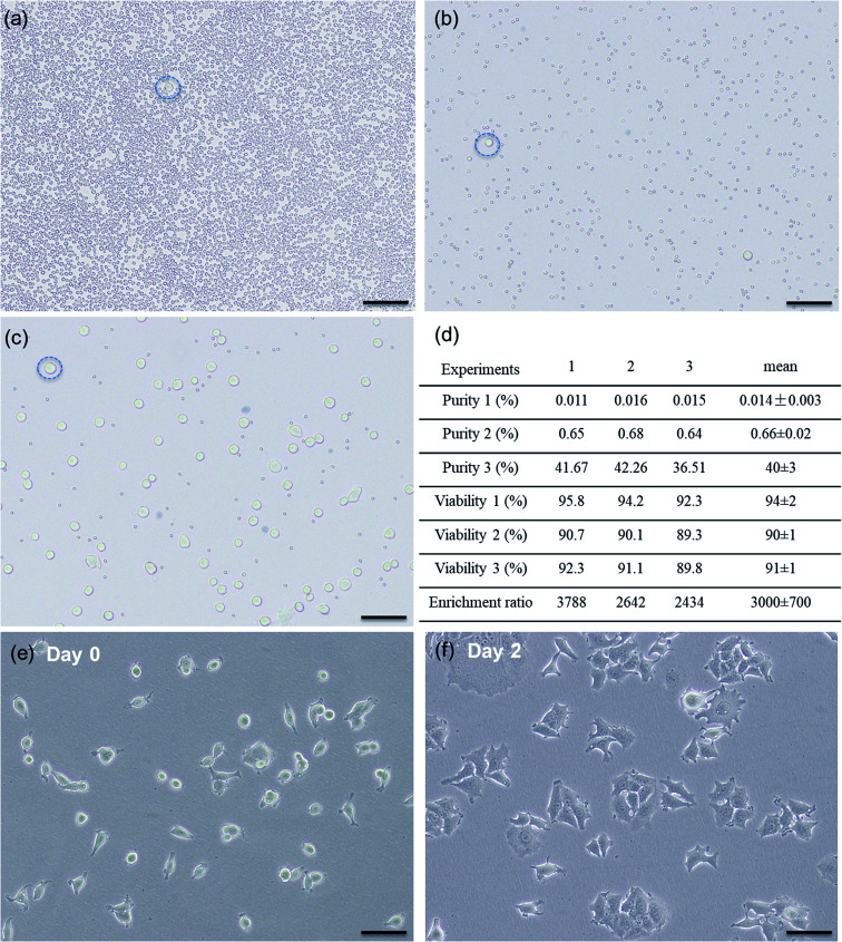
The ability to isolate rare circulating tumor cells (CTCs) from blood samples is essential to perform liquid biopsy as a routine diagnostic and prognostic test. Both label-free and surface biomarker-based cell sorting technologies have been developed to address the demand in high-integrity isolation of rare CTCs for cancer research. Label-free cell sorting mainly relies on the size difference between CTCs and blood cells; thus, it lacks sufficient sorting specificity. Surface biomarker-based cell sorting is highly specific; however, it requires expensive, labor-intensive, and time-consuming labeling due to the use of multiple sets of surface biomarkers. Because of the complex nature and high heterogeneity of tumorigenesis, it is difficult to rely on a single sorting process for high-integrity rare cell isolation. In this study, for the first time, we present a hybrid microfluidic cell sorting method combining high throughput size-dependent inertial focusing for size-based pre-enrichment and high accuracy fluorescence activated acoustic sorting for single cell isolation. After one single hybrid sorting process, we have demonstrated at least 2500-fold purity enrichment of MCF-7 breast cancer cells spiked in diluted whole blood samples with cell viability maintained at 91 ± 1% (viability before sorting was 94 ± 2%). This developed hybrid microfluidic cell sorting technique provides a promising solution for rare cell isolation needed in a variety of biological research and clinical applications.
This journal is © The Royal Society of Chemistry.
Conflict of interest statement
There are no conflicts to declare.
Figures
Similar articles
-
High-Throughput Isolation of Circulating Tumor Cells Using Cascaded Inertial Focusing Microfluidic Channel.Anal Chem. 2018 Apr 3;90(7):4397-4405. doi: 10.1021/acs.analchem.7b04210. Epub 2018 Mar 20. Anal Chem. 2018. PMID: 29537252
-
An integrated enrichment system to facilitate isolation and molecular characterization of single cancer cells from whole blood.Cytometry A. 2018 Dec;93(12):1226-1233. doi: 10.1002/cyto.a.23599. Cytometry A. 2018. PMID: 30549400 Free PMC article.
-
Acoustic separation of circulating tumor cells.Proc Natl Acad Sci U S A. 2015 Apr 21;112(16):4970-5. doi: 10.1073/pnas.1504484112. Epub 2015 Apr 6. Proc Natl Acad Sci U S A. 2015. PMID: 25848039 Free PMC article.
-
Developments in label-free microfluidic methods for single-cell analysis and sorting.Wiley Interdiscip Rev Nanomed Nanobiotechnol. 2019 Jan;11(1):e1529. doi: 10.1002/wnan.1529. Epub 2018 Apr 24. Wiley Interdiscip Rev Nanomed Nanobiotechnol. 2019. PMID: 29687965 Free PMC article. Review.
-
Microfluidics for label-free sorting of rare circulating tumor cells.Analyst. 2020 Nov 9;145(22):7103-7124. doi: 10.1039/d0an01148g. Analyst. 2020. PMID: 33001061 Review.
Cited by
-
Advances in single-cell metabolomics to unravel cellular heterogeneity in plant biology.Plant Physiol. 2023 Sep 22;193(2):949-965. doi: 10.1093/plphys/kiad357. Plant Physiol. 2023. PMID: 37338502 Free PMC article. Review.
-
Recent advances in acoustofluidic separation technology in biology.Microsyst Nanoeng. 2022 Sep 1;8:94. doi: 10.1038/s41378-022-00435-6. eCollection 2022. Microsyst Nanoeng. 2022. PMID: 36060525 Free PMC article. Review.
-
Design of a Hybrid Inertial and Magnetophoretic Microfluidic Device for CTCs Separation from Blood.Micromachines (Basel). 2021 Jul 26;12(8):877. doi: 10.3390/mi12080877. Micromachines (Basel). 2021. PMID: 34442499 Free PMC article.
-
Enhancing cell characterization with microfluidics and AI: a comprehensive review of mechanical, electrical, and hybrid techniques.Biotechnol Rep (Amst). 2025 Jul 22;47:e00905. doi: 10.1016/j.btre.2025.e00905. eCollection 2025 Sep. Biotechnol Rep (Amst). 2025. PMID: 40735651 Free PMC article. Review.
-
Manipulation with sound and vibration: A review on the micromanipulation system based on sub-MHz acoustic waves.Ultrason Sonochem. 2023 Jun;96:106441. doi: 10.1016/j.ultsonch.2023.106441. Epub 2023 May 13. Ultrason Sonochem. 2023. PMID: 37216791 Free PMC article. Review.
References
-
- Ashworth T. R. A case of cancer in which cell similar to those in the tumours were seen in the blood after death. Med. J. Aust. 1869;14:146–149.
LinkOut - more resources
Full Text Sources
Other Literature Sources