. 2022 May 3:10:e13319.
doi: 10.7717/peerj.13319. eCollection 2022.

Glucocorticoids induce osteonecrosis of the femoral head in rats via PI3K/AKT/FOXO1 signaling pathway

Affiliations

Glucocorticoids induce osteonecrosis of the femoral head in rats via PI3K/AKT/FOXO1 signaling pathway

Fei Sun et al. PeerJ. .

Abstract

Background: Steroid-induced osteonecrosis of the femoral head (SONFH) is a disorder that causes severe disability in patients and has a high incidence worldwide. Although glucocorticoid (GC)-induced apoptosis of osteoblasts is an important cytological basis of SONFH, the detailed mechanism underlying SONFH pathogenesis remains elusive. PI3K/AKT signaling pathway was reported to involve in cell survival and apoptosis.

Objective: We explored the role of PI3K/AKT/FOXO1 signaling pathway and its downstream targets during glucocorticoid -induced osteonecrosis of the femoral head.

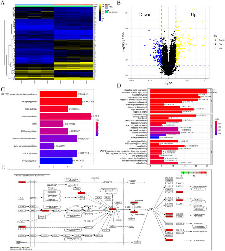
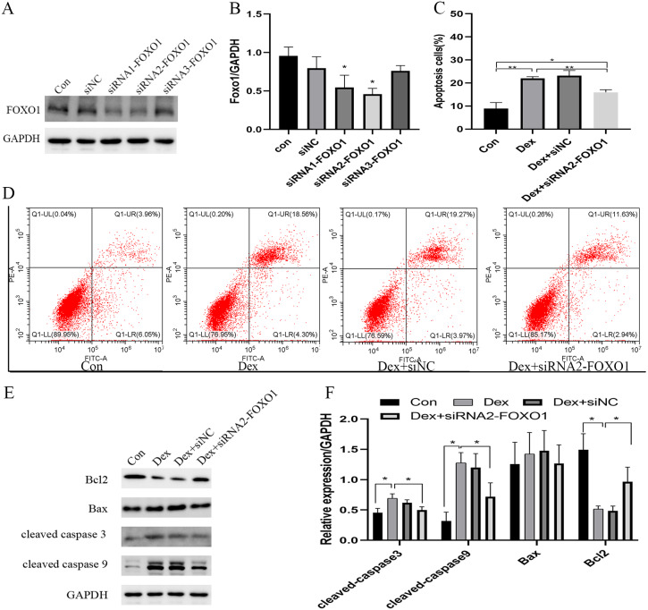
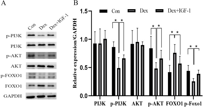
Methods: We obtained gene expression profile of osteoblasts subjected to dexamethasone (Dex) treatment from the Gene Expression Omnibus (GEO) database. Differentially expressed genes (DEGs) were screened out and functional enrichment analysis were conducted by bioinformatics analysis. In vitro, we analyzed Dex-induced apoptosis in MC3T3-E1 cells and explored the role of PI3K/AKT/FOXO1 signaling pathway in this phenomenon by employing siRNA-FOXO1 and IGF-1(PI3K/AKT agonist). Finally, we verified our results in a rat model of SONFH.

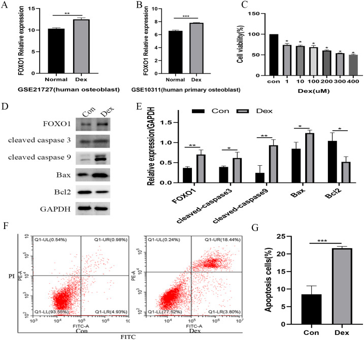
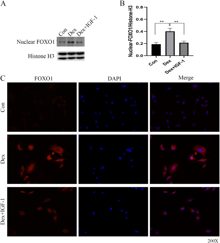
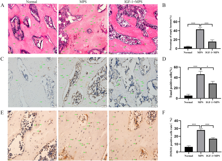
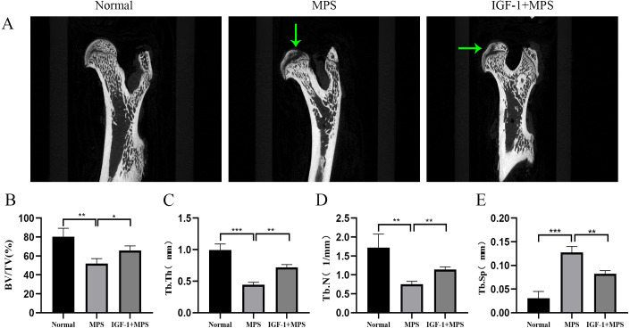
Results: In Dex-treated osteoblasts, DEGs were mainly enriched in the FOXO signaling pathway. Dex inhibited MC3T3-E1 cell viability in a dose-dependent effect and induced apoptosis by increasing the expression levels of FOXO1, Bax, cleaved-Caspase-3, and cleaved-Caspase-9, while reducing the expression of Bcl-2. Notably, these results were reversed by siRNA-FOXO1 treatment. Dex inhibited PI3K/AKT signaling pathway, upregulated FOXO1 expression and increased FOXO1 nuclear translocation, which were reversed by IGF-1. Compared to normal rats, the femoral head of SONFH showed increased expression of FOXO1, increased number of apoptotic cells, and empty osteocytic lacunas, as well as decreased bone tissue content and femoral head integrity. Significantly, the effects of GC-induced SONFH were alleviated following IGF-1 treatment.

Conclusion: Dex induces osteoblast apoptosis via the PI3K/AKT/FOXO1 signaling pathway. Our research offers new insights into the underlying molecular mechanisms of glucocorticoid-induced osteonecrosis in SONFH and proposes FOXO1 as a therapeutic target for this disease.

Keywords: Apoptosis; Dexamethasone; FOXO1; Glucocorticoids; Osteoblast; PI3K/AKT; SONFH.


Conflict of interest statement

The authors declare there are no competing interests.

Figures

Figure 1
Figure 1. Identification of DEGs caused by Dex from GSE21727.
(A) Volcano-plot visualized the DEGs with cut-off criterion as —logFC— >1 and adjusted P < 0.05 (red dots means upregulated genes and green dots means downregulated genes). (B) A heatmap of DEGs clustering relationship between control and Dex-treated groups. (C–D) GO and KEGG functional enrichment analysis of DEGs. (E) The function enrichment of the FOXO signaling pathway.
Figure 2
Figure 2. Evaluate the effect of Dex on MC3T3-E1 cells apoptosis.
(A–B) The relative expression level of FOXO1 in GSE21727 and GSE10311 from GEO database. (C) Cell viability of MC3T3-E1 after Dex treatment for 48 h. (D–E) Results of western blot for the protein levels of FOXO1, cleaved-Caspase 3, cleaved-Caspase 9, Bax and Bcl-2 after different treatment. (F–G) Flow cytometric analysis of apoptosis rates of MC3T3-E1 cells upon Dex treatment. Con: control group. All data in vivo and in vitro experiments are triplicate biological replicates. * P < 0.05, ** P < 0.01, *** P < 0.0001.
Figure 3
Figure 3. Effect of siRNA-mediated knockdown of Foxo1 on Dex-induced apoptosis.
(A–B) MC3T3-E1 cells were transfected with Foxo1 siRNA, and western blotting was performed to analyze the protein levels of FOXO1. (C–D) MC3T3-E1 cell apoptosis after 48 h Dex and siRNA-FOXO1 treatment as determined using FITC-PI/Annexin V staining. (E–F) Protein expression levels of Bax, cleaved-Caspase-3, and Bcl-2 after Dex and siRNA-FOXO1 treatment. GAPDH was used as a loading control. Con: control group. All data in vivo and in vitro experiments are triplicate biological replicates. * P < 0.05, ** P < 0.01, *** P < 0.0001.
Figure 4
Figure 4. Dex inhibited the activities of PI3K/AKT signaling pathways in MC3T3-E1 cells.
(A–B) Western blotting was performed to analyze the expression levels of PI3K, p-PI3K, AKT, p-AKT, FOXO1, and p-FOXO1 in MC3T3-E1 cells after Dex treatment (200 µM, 48 h). Con: control group. All data in vivo and in vitro experiments are triplicate biological replicates. * P < 0.05, ** P < 0.01, *** P < 0.0001.
Figure 5
Figure 5. Immunofluorescence studies showing the nuclear translocation of FOXO1.
(A–B) Western blotting indicated the expression of FOXO1 in the nucleus after Dex treatment. (C) The distribution and expression of FOXO1 protein in the MC3T3-E1 treated with Dex were evaluated by immunofluorescence. The FOXO1 was stained as red granular dots, while the nucleus was stained with blue. Con: control group. All data in vivo and in vitro experiments are triplicate biological replicates. * P < 0.05, ** P < 0.01, *** P < 0.0001.
Figure 6
Figure 6. Evaluation of osteonecrosis and apoptosis in animal module.
(A–B) HE staining of femoral heads of rats in the Normal, MPS, and IGF-1+ MPS group; green arrows indicate empty osteocytic lacunas. (C–D) Tunel staining to detect apoptosis cells; green arrows indicate positive apoptosis cells. (E–F) Immunohistochemistry to evaluate the expression of FOXO1 in femoral heads of rats; green arrows indicate FOXO1-positive cells that are stained as brown dots. * P < 0.05, ** P < 0.01, *** P < 0.0001.
Figure 7
Figure 7. Micro-CT for quantitative evaluation of bone microstructure.
(A) Micro-CT scaning for the femoral heads of rats in the Normal, MPS, and IGF-1+ MPS group. green arrows indicate cavity under subchondral bone in the femoral heads of rats. (B–E) Quantitative evaluation of BV/TV(B), Tb.Th(C), Tb.N(D), and Tb.Sp(E) in the three groups. * P < 0.05, ** P < 0.01, *** P < 0.0001.

Similar articles

Cited by

References

    1. Al-Omari AA, Aleshawi AJ, Marei OA, Younes HMB, Alawneh KZ, Quran EAL, Mohaidat ZM. A vascular necrosis of the femoral head after single steroid intra-articular injection. European Journal of Orthopaedic Surgery & Traumatology. 2020;30(2):193–197. doi: 10.1007/s00590-019-02555-8. - DOI - PubMed
    1. An YM, Li YJ, Zhang CL, Cong X, Gao YS, Wu LL, Dou D. Decreased PKG transcription mediated by PI3K/Akt/FoxO1 pathway is involved in the development of nitroglycerin tolerance. Biochemical and Biophysical Research Communications. 2019;508(4):1195–1201. doi: 10.1016/j.bbrc.2018.12.065. - DOI - PubMed
    1. Arbab D, Konig DP. Atraumatic femoral head necrosis in adults. Deutsches Ärzteblatt International. 2016;113(3):31–38. - PMC - PubMed
    1. Caplan A, Fett N, Rosenbach M, Werth VP, Micheletti RG. Prevention and management of glucocorticoid-induced side effects: a comprehensive review: a review of glucocorticoid pharmacology and bone health. Journal of the American Academy of Dermatology. 2017;76(1):1–9. doi: 10.1016/j.jaad.2016.01.062. - DOI - PubMed
    1. Chang C, Greenspan A, Gershwin ME. The pathogenesis, diagnosis and clinical manifestations of steroid-induced osteonecrosis. Journal of Autoimmunity. 2020;110:102460. doi: 10.1016/j.jaut.2020.102460. - DOI - PubMed

Publication types

MeSH terms